
Scalopidia spinosipes, 1858
|
publication ID |
https://doi.org/ 10.11646/zootaxa.3731.1.2 |
|
publication LSID |
lsid:zoobank.org:pub:ECD3423E-FB05-4783-A08A-64EF16C1A57F |
|
DOI |
https://doi.org/10.5281/zenodo.6152191 |
|
persistent identifier |
https://treatment.plazi.org/id/F5548796-1D4C-D86E-59BA-B951FF73D3F7 |
|
treatment provided by |
Plazi |
|
scientific name |
Scalopidia spinosipes, 1858 |
| status |
|
Scalopidia spinosipes, 1858 View in CoL
( Figs. 1 View FIGURE 1 , 2 View FIGURE 2 A–D, 3, 5A, 6A, 7A–D, 8A, B, 9A, B, 10A, B, 11A, B, 12A–D, 14A)
Scalopidia spinosipes Stimpson 1858: 95 .—Rathbun 1910: 344, pl. 2 fig. 2.—Tesch 1918: 224, pl. 14 fig. 3.—964: 235, fig. 14; 1968: 92.—Evans 1967: 407.—Fang 1991: 352.—Huang 1994: 592.—Ng et al. 2001: 74, pl. 7f. —Ng et al. 2008: 85.— Ya ng et al. 2008: 771.—Guinot et al. 2013: 118, 294 (part).
Hypophthalmus leucochirus Richters , in Lenz & Richters, 1881: 429, pls. 1–10.
Scalopidia leucochirus .—Serène 1964: 235.
Scalopidia leuchochirus [sic].—Serène 1968: 92.—Ng et al. 2008: 85.
Material examined. Hong Kong: lectotype male (here designated) (9.3× 12.1 mm), paralectotype female (13.3× 17.7 mm) (Stimpson Collection, NHM 61.44).— 1 male (14.8× 19.2 mm) (ZRC 2006.0073), among shellfish, Tai Po Wet market, Hong Kong, coll. P.K.L. Ng, 2006. Singapore: 1 male (ZRC 1989.3671), off Bedok, in mud, dredge, coll. P.K.L. Ng, 27 October 1982.— 1 male (12.2× 16.7 mm) (ZRC 2000.1604), Southern Islands, dredge, coll. D. Lane, 1992.— 1 female (ZRC 2000.1455), off Tanah Merah Jetty, 26 m, 118’13.8” N 10359 View Materials ’46.9”E, coll. L.W. Loh et al., 29 August 1997. Indonesia: 1 male (16.1× 11.8 mm) (ZRC 1969.10.13.2), coll. RV Djalanidhi, R. Serène, 6 October 1967. South China Sea: 1 male (ZRC 1985.1627), near Horsburgh Lighthouse, South China Sea, coll. H. Huat, June 1983.— 6 males (largest 14.8× 20.4 mm), 11 females (largest 14.2× 19.2 mm), 1 juvenile (ZRC 2000.1343), about 30 miles from Horsburgh Lighthouse, South China Sea, coll. H. Huat, 10 September 1983. Gulf of Thailand: 3 males, 1 female (ZRC 2013.1414), Sri Racha Fish Port, 20 m, near Chonburi, Thailand, coll. trawlers, J. Lai & N.K. Ng, 17 March 2005.— 1 female (ZRC 2008. 0190), Sri Racha Fish Port, 20 m, near Chonburi, Thailand, coll. trawlers, N.K. Ng et al., 18 March 2005.— 2 males (larger 19.1× 26.3 mm) (ZRC 2013.1413), Sri Racha Fish Port, 20 m, near Chonburi, Thailand, coll. trawlers, J. Lai & N.K. Ng, 19 March 2005.— 4 males, 1 female (ZRC 1997.672), Chonburi Port, 20 m, Thailand, coll. trawlers, P. Naiyanetr, 1996. — 1 male (ZRC 2000.23), Angsila Port, near Chonburi, 20 m, Thailand, coll. trawlers, P.K.L. Ng, November 1999.— 1 male (ZRC 2013.1410), Angsila Port, near Chonburi, 20 m, Thailand, coll. trawlers, P.K.L. Ng, 20 February 2000.— 4 males, 2 ovigerous females, 3 females (ZRC 2000.0886), Angsila Port, near Chonburi, 20 m, Thailand, coll. trawlers, P.K.L. Ng, 20 February 2000. Vietnam: 1 male (17.8×13.0 mm) (ZRC 1969.11.25.1), coll. Fisheries UNDP Vietnam, R. Serène, 25 November 1969. Philippines: 1 juvenile male (ZRC 2013.1423), station D10, Bohol Sea, coll. PANGLAO 2004 Expedition, 20 June 2004.— 1 juvenile female (ZRC 2013.1424), station S25, Bohol Sea, coll. PANGLAO 2004 Expedition, 23 June 2004.— 1 female (12.5× 9.2 mm) (ZRC 2013.1425), station T23, Bohol Sea, coll. PANGLAO 2004 Expedition, 21 June 2004.
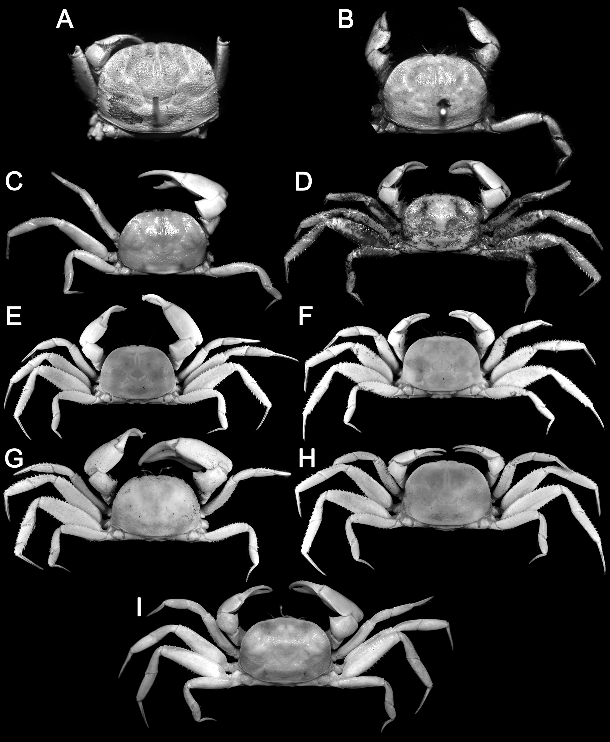
Diagnosis. Carapace subovate, lateral margins convex ( Fig. 2 View FIGURE 2 A–D); dorsal carapace surface uneven with regions of different convexities ( Figs. 2 View FIGURE 2 A–D, 5A); cornea with pigmentation in centre ( Figs. 3 View FIGURE 3 B, 5A); cheliped carpus with long sharp tooth on inner dorsal angle, distal margin very finely granular or almost smooth; P2 merus dorsal margin with sharp granules and/or short spines, ventral margin with row of sharp spines ( Figs. 2 View FIGURE 2 C, D, 3A); P2 carpus unarmed ( Figs. 2 View FIGURE 2 C, D, 3A); P2 propodus with row of spines on dorsal margin ( Figs. 2 View FIGURE 2 C, D, 3A); P3, P4 merus dorsal margins with strong spines, ventral margin with 2 rows of sharp spines ( Figs. 2 View FIGURE 2 C, D, 3A); P3, P4 carpus each with rows of strong spines on dorsal margin, with low subdorsal ridge; P3, P4 propodus each with strong spines on dorsal, ventral margins ( Figs. 2 View FIGURE 2 C, D, 3A); P5 merus with strong spines on dorsal margin, ventral margin with numerous strong spines on proximal half ( Figs. 2 View FIGURE 2 B–D, D, 3A, 8A, B); surface of thoracic sternum mostly smooth or pitted, surface near sterno-abdominal cavity with low, flattened granules ( Fig. 9 View FIGURE 9 A, B); episternite 7 as tranversely elongated subrectangular plate ( Fig. 9 View FIGURE 9 B); male abdominal somite 6 as wide as long ( Figs. 9 View FIGURE 9 A, 10A, B); G1 distal part elongated, tip prominently open distally, slightly flared ( Fig. 12 View FIGURE 12 A-C).
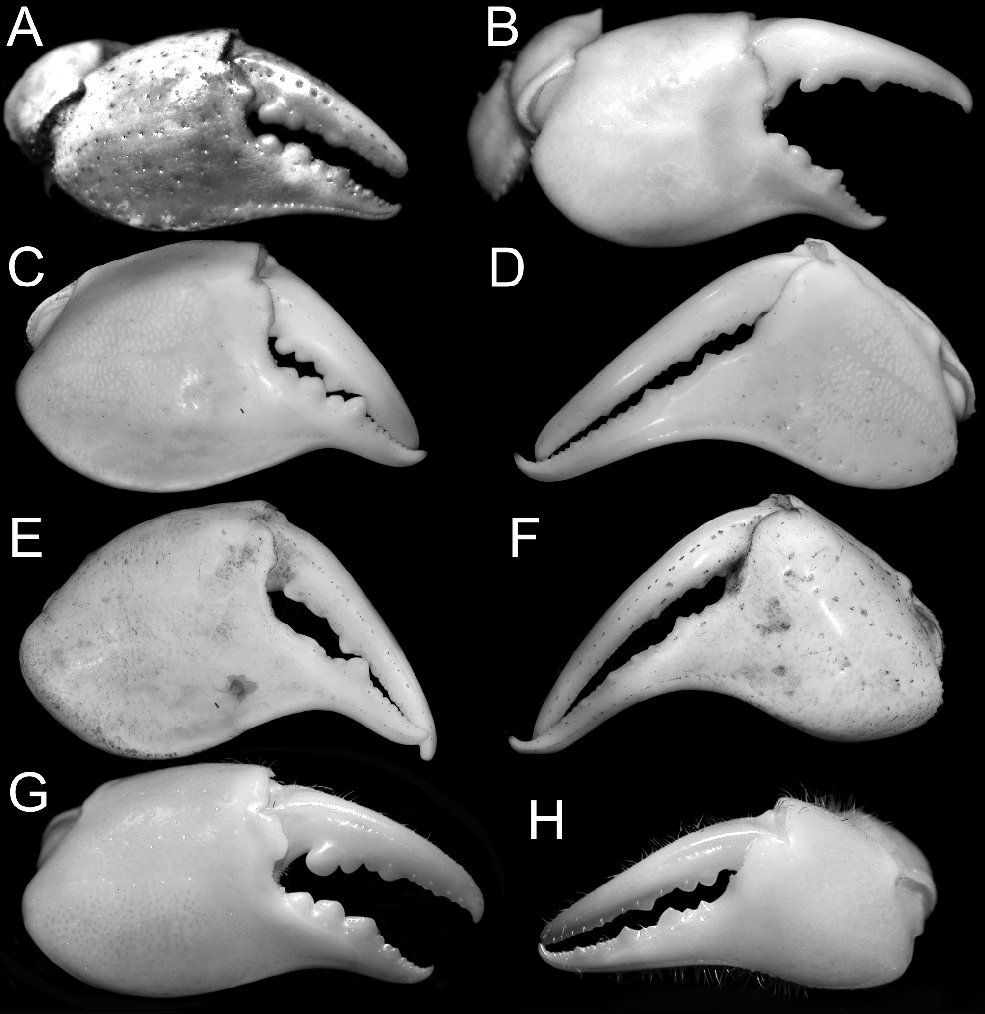
Variation. Females are similar to males in non-sexual characters. Their chelipeds are almost equal but the two chelae do not differ substantially in structure except the difference in size. The same is true for young male specimens. Large male specimens, however, have very unequal and heteromorphic chelipeds; with the pollex prominently curved, as well as very different palm and finger structures and dentition ( Fig. 7 View FIGURE 7 C, D). Small males ( Fig. 7 View FIGURE 7 A) and females have the outer surface of the chela pitted, setose to different degrees, the fingers relatively shorter, and less curved with the teeth well developed ( Fig. 7 View FIGURE 7 A). As males become larger, the pollex curves more substantially ( Figs. 3 View FIGURE 3 D, 7B).
Remarks. Evans (1967: 407) listed two dried syntype specimens of Scalopidia spinosipes as NHM 61.44. Both specimens were examined ( Fig. 2 View FIGURE 2 A, B), and although in poor condition, they are easily identifiable with Stimpson’s species. The male is here designated as the lectotype. They agree very well with a fresh male specimen from Hong Kong (ZRC 2006.0073).
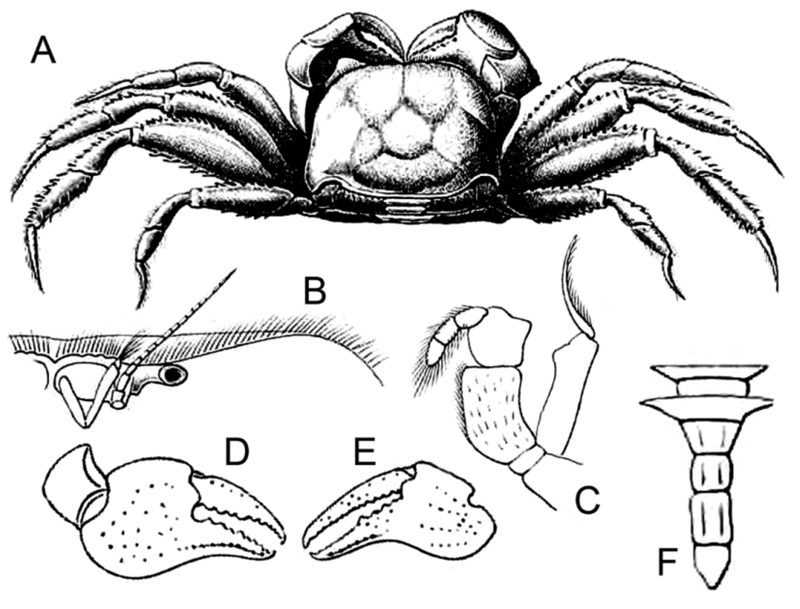
We disagree with Serène (1964, 1968) in treating S. leucochirus as a distinct species. Scalopidia leucochirus was described on the basis of one male measuring 19 × 14 mm from Hong Kong infected with the rhizocephalan Sacculina . The figures of the species by Richters (in Lenz & Richters 1881: pls. 1–10) ( Fig. 3 View FIGURE 3 ) show no major differences with the large series of specimens of S. spinosipes we have from East and Southeast Asia, particularly in the armature of ambulatory legs. There is one discrepancy. The figure of the male abdomen of Hypophthalmus leucochirus by Richters (in Lenz & Richters 1881: pl. 10) ( Fig. 3 View FIGURE 3 F) is unusual because somite 6 is depicted as being much longer than wide. It is quite unlike any seen in any Scalopidia species and is almost certainly incorrectly drawn.


No known copyright restrictions apply. See Agosti, D., Egloff, W., 2009. Taxonomic information exchange and copyright: the Plazi approach. BMC Research Notes 2009, 2:53 for further explanation.