
Selwynia sibogae (Tesch, 1918)
|
publication ID |
https://doi.org/ 10.11646/zootaxa.4092.3.2 |
|
publication LSID |
lsid:zoobank.org:pub:547625D1-70CC-48A1-9C36-3A0DD41B83A5 |
|
DOI |
https://doi.org/10.5281/zenodo.6081358 |
|
persistent identifier |
https://treatment.plazi.org/id/F56987A9-FFA0-FFF9-FB8F-F98FFBF27F90 |
|
treatment provided by |
Plazi |
|
scientific name |
Selwynia sibogae (Tesch, 1918) |
| status |
|
Selwynia sibogae (Tesch, 1918)
( Figs. 1–4 View FIGURE 1 View FIGURE 2 View FIGURE 3 View FIGURE 4 )
Aphanodactylus sibogae Tesch, 1918: 283 , pl. 18, fig. 2; Schmitt et al. 1973: 128; Ng et al. 2008: 247; Ng & Naruse 2009: 287, fig. 5; Ahyong & Ng 2009: 36.
Not Aphanodactylus sibogae Cases & Storch 1981 , fig. 9 = A. panglao Ng & Naruse, 2009.
Material examined. Lectotype (herein designated): ovigerous female (10.9 × 6.1 mm) (RMNH ZMA DE 103.11), stn. 313, in tubes of terebellid worm ( Loimia sp.), east of Dangar Besar, Sapeh Bay, north coast of Sumbawa, Lesser Sunda Islands, Indonesia, up to 36 m depth, coll. SIBOGA Expedition, 14–16 February 1900. Others: 1 ovigerous female (8.8 × 5.4 mm) (RMNH 2162), in tubes of terebellid worm ( Loimia sp.), east of Dangar Besar, Sapeh bay, north coast of Sumbawa, Lesser Sunda Islands, Indonesia, up to 36 m depth, coll. SIBOGA Expedition, 14–16 February 1900; 1 male (7.7 × 5.3 mm) (ZRC 2010.0298), Sekotong, western Lombok, Indonesia, coll. D.L. Rahayu, 16 May 2007.
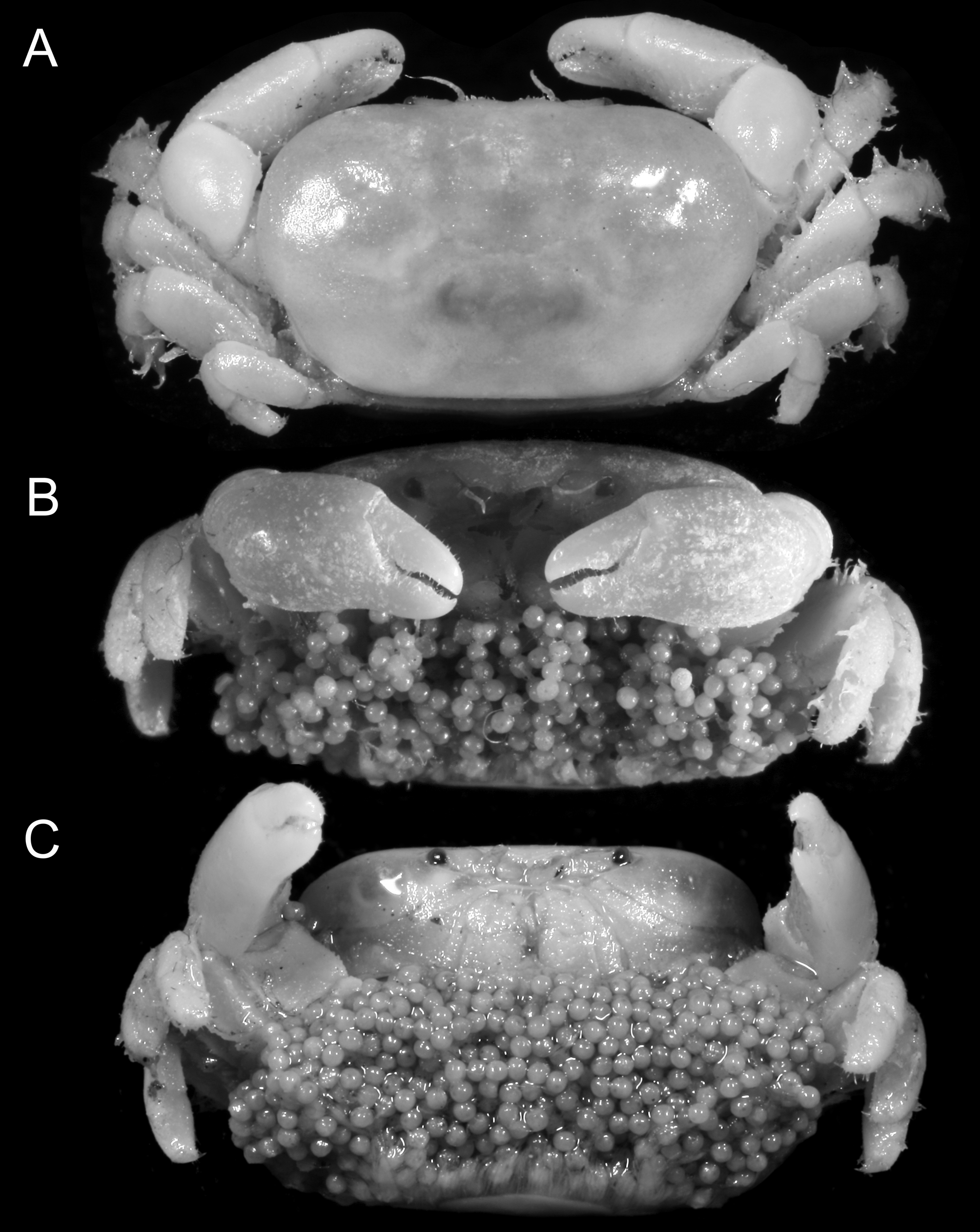
Redescription (female lectotype). Carapace ( Fig. 1 View FIGURE 1 A) oval, about 1.8 broader than long; dorsal surface smooth, region poorly demarcated, flattened to gently concave on mesogastric region, shallow pit on each hepatic region. Front ( Fig. 1 View FIGURE 1 B) deflexed, medially concave in dorsal view. Orbital margin ( Fig. 1 View FIGURE 1 B) entire, slightly narrower distally, narrow gap between cornea, external orbital angle. Anterolateral margin entire, convex laterally, not cristate, continuous with posterolateral margin. Epistome very short, medially sunken. Antennule folding transversely in fossa. Antenna with stout basal antennal article, not reaching distolateral angle of carapace; antenna enters orbit. Ocular peduncles movable, short, stout, subconical, cornea small, well pigmented.
Third maxillipeds ( Fig. 1 View FIGURE 1 C) covering approximately four-fifths of buccal cavern when closed; ischium longer, broader than merus; palp attached to distolateral angle of merus; exopod slender, reaching proximal half of merus; flagellum long.
Thoracic sternites 1, 2 fused, 2/3, 3/4 each demarcated by shallow ridge; sternites 4–8 laterally demarcated by shallow sutures, each medially interrupted; sutures between sternites 4/ 6/7 longer than others, vulva on distal twothirds of sternite 6, between mesial ends of sutures 5/6, 6/7.
Chelipeds ( Fig. 1 View FIGURE 1 C) equal. Merus triangular in cross section, dorsal margin rugose, dorsal margin, outer surface setose. Carpus smooth, inner angle absent. Chela with smooth surfaces, palm about 1.2 times longer than dactylus; immovable finger with gently sinuous cutting edge, cutting edge convex medially, lined with small teeth; movable finger with cutting edge concave on distal half.
Ambulatory legs (P2–P5) relatively short ( Figs. 1 View FIGURE 1 A, 4G–K); P3 longest, P5 shortest; extensor margin, surface glabrous, sparse setae on flexor margins, surfaces of carpus, propodus. Merus as long as or slightly longer than combined lengths of propodus, dactylus, extensor margin of P3 with few tubercles proximally, P3 serrated, flexor margin armature of P2–P5 as follows: P2 2+1/3+2, P3 3+1/3+0, P4 2+3/3+1, P5 0+0/0+0. Flexor margin of basisischium of P5 unarmed. Dactylus very short, claw-like.
Abdomen ( Figs. 1 View FIGURE 1 C, 4F) with all somites, telson distinct, mobile; somites wide.
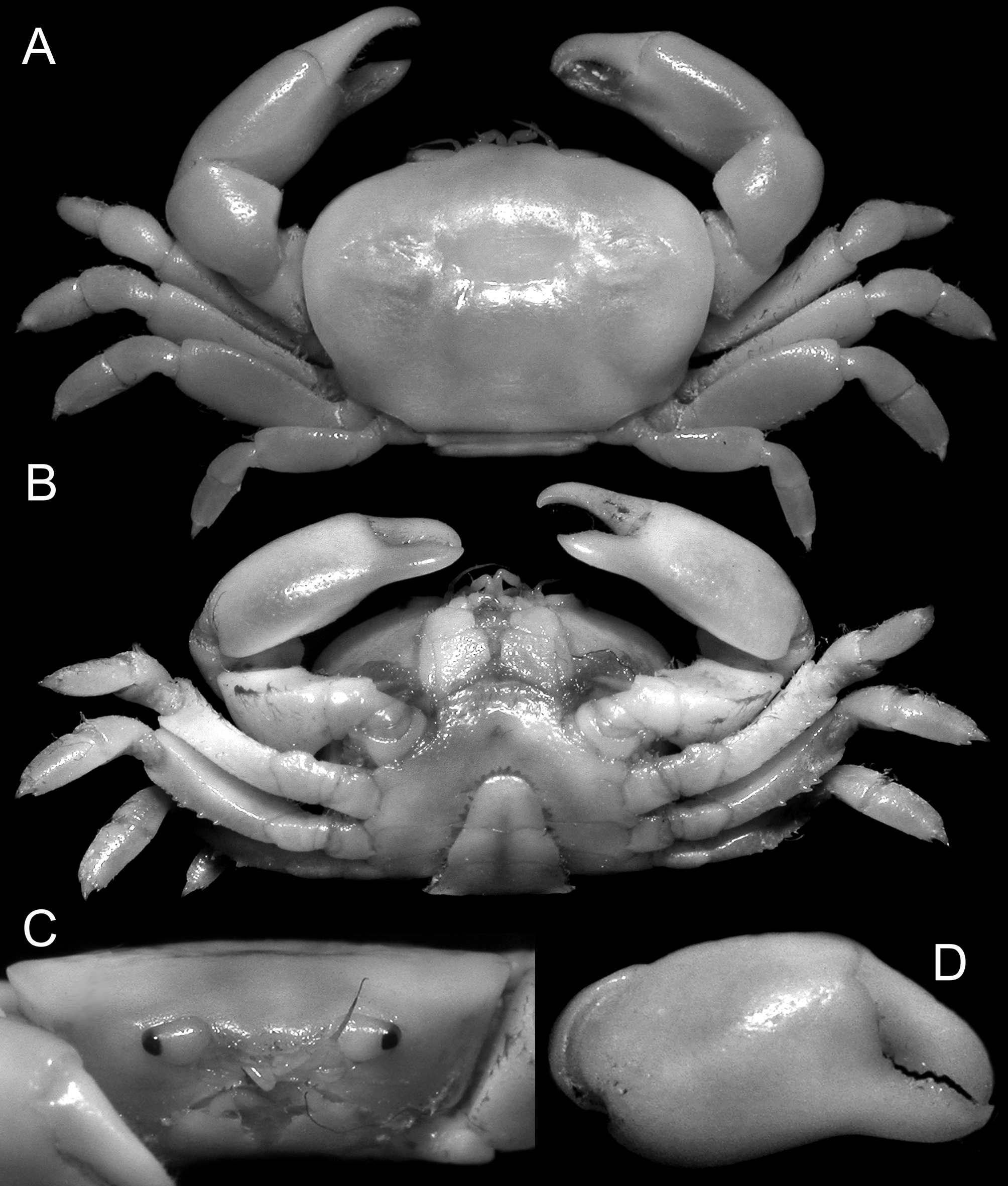
Description (male). Carapace ( Fig. 3 View FIGURE 3 A) subquadrate, about 1.3 broader than long; dorsal surface smooth, regions poorly demarcated, shallow depression on each hepatic region. Front ( Fig. 3 View FIGURE 3 C) deflexed, medially concave in dorsal view. Orbital margins ( Fig. 3 View FIGURE 3 C) entire, oval, no gap between cornea, external orbital angle. Epistome longer than that of female, posterior margin slightly concave. Antennule, antenna as in female. Eyes ( Fig. 3 View FIGURE 3 C) short, stout, with distinct cornea. Third maxillipeds ( Figs. 3 View FIGURE 3 B, 4A) similar to female third maxillipeds except for more rounded distomesial angle.
Thoracic sternites 1, 2 fused, sternites 2/3 demarcated by distinct suture, 3/4 demarcated by low ridge, sternites 4–8 demarcated by shallow, narrow sutures, press button of abdominal locking mechanism present on distal quarter of sternite 5; penis emerging from near anterior border of sternite 8.
Chelipeds relatively strong, slightly unequal in size. Merus triangular in cross section, ventro-outer margin granulated, dorsal margin rugose, dorsal margin, outer surface setose. Carpus smooth, inner angle rounded. Chelae ( Fig. 3 View FIGURE 3 D), ambulatory legs ( Figs. 3 View FIGURE 3 A, 4C) as in female, except for armature of flexor margins of ambulatory merus.
Flexor margin armature of P2–P5 as follows: P2 3+5/2+9; P3 3+5/3+4, P4 2+3/2+4, P5 0+0/0+0. Flexor margin of basis-ischium of P5 unarmed. Dactylus very short, claw-like.
Abdomen ( Fig. 4 View FIGURE 4 B) with all somites, telson distinct, mobile, relatively narrow, first, second somites short, third somite to telson forming straight lateral margins. G1 ( Fig. 4 View FIGURE 4 D, E) simple, proximal five-sixths straight, distal onesixth bent inwards towards median part of sternum.
Colour. The present specimen from Lombok (ZRC 2010.0298) was porcelain white in life, without any other colours.
Remarks. Tesch (1918) gave a detailed description and figures of one male (7.8 × 5.3 mm) and one female specimen (11.25 × 6.0 mm). Ng & Naruse (2009: fig. 5) located one female specimen in RMNH which had been labelled as a syntype (see Fransen et al. 1997) ( Fig. 2 View FIGURE 2 ). This specimen was an ovigerous female (RMNH 2162) measuring 8.8 × 5.4 mm, and although it did not exactly match the measurements given by Tesch (1918: 285), since only one pair was known, Ng & Naruse (2009) supposed the female they examined must be the one cited by Tesch (1918); and assumed the syntype male was in the Zoological Museum University of Amsterdam (ZMA). Accordingly, they designated and figured this RMNH female specimen (Ng & Naruse 2009: fig. 5) as the lectotype of Aphanodactylus sibogae Tesch, 1918 , arguing that the name needed stability since they were describing an allied species from the Philippines, A. panglao . The ZMA material has since been transferred to the RMNH and in 2013, the first author searched the Siboga material there in an attempt to locate the paralectotype male. The type bottle for the species (ZMA DE 103.11) was found but surprisingly, it contained one ovigerous female measuring 10.9 × 6.1 mm instead of the expected male. This ZMA specimen actually agrees more closely with the specimen examined and cited by Tesch (1918: 285) (11.25 × 6.0 mm) and is certainly one of the two syntypes. The RMNH specimen is thus not a type as had been supposed by Fransen et al. (1997) and Ng & Naruse (2009). Ng & Naruse’s (2009) designation of a lectotype is thus invalid. We here select the ZMA specimen now transferred to RMNH as the correct lectotype of Aphanodactylus sibogae Tesch, 1918 ( Fig. 1 View FIGURE 1 ).
As to where the paralectotype male specimen is, we do not know. It is not among the ZMA material we have searched, and may be lost or misplaced. We are also puzzled as to where the second smaller ovigerous female (RMNH 2162) came from. It is possible that Tesch found the third specimen after his publication was in press, and had simply kept it with the remaining specimens before the material was shared between RMNH and ZMA. This seems likely as its label is identical to that of the lectotype female. The recent male specimen collected from Lombok, Indonesia) (ZRC 2010.0298), fortunately agrees well with the description of the male A. sibogae by Tesch (1918).
Selwynia sibogae is arguably closest to S. loimiae and S. panglao , with similar carapace and leg proportions as well as armature; and females of these species are not easy to separate. The males differ in the form of their G1s as well as proportions and armature of the ambulatory legs.
Biology. The types were obtained from the tubes of a terebellid worm ( Loimia sp.) at a depth of about 36 m by dredging. The recent male specimen (ZRC 2010.0298) was collected without any host data, but it may have been dislodged from the worm tube as it was obtained as a part of an intertidal survey.
Distribution. Sumbawa and Lombok, Lesser Sunda Islands, Indonesia. Intertidal.


No known copyright restrictions apply. See Agosti, D., Egloff, W., 2009. Taxonomic information exchange and copyright: the Plazi approach. BMC Research Notes 2009, 2:53 for further explanation.
|
Kingdom |
|
|
Phylum |
|
|
Class |
|
|
Order |
|
|
Family |
|
|
Genus |
|
Kingdom |
|
|
Phylum |
|
|
Class |
|
|
Order |
|
|
Family |
|
|
Genus |