
Echinoderes lupherorum, Sørensen & Rohal & Thistle, 2018
|
publication ID |
https://doi.org/ 10.5852/ejt.2018.456 |
|
publication LSID |
lsid:zoobank.org:pub:DE1B1DEE-9871-4803-9F67-025F2B439560 |
|
DOI |
https://doi.org/10.5281/zenodo.3818840 |
|
persistent identifier |
https://treatment.plazi.org/id/B3E96033-83C3-4924-8FFE-3592E427620C |
|
taxon LSID |
lsid:zoobank.org:act:B3E96033-83C3-4924-8FFE-3592E427620C |
|
treatment provided by |
Valdenar |
|
scientific name |
Echinoderes lupherorum |
| status |
sp. nov. |
Echinoderes lupherorum sp. nov.
urn:lsid:zoobank.org:act:B3E96033-83C3-4924-8FFE-3592E427620C
Figs 18–20 View Fig View Fig View Fig , Tables 12–13
Diagnosis
Echinoderes with middorsal spines on segments 4 to 8, and spines in lateroventral positions on segments 6 to 9. Tubes present in lateroventral positions on segments 2 and 5, and in midlateral positions of segment 10. Very minute glandular cell outlets type 2 present in subdorsal positions on segment 2 and in laterodorsal positions on segments 8 and 9. Tergal plate of segment 11 terminates in pointed, acicular tergal extensions, constituting 6 to 8% of trunk length. Males with three pairs of penile spines; females with long lateral terminal accessory spines, and papillae in ventrolateral positions of segment 7 and ventromedial positions of segment 8.
Etymology
The second author (MR) dedicates this species to her fiancé Brach Lupher and the Lupher family – her family to be.
Material examined
Holotype
UNITED STATES OF AMERICA: adult ♀, US West Coast , off southern Oregon, 42°33′50″ N, 131°59′48″ W, St. 2, 3601 m deep, collected from mud, 17 Sep. 2008, mounted in Fluoromount G on an H-S slide (NHMD-225220). See Fig. 1 View Fig for localities and Table 1 View Table 1 for detailed station data.
GoogleMapsParatypes
UNITED STATES OF AMERICA: 4 ♀♀, 1 ♂, same collecting data as for holotype; 1 ♀, St. 3 (NHMD- 225221–225226). All paratypes are mounted in Fluoromount G, three on H-S slides and three on glass slides.
Additional non-type material
UNITED STATES OF AMERICA: 3 ♀♀, 4 ♂♂, St. 6, mounted for SEM and stored in the first author’s personal reference collection.
Description
Adults with head, neck and eleven trunk segments ( Figs 18 View Fig A–B, 19A, 20A–B). The trunk is relatively large, appears slender, tapered in both ends, and broadest from segment 5 to 8. Lateral terminal spines are long and slender ( Fig. 18C View Fig ), from 80% and up to same length as the trunk, and lateral terminal accessory spines in females are shorter ( Figs 18 View Fig A–B), around 20% of the lateral terminal spine length. For a complete overview of measurements and dimensions, see Table 12. Distribution of cuticular structures, i.e., sensory spots, glandular cell outlets, spines and tubes, is summarized in Table 13.
The head consists of a retractable mouth cone and an introvert. Inner oral styles are present, but their exact number and arrangement could not be determined. The external mouth cone armature consists of nine outer oral styles, each with four or five basal fringe tips. The introvert sectors are defined by ten primary spinoscalids in Ring 01. Each primary spinoscalid consists of a basal sheath and a distal end piece with a blunt tip ( Fig. 20C View Fig ). The basal sheaths have marginal extensions forming two layers of transverse fringes. End pieces are smooth. The arrangement of scalids follows the same pattern as in Echinoderes hviidarum sp. nov. described above; hence, see Fig. 11 View Fig for a complete overview. All spinoscalids consist of a basal sheath and a pointed end piece. The basal sheaths terminate in fringed margins in spinoscalids of Rings 02 and 04, whereas the sheaths of Ring 03 have a median spike only. Spinoscalids of Rings 05 to 07 are composed as those in the preceding ring, but with shorter end pieces ( Figs 11 View Fig , 20C View Fig ).
The neck has 16 placids, measuring 18 µm in length, except for the slightly longer midventral ones, measuring 20 µm in length. The midventral placid is also broadest, measuring 16 µm in width at its base, whereas all others are narrower, measuring 10 µm in width at their bases. Four dorsal and two ventral trichoscalid plates are present; trichoscalid plates are fairly small, and rounded to oval.
Segment 1 consists of a complete cuticular ring. Sensory spots are located on the anterior half of the segment, but not at the anterior margin in subdorsal and laterodorsal positions ( Figs 18A View Fig , 19B View Fig , 20E View Fig ); sensory spots are small and rounded, with a few papillae. Their minute size, combined with the dense hair covering on most segments, makes it very difficult to observe the sensory spots in SEM, and often they are more easily observed in LM. Glandular cell outlets type 1 only observed in lateroventral positions. Dorsal and lateral sides have scattered cuticular hairs, whereas the ventral side is nearly devoid of hairs ( Fig. 20 View Fig D–E). The posterior segment margin is nearly straight, terminating in a pectinate fringe with long and flexible fringe tips.
Segment 2 consists of a complete cuticular ring. The pachycyclus of the anterior segment margin is relatively thin and not interrupted. Very minute glandular cell outlets type 2 are present in subdorsal positions ( Figs 18A View Fig , 19B View Fig , 20 View Fig E–F), and well-developed tubes in lateroventral positions ( Figs 18B View Fig , 19C View Fig , 20D View Fig ). Sensory spots are located in middorsal ( Fig. 20 View Fig E–F), subdorsal, laterodorsal ( Fig. 19B View Fig ) and ventromedial positions. Glandular cell outlets type 1 are not observed. A secondary pectinate fringe is present near the anterior segment margin of this and the following segments, but it is usually covered by the preceding segment. On this and the following eight segments, the cuticular hairs are bracteate and densely cover the dorsal and lateral areas. The posterior segment margin is as on the preceding segment.
Segment 3, and the remaining segments, consist of one tergal and two sternal plates ( Figs 18B View Fig , 19A, C, E, G View Fig ). The pachycyclus of the anterior segment margin is well-developed and interrupted at the tergosternal junctions and middorsally. Sensory spots are present in subdorsal ( Fig. 19D View Fig ) and sublateral positions, as well as glandular cell outlets type 1 in ventromedial positions. Cuticular hairs densely cover the tergal plate and the lateral halves of the sternal plates. The posterior segment margin is nearly straight, with fringe tips as on the preceding segment.
Segment 4 with a middorsal acicular spine ( Figs 18A View Fig , 19D View Fig ). There are sensory spots and glandular cell outlets type 1 present in ventromedial positions ( Fig. 19E View Fig ). Pachycycli, pectinate fringe of posterior margin and cuticular hairs as on preceding segment.
Segment 5 with a middorsal acicular spine and well-developed tubes in lateroventral positions ( Figs 18 View Fig A–B, 19D–E); tubes have weakly developed, yet visible bases and lateral wings. Sensory spots are present in subdorsal ( Fig. 19D View Fig ) and midlateral positions. Cuticular hairs, glandular cell outlets type 1, pachycycli and pectinate fringe of posterior margin as on preceding segment.
Segment 6 with acicular spines in middorsal and lateroventral positions ( Figs 18 View Fig A–B, 19D–E). Sensory spots present in paradorsal ( Fig. 19D View Fig ), midlateral and ventromedial positions. Glandular cell outlets type 1, pachycycli, pectinate fringe of posterior margin and cuticular hairs as on preceding segment.
Segment 7 with acicular spines in middorsal and lateroventral positions ( Figs 18 View Fig A–B, 19F–G). Sensory spots present in paradorsal and midlateral positions, and males in addition with sensory spots in ventromedial positions. Females with papillary structures in ventrolateral positions ( Figs 18B View Fig , 19G View Fig ); the structures are quite prominent in LM ( Fig. 19G View Fig ), but hardly visible in SEM. Glandular cell outlets type 1, pachycycli, pectinate fringe of posterior margin and cuticular hairs as on preceding segment.
Segment 8 with acicular spines in middorsal and lateroventral positions ( Figs 18 View Fig A–B, 19F–G). Very minute glandular cell outlets type 2 present in laterodorsal positions ( Figs 18A View Fig , 19F View Fig , 20 View Fig G–H). Sensory spots present in paradorsal ( Fig. 20G View Fig ), subdorsal ( Fig. 19F View Fig ) and laterodorsal ( Fig. 20 View Fig G–H) positions (one specimen without subdorsal sensory spots though ( Fig. 20G View Fig ), and one too dirty to confirm their presence). Female specimens with papillary structures in ventromedial positions ( Figs 18B View Fig , 19G View Fig ). Glandular cell outlets type 1 present in subdorsal ( Fig. 19F View Fig ) and ventromedial positions. Pachycycli, pectinate fringe of posterior margin and cuticular hairs as on preceding segment.
Segment 9 with acicular spines in lateroventral positions ( Figs 18B View Fig ). Very minute glandular cell outlets type 2 present in laterodorsal positions ( Figs 18A View Fig , 19F View Fig , 20I View Fig ). Sensory spots present in paradorsal ( Fig. 19F View Fig ), subdorsal ( Fig. 19F View Fig ), laterodorsal ( Fig. 20I View Fig ) and ventrolateral positions. Small rounded sieve plates are present in lateral accessory positions ( Fig. 18B View Fig ). Glandular cell outlets type 1 are present in subdorsal and ventromedial positions. Cuticular hair covering on tergal plate is less dense than on the preceding segments, and the subdorsal to middorsal areas have no hair at all. Pachycycli and pectinate fringe of posterior margin as on preceding segment.
Segment 10 with tubes in midlateral positions ( Figs 18 View Fig , 19H, J View Fig , 20 View Fig I–J); tubes appear fairly stout and are usually much easier to observe in LM ( Fig. 19H View Fig ) than tubes on segment 10 in other species of Echinoderes ; differentiated bases and lateral wings apparently not present. Sensory spots are present in paradorsal and ventrolateral positions. Glandular cell outlets type 1 are present as two middorsal ones and a pair in paraventral positions. Cuticular hairs are scattered over the tergal plate and lateral parts of the sternal plates, but not as densely as on preceding segments. The posterior segment margin of the tergal plate is straight, whereas the margins of the sternal plates are broadly concave. Pachycycli as on preceding segment.
Segment 11 with long lateral terminal spines ( Fig. 18C View Fig ). Males with three pairs of penile spines ( Figs 18 View Fig D–E, 19J); dorsal and ventral penile spines are thin, and quite long flexible tubes, whereas the median one is thick, conical, and stout. Females with lateral terminal accessory spines ( Figs 18 View Fig A– B, 19H). Sensory spots present in subdorsal positions. The segment is completely devoid of cuticular hairs, besides the short hair-like extensions in the mid- to subdorsal areas. Tergal extensions are long and slender, extending into long but yet delicate spinous tips ( Figs 18 View Fig , 19 View Fig H–I, 20J); tergal extensions measure 24 to 28 µm, equal to 6 to 8% of trunk length. Sternal extensions shorter and rounded.
Remarks
Due to their close resemblance, diagnostic remarks for E. lupherorum sp. nov. will follow below together with remarks for E. yamasakii sp. nov.
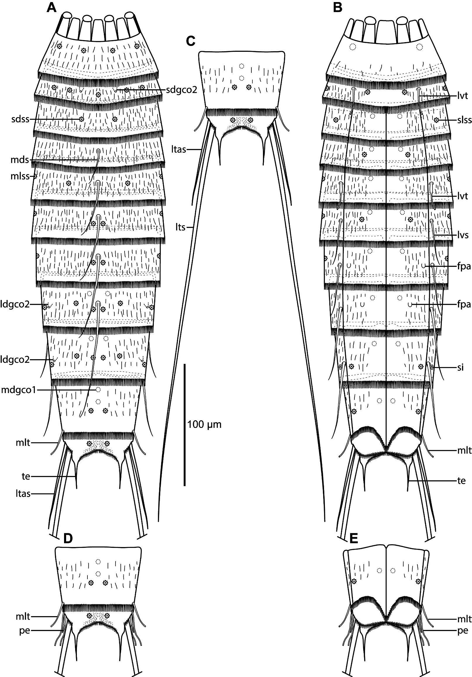
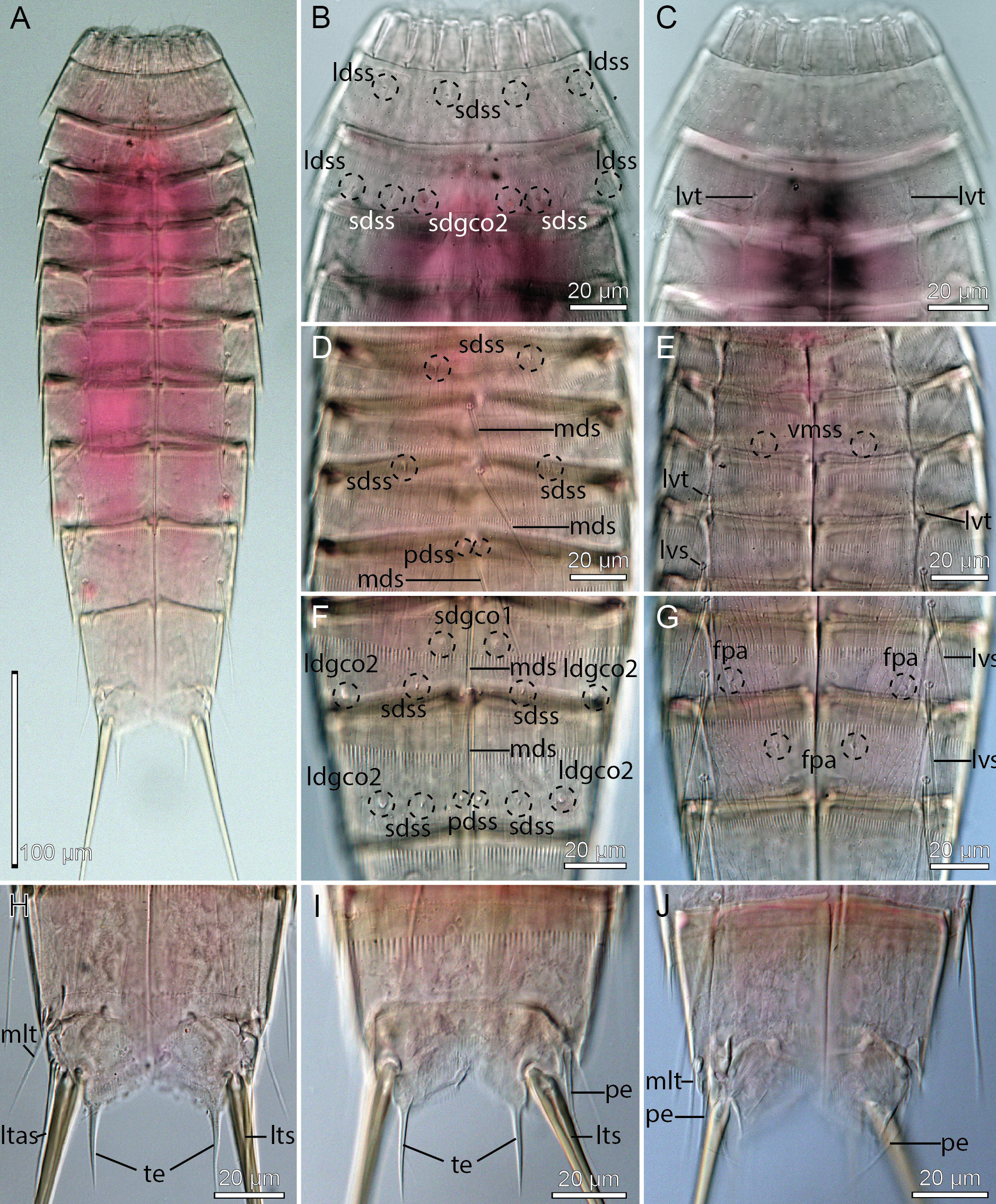
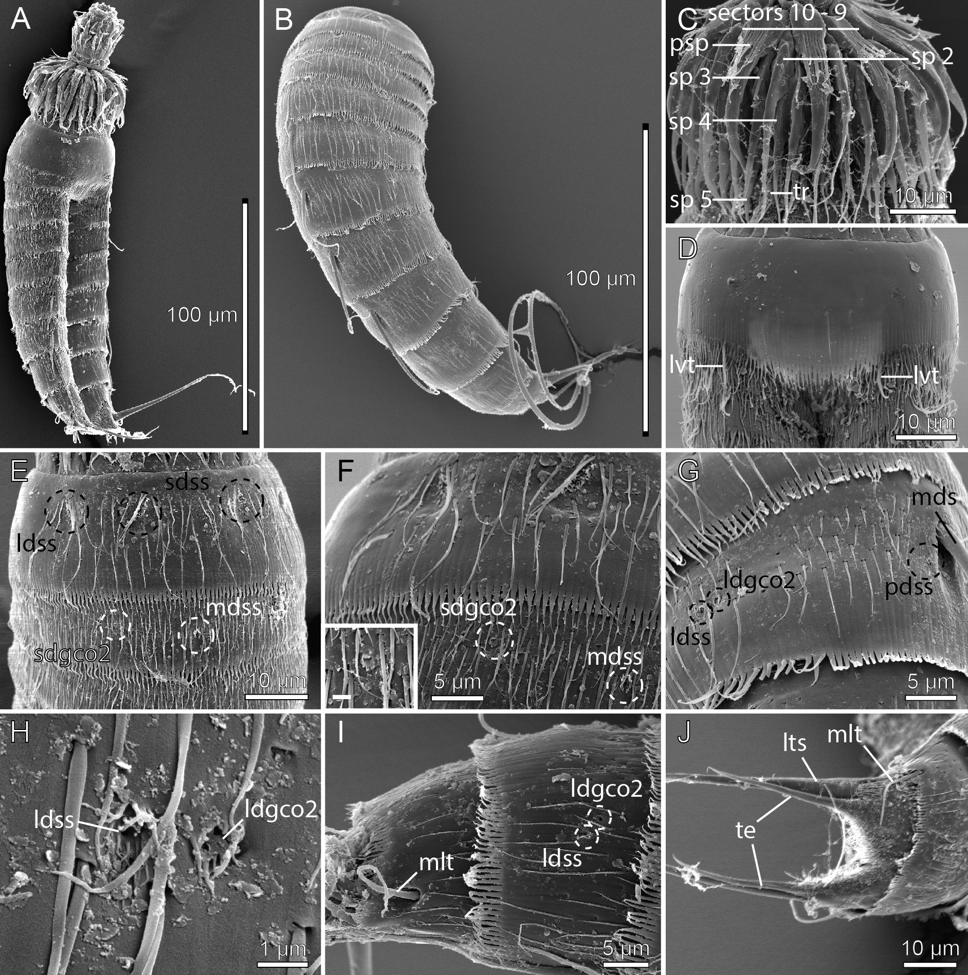
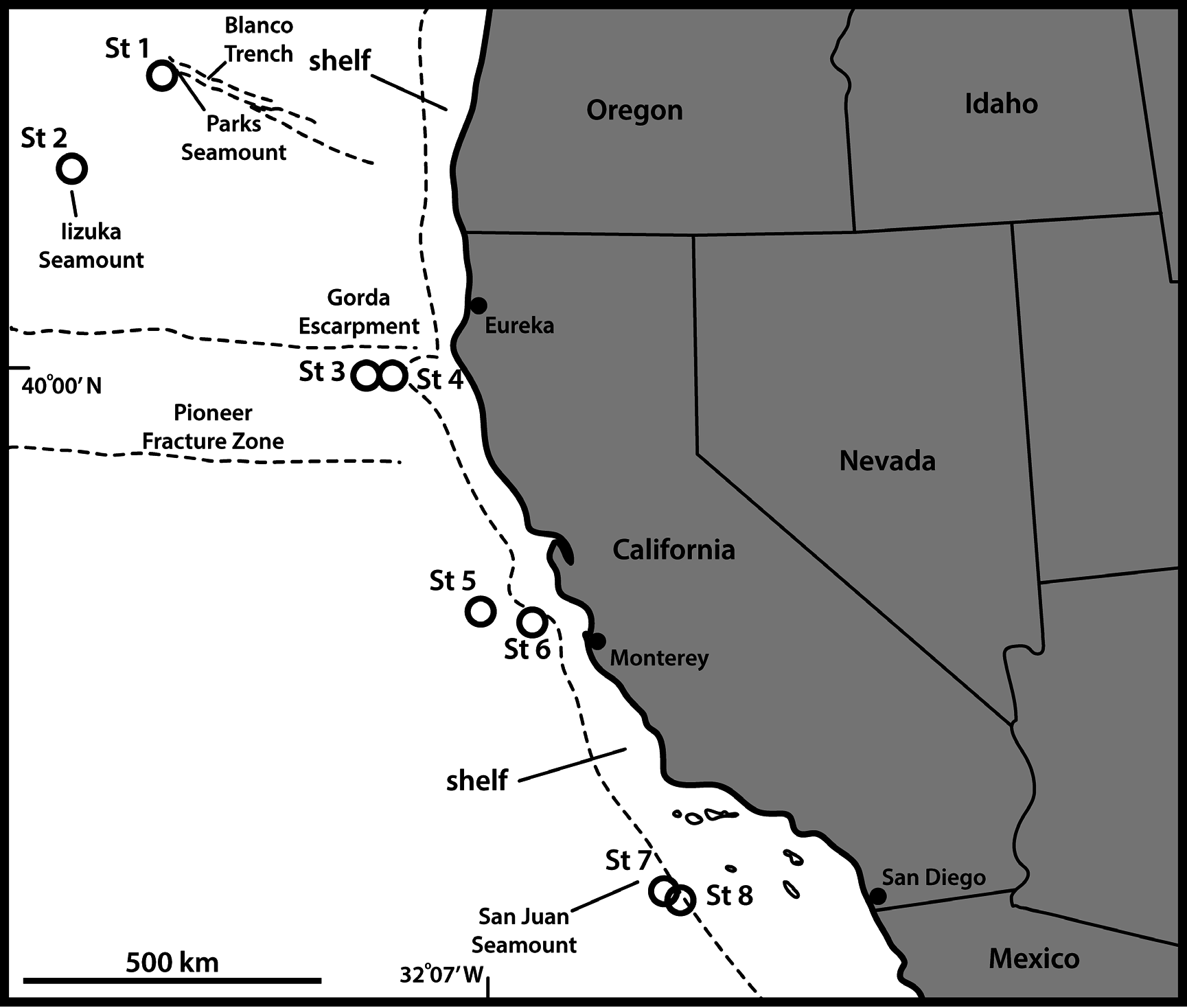

Table 12. Measurements from light microscopy of Echinoderes lupherorum sp. nov. (in µm), including number of measured specimens (n) and standard deviation (SD). Abbreviations: (ac) = acicular spine; LTAS = lateral terminal accessory spine; LTS = lateral terminal spine; LV = lateroventral; MD = middorsal; ML = midlateral; MSW-8 = maximum sternal width, measured on segment 8 in this species; S = segment lengths; SW-10 = standard width, always measured on segment 10; TE = tergal extension; TL = trunk length; (tu) = tube.
| Character | n | Range | Mean | SD |
|---|---|---|---|---|
| TL | 7 | 331–415 | 371 | 36.42 |
| MSW-8 | 6 | 75–81 | 78 | 2.16 |
| MSW-8/TL | 6 | 19.3–23.9% | 21.0% | 1.83% |
| SW-10 | 6 | 65–71 | 67 | 2.58 |
| SW-10/TL | 6 | 15.7–20.2% | 18.1% | 1.77% |
| S1 | 7 | 35–42 | 39 | 2.54 |
| S2 | 7 | 33–37 | 35 | 1.46 |
| S3 | 7 | 39–43 | 41 | 1.50 |
| S4 | 7 | 42–47 | 45 | 1.90 |
| S5 | 7 | 42–52 | 48 | 3.78 |
| S6 | 7 | 50–57 | 54 | 2.70 |
| S7 | 7 | 54–60 | 57 | 2.34 |
| S8 | 7 | 54–62 | 59 | 2.82 |
| S9 | 7 | 59–62 | 61 | 1.38 |
| S10 | 7 | 44–51 | 49 | 2.51 |
| S11 | 7 | 53–58 | 56 | 1.91 |
| MD4 (ac) | 7 | 31–36 | 33 | 1.95 |
| MD5 (ac) | 6 | 40–48 | 44 | 3.27 |
| MD6 (ac) | 7 | 50–67 | 54 | 6.00 |
| MD7 (ac) | 6 | 54–83 | 70 | 11.15 |
| MD8 (ac) | 6 | 90–128 | 112 | 16.97 |
| LV6 (ac) | 6 | 32–43 | 38 | 4.32 |
| LV7 (ac) | 7 | 45–53 | 48 | 3.86 |
| LV8 (ac) | 7 | 53–68 | 60 | 5.77 |
| LV9 (ac) | 7 | 54–74 | 64 | 7.63 |
| ML10 (tu) | 6 | 29–34 | 31 | 1.75 |
| TE | 7 | 24–28 | 26 | 1.40 |
| TE/TL | 7 | 6.0–8.2% | 7.2% | 0.94% |
| LTS | 6 | 324–355 | 341 | 12.45 |
| LTS/TL | 6 | 80.5–100.9% | 91.5% | 7.59% |
| LTAS | 6 | 62–83 | 75 | 7.40 |
| US |
University of Stellenbosch |
No known copyright restrictions apply. See Agosti, D., Egloff, W., 2009. Taxonomic information exchange and copyright: the Plazi approach. BMC Research Notes 2009, 2:53 for further explanation.
|
Kingdom |
|
|
Phylum |
|
|
Class |
|
|
Order |
|
|
Family |
|
|
Genus |