
Berosus decolor Knisch, 1924
|
publication ID |
https://doi.org/ 10.11646/zootaxa.3981.4.8 |
|
publication LSID |
lsid:zoobank.org:pub:8385E8B9-26D2-425D-8E5A-A9B29C88270B |
|
DOI |
https://doi.org/10.5281/zenodo.5657641 |
|
persistent identifier |
https://treatment.plazi.org/id/F66D87AC-107B-2E2C-62C1-FADD29D73B73 |
|
treatment provided by |
Plazi |
|
scientific name |
Berosus decolor Knisch, 1924 |
| status |
|
Berosus decolor Knisch, 1924 View in CoL
( Figs. 1–14 View FIGURES 1 – 2 View FIGURES 3 – 4 View FIGURES 5 – 7 View FIGURES 8 – 10 View FIGURES 11 – 12 View FIGURES 13 – 14 )
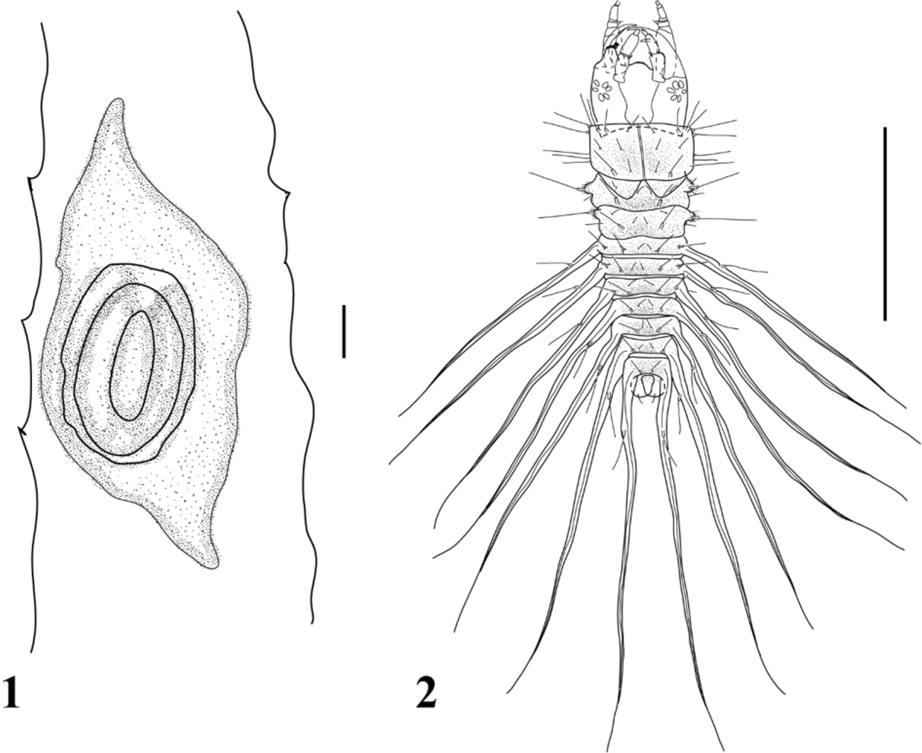
Diagnosis. Egg case: oval-shape, mast lacking and bearing 2‒ 3 eggs. Larvae: Frontal sulcus well defined in all instars; cervical sclerites absent, anterior margin of the nasale with six teeth; left epistomal lobe with 14‒16 strong and curved setae (gFR2) on anterior margin except for first outer seta that is short and stout, inner eight setae bearing a small tooth; first antennomere with a digitiform membranous projection on distal inner margin; right mandible with three teeth; abdominal segments I‒VII with a pair of tracheal gills, lacking sclerotized basal ring.
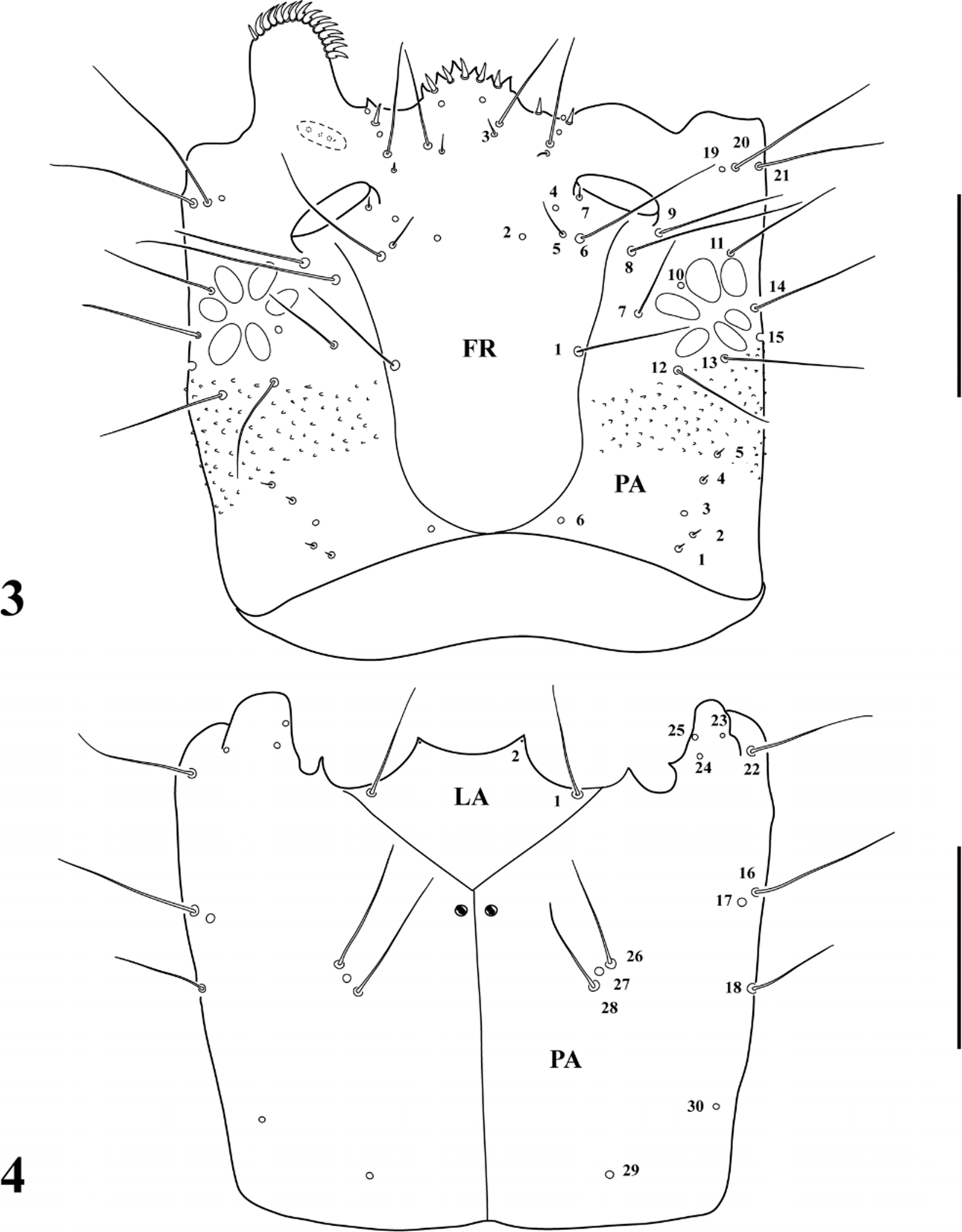
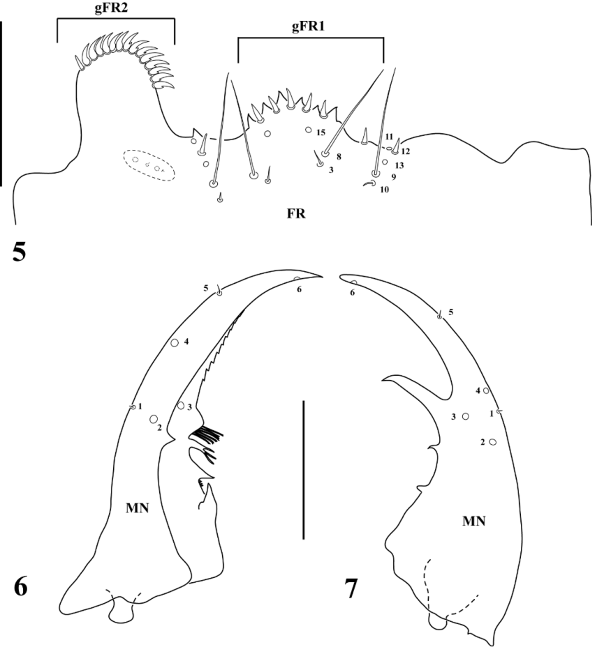
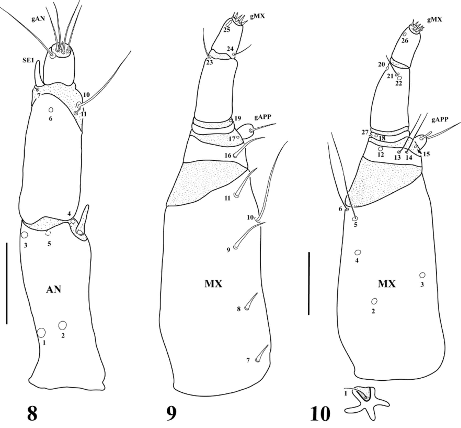
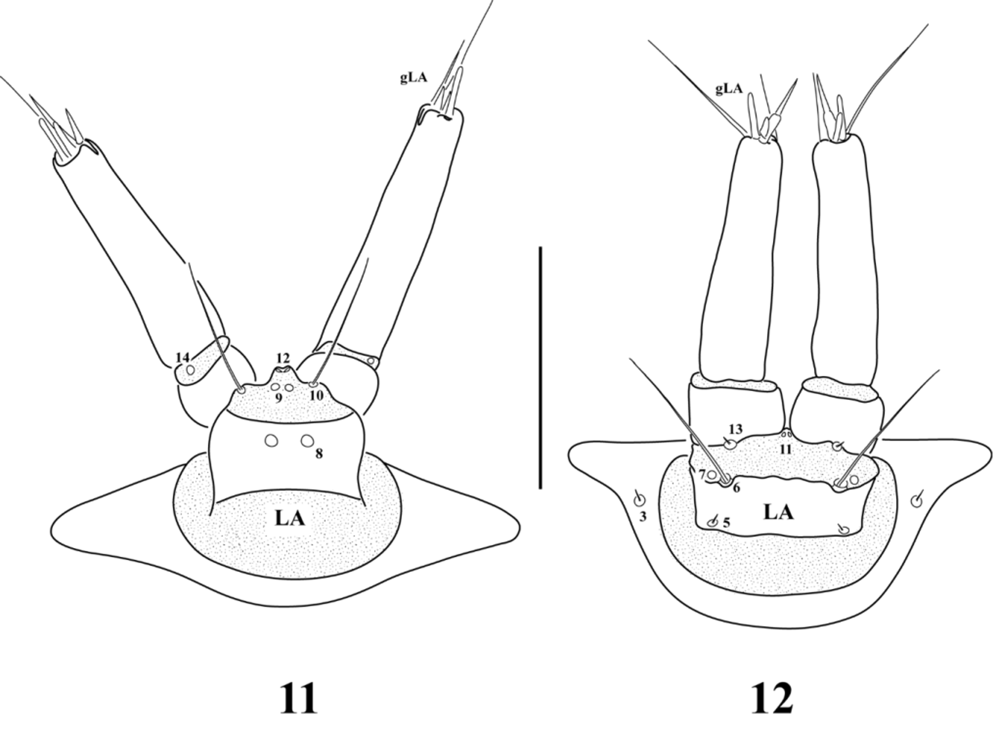
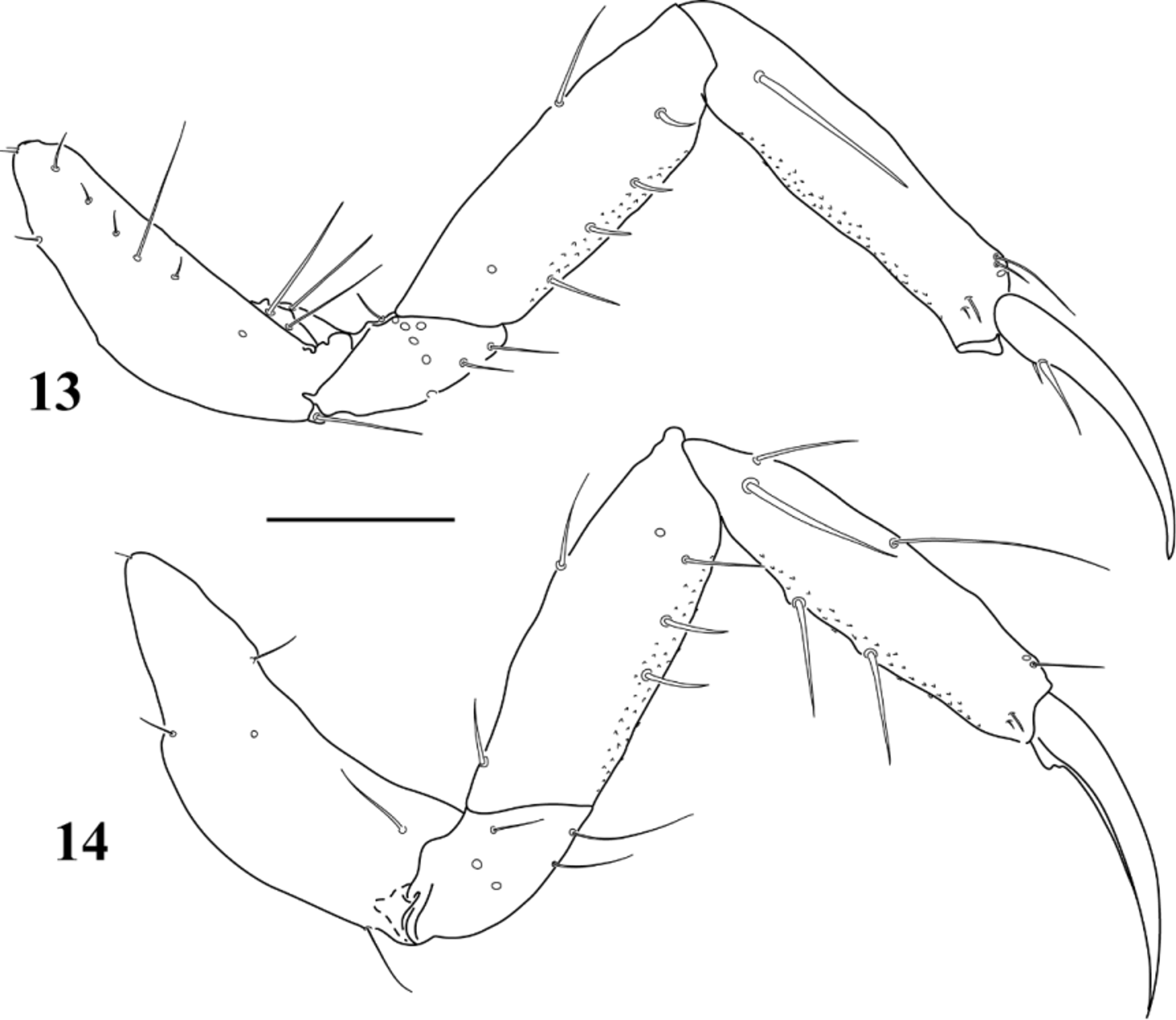
Description, egg case ( Fig. 1 View FIGURES 1 – 2 ). Whitish, oval-shape, sometimes elongate. Attached to stems and leaves of aquatic plants. Made of two layers: inferior layer very thin, weaved onto the substrate, superior layer thicker, covering the eggs. Mast lacking. Two eggs per case, occasionally three. Egg case length: 1.60‒3.65 mm, width: 1.00‒1.55 mm. Description, instar I ( Figs. 2‒14 View FIGURES 1 – 2 View FIGURES 3 – 4 View FIGURES 5 – 7 View FIGURES 8 – 10 View FIGURES 11 – 12 View FIGURES 13 – 14 ). Color: creamy-yellow, sclerotized areas light brown, lacking color pattern. Body ( Fig. 2 View FIGURES 1 – 2 ): morphometric measurements and ratios shown in Table 1 View TABLE 1 . Head capsule ( Figs. 3‒5 View FIGURES 3 – 4 View FIGURES 5 – 7 ): subquadrangular, epicraneal sulcus well developed, frontal sulci inverse bell shaped, coronal sulcus absent; cervical sclerites absent; area between setae PA5 and PA12 entirely covered by spinulae. Labroclypeus ( Fig. 5 View FIGURES 5 – 7 ): asymmetrical, nasale slightly projected forward with 6 anterior teeth; lateral lobes of the epistome strongly asymmetrical, right one weakly developed, left one large, covering the base of the mandible; one small tooth present between left epistomal lobe and nasale. Antenna ( Fig. 8 View FIGURES 8 – 10 ): composed of three antennomeres, A1 shorter than A2+A3, with a membranous digitiform projection on distal inner margin; sensorium located on outer apical margin of A2, slightly shorter than A3. Mandibles ( Figs. 6‒7 View FIGURES 5 – 7 ): asymmetrical, right mandible with three teeth on inner margin, distal one much larger than the other two; left mandible with a shallow groove and four teeth, basal tooth sharp and pointing to the apex, second tooth pointing mediad with three sharp points on base, third tooth trifid, distal tooth triangular, pointing mediad, with six long bifid projections; distal inner margin serrate. Maxilla ( Figs. 9‒10 View FIGURES 8 – 10 ): longer than the antenna, composed of three articles. Cardo small, irregular. Stipes slightly longer than palpus. Palpus composed of four palpomeres, MP1 and MP4 subequal in length, MP2 shortest, MP3 longest. MP1 with a short projection on distal inner margin, MP4 subconical. Labium ( Figs. 4 View FIGURES 3 – 4 , 11‒12 View FIGURES 11 – 12 ): submentum wide, subtrapezoidal. Mentum reduced, subpentagonal. Prementum subrectangular, wider than long. Ligula reduced to a membranous lobe between palpi. Palpus composed of two palpomeres, LP2 much longer than LP1. Thorax ( Fig. 2 View FIGURES 1 – 2 ): pro-, meso- and metathorax covered with setae and fine spinulae. Pronotal plate large, covering most of prothorax, subrectangular with posterior margin rounded, with fine sagittal line. Mesonotum with one pair of subtriangular sclerites on anterior margin (we were unable to find the inner pair, although we cannot confirm if it is really absent). Metanotum without sclerites. Meso- and metathorax shorter than prothorax, with a pair of lateral tubercles bearing one long and several short spinula. Legs ( Figs. 13–14 View FIGURES 13 – 14 ): five-segmented, long, visible in dorsal view. Metathoracic leg longest, prothoracic leg shortest. Coxa large, cylindrical; trochanter short; femur slender, slightly longer than coxa; tibiotarsus subcylindrical, slender, almost as long as femur; claw long, slender, curved, about 3/4 times as long as tibiotarsus. Abdomen ( Fig. 2 View FIGURES 1 – 2 ): Ten-segmented, covered by a dense layer of fine spinulae. Segments I-VII similar in shape, tapering towards posterior end, subdivided by a transverse fold, each with a pair of tracheal gills, segments I-VII with two pairs of tubercles bearing one long seta and several spinulae (one pair dorsal, one pair at the base of each tracheal gill). Segment VIII small, with two posterior lobes and a subcircular sclerite dorsally. Segments IX and X strongly reduced. Tracheal gills filiform, as long as total length of body, with a long seta near the base. Sclerotized ring at the base of gills absent. Nine pairs of non-functional spiracles, one mesothoracic and eight abdominal. Spiracular atrium absent. Chaetotaxy. Head capsule ( Figs. 3‒5 View FIGURES 3 – 4 View FIGURES 5 – 7 ): Frontale with 49 dorsal sensilla. Central part with 2 long setae (FR1) near frontal sulci and 2 pores (FR2) situated more anteriorly and mesally, seta FR3 short and displaced anteriorly close to seta FR8. Two setae (FR5 long, FR6 short) and 1 pore (FR4) near the base of each antenna. One short seta (FR7) close to the inner margin of the antennal socket. Nasale with a group of 6 stout and short setae (gFR1) on anterior margin, 5 setae between nasale teeth and 1 seta displaced between right epistomal lobe and nasale; 1 pore (FR15) and 1 long seta (FR8) on median portion of the nasale. Right epistomal lobe lacking sensilla; left epistomal lobe with 14‒16 strong and curved setae (gFR2) on anterior margin except for first outer seta which is straight and somewhat shorter, inner 8 setae bearing a small tooth. A group of 5 sensilla placed in a row in the area between nasale and epistomal lobes; 1 pore (FR11) on anterior margin, 1 stout short seta (FR12) and 1 pore (FR13) followed by 1 long seta (FR9) and 1 short seta (FR10) on posterior margin of the nasale; pore FR14 absent. Ventral surface of left epistomal lobe with a suboval sclerotized area bearing 1 minute seta, 1 spinula and 2 pores that could be easily confused with FR14. Each parietale with 30 sensilla; dorsal surface with a group of 4 short setae (PA1, PA2, PA4, PA5) and 1 pore (PA3) placed in a row posteriorly; 1 pore (PA6) near frontal sulci, close to posterior margin of cephalic capsule; 7 long setae (PA7, PA8, PA9, PA11, PA12, PA13, PA14) and 2 pores (PA10, PA15) on surrounding ocular area; anterolateral corner of epicranium with 2 long setae (PA20, PA21) and 1 pore (PA19) on anterolateral position. Ventral surface with 1 long seta (PA22) and 3 pores (PA23, PA24, PA25) near mandibular acetabulum; 2 long setae (PA16, PA18) and 1 pore (PA17) on lateral surface at mid-length; 2 long setae (PA26, PA28) and 1 pore (PA27) near midline, posterior to the tentorial pit; 2 pores (PA29, PA30) on basal third of parietal. Antenna ( Fig. 8 View FIGURES 8 – 10 ): A1 with 2 dorsal pores (AN1, AN2) at basal third, 2 lateral pores (AN3, AN4) and 1 ventral pore (AN5) at the tip; A2 with 1 dorsal pore (AN6) on distal margin, 1 long seta (AN10) and 1 short seta (AN11) apically on inner margin, and 1 minute seta (AN7) and the sensorium (SE1) apically on outer margin; setae AN8 and AN9 absent; A3 with at least 5 long and 1 short setae apically (gAN). Mandibles ( Figs. 6‒7 View FIGURES 5 – 7 ): with 1 minute seta (MN1) and 3 pores (MN2, MN3, MN4) at mid-length, MN3 on inner side of the groove and MN4 displaced anteriorly on left mandible, MN4 closer to MN 1 in right mandible; 1 minute seta (MN5) subapically and 1 pore (MN6) distally. Maxilla ( Figs. 9‒10 View FIGURES 8 – 10 ): Cardo with 1 long and thin seta ( MX 1) on a membranous area. Stipes with 10 sensilla: 5 unifid setae ( MX 7- MX 11) on inner margin, seta MX 10 hair-like and MX 11 with an intermediate shape between hair-like and spinelike, the others short and stout; 2 long distal setae ( MX 5, MX 6) on outer lateral surface and 3 ventral pores ( MX 2- MX 4). MP1 with 1 long seta ( MX 16) on inner basal margin, similar to MX 11, 1 pore ( MX 12) and 2 long setae ( MX 13, MX 14) ventrally along apical margin of sclerite; inner process of MP1 with 2 pores at the base ( MX 15 ventral, MX 17 dorsal), and 2 long and 1 short setae at the tip (gAPP). MP2 with 2 pores ( MX 18 ventral, MX 19 dorsal) and 1 minute lateral seta ( MX 27). MP3 with 2 subapical setae ( MX 21 ventral, MX 23 dorsal) and 2 distal pores ( MX 20, MX 22). MP4 with 1 basal seta ( MX 24) on inner margin, 1 digitiform sensillum ( MX 25) and 1 pore ( MX 26) subapically on outer margin, and several short sensilla on apex (gMX). Labium ( Figs. 4 View FIGURES 3 – 4 , 11‒12 View FIGURES 11 – 12 ): Submentum with 1 long (LA1) and 1 minute (LA2) seta on each side. Mentum with 1 short seta (LA3) at distal third of ventral surface; pore LA4 absent. Prementum with 2 pores: LA8 at mid-length and LA9 distally, and 1 long seta (LA10) on dorsal surface; 1 minute seta (LA5), 1 long seta (LA6) and 1 pore (LA7) on ventral surface; ligula with 2 pores (LA11, LA12) apically. LP1 with 1 pore (LA14) dorsally on distal margin and 1 minute seta (LA13) at the base on ventral surface; LP2 with several apical setae (gLA), LA15 absent. Legs ( Figs. 13‒14 View FIGURES 13 – 14 ): The number and position of pores are the same in pro- meso- and metathoracic legs. Coxa with 2 pores (1 anterodorsal, 1 posterior), trochanter with 8 pores (6 anterior, 2 posterior), femur with 2 pores (1 anteroproximal, 1 posterodorsal), tibiotarsus with 2 pores (1 anterodorsal, 1 posterodorsal). Tibiotarsus and femur with a ventral patch of spinulae on anterior and posterior surfaces. For setae distribution see Table 2.
Description, instar II. As instar I except for the following features.
Body. Morphometric measurements and ratios shown in Table 1 View TABLE 1 . Chaetotaxy. Head capsule: each parietale with 5 secondary setae, 1 short seta between PA8 and PA9, 1 close to frontal sulcus at mid-length between PA1 and PA6; and a group of 3 setae distributed between PA13 and PA14, PA14 and PA16 and ventrally near PA16. Mandibles: with 3 minute secondary setae distributed on basal half along outer margin. Antenna: ventral surface of A2 with 1 minute secondary seta at the base. Maxilla: cardo with 1 secondary seta close to MX 1; stipes with 1 long secondary seta near MX 5 and 2 minute secondary setae on outer margin (extremely difficult to observe in instar II but clearly visible in instar III). Labium: Mentum with 1 long secondary seta at each side on dorsal surface. Legs: secondary sensilla absent ( Table 2).
Description, instar III. As instar II except for the following features.
Body. Morphometric measurements and ratios shown in Table 1 View TABLE 1 . Chaetotaxy. Head capsule: each parietale with 1 long secondary seta close to PA19. Legs: distribution of secondary setae shown in Table 2.
No known copyright restrictions apply. See Agosti, D., Egloff, W., 2009. Taxonomic information exchange and copyright: the Plazi approach. BMC Research Notes 2009, 2:53 for further explanation.