
Entomobrya manuhoko Bernard, Soto-Adames & Wynne
|
publication ID |
https://doi.org/10.11646/zootaxa.3949.2.6 |
|
publication LSID |
lsid:zoobank.org:pub:F678EF65-50F2-4B30-BDFD-A1DF3295D144 |
|
DOI |
https://doi.org/10.5281/zenodo.5631729 |
|
persistent identifier |
https://treatment.plazi.org/id/A783DDB5-7FBB-4C30-9ACE-B00FB1C74034 |
|
taxon LSID |
lsid:zoobank.org:act:A783DDB5-7FBB-4C30-9ACE-B00FB1C74034 |
|
treatment provided by |
Plazi |
|
scientific name |
Entomobrya manuhoko Bernard, Soto-Adames & Wynne |
| status |
sp. nov. |
Entomobrya manuhoko Bernard, Soto-Adames & Wynne , n. sp.
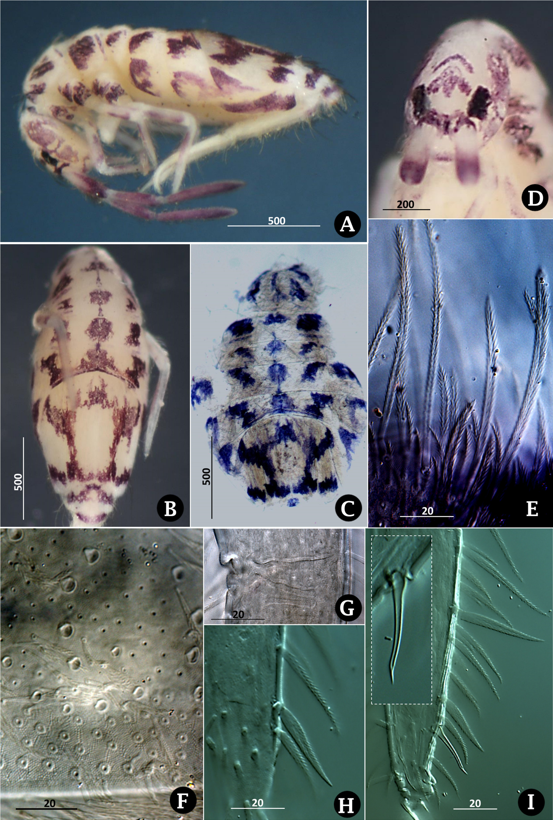
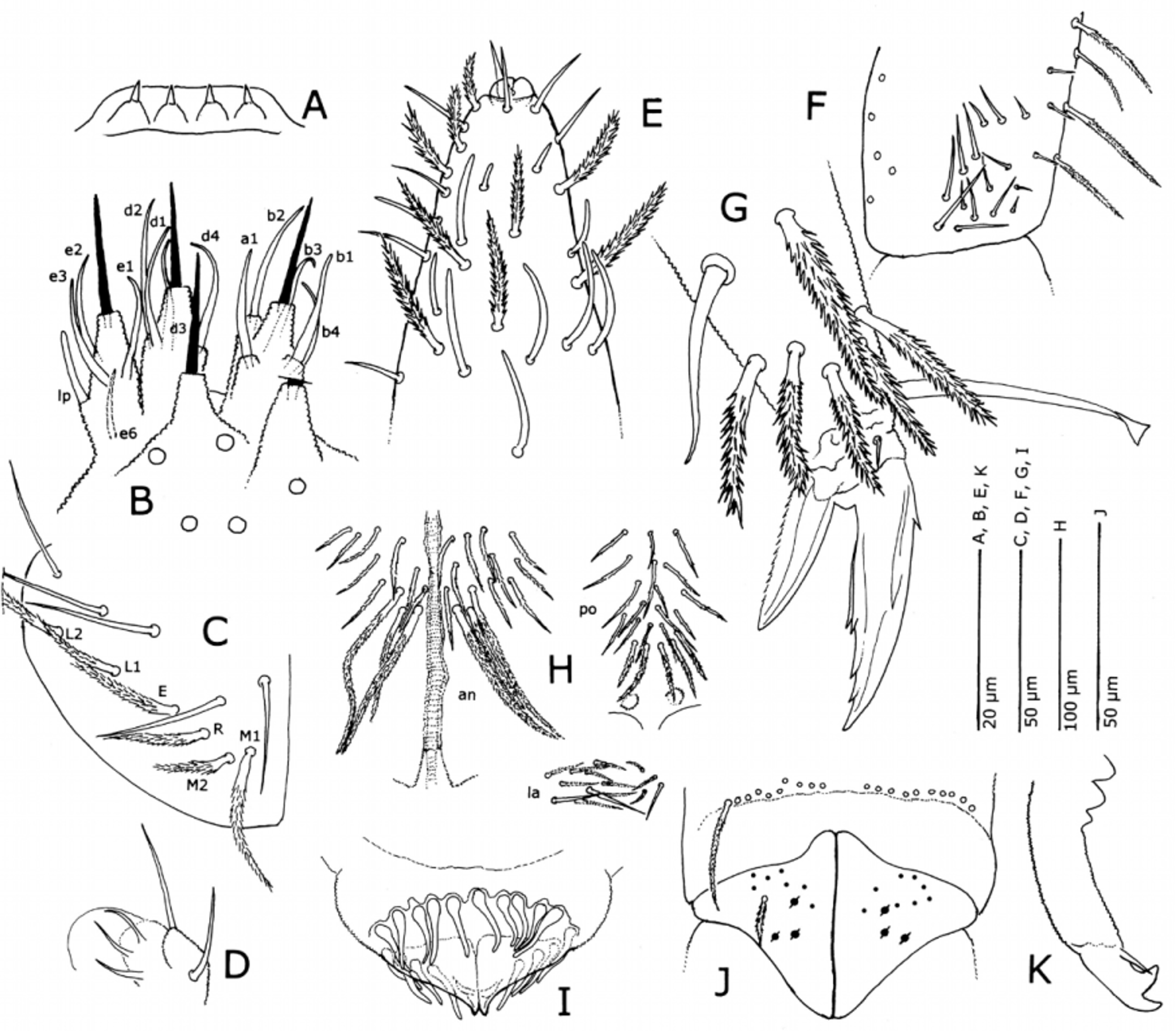
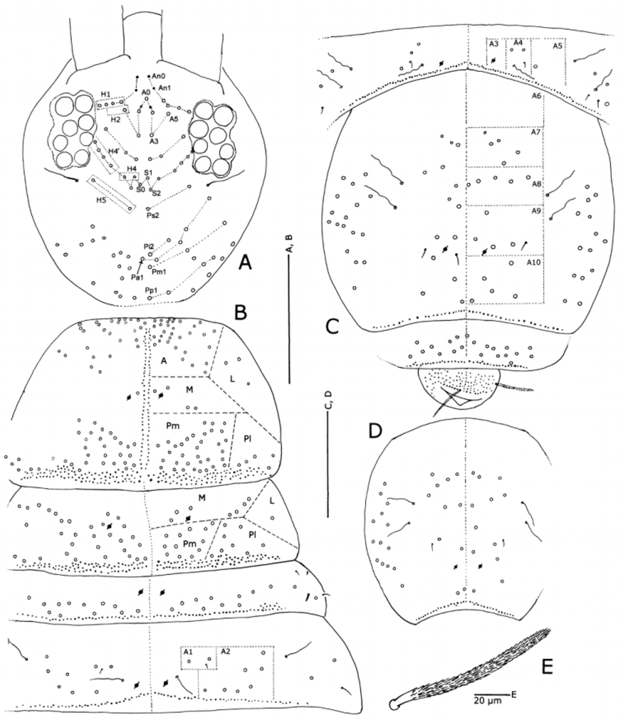
Figures 1‒3 View FIGURE 1 View FIGURE 2 View FIGURE 3
Material examined. CHILE, Rapa Nui, Maunga Hiva Hiva region, holotype female dissected and mounted on three slides, 1 female paratype, 1 male paratype, 5 juvenile paratypes, Cave Q15-074, direct search of fern-moss gardens, 13 July 2009, J. Wynne, coll. Additional paratypes: 1 male, 1 female, and 1 juvenile, same data as holotype except collected at fig tree in entrance; 3 specimens, sex undetermined, from Cave Q15-076/078, direct search of fern-moss gardens in entrance 3, 0 4 July 2009, J. Wynne, coll.; 2 juveniles from Cave Q15-127, opportune collection within entrance, 0 2 July 2009, J. Wynne, coll.
Description. Males and females similar except where noted. Length up to 2.3 mm. macrosetae cylindrical, rounded at tip ( Fig. 3 View FIGURE 3 E). In ethanol background color light cream-yellow, markings primarily reddish to brownish violet ( Figs. 1 View FIGURE 1 A‒D). Ant. I pale, Ant. II pale proximally, red-violet distally, Ant. III pale at base otherwise pigmented, Ant. IV completely violet. Head ( Fig. 1 View FIGURE 1 B) with strong interantennal band, trident-shaped spot between eyepatches, and lunate marking posteriorly; genae with prominent light violet patches extending nearly head length ( Fig. 1 View FIGURE 1 A). Prothorax with violet spots dorsally. Mesothorax, metathorax and Abd. I with three broken bands of pigment, forming large spots. Abd. II and III with five broken longitudinal bands, including a lateral band on each side. Median band of spots on mesonotum, metanotum and Abd. I‒III tergites connected by thin line of pigment. Lateral pigment on Abd. IV consisting of anterior V-shaped marking and compact posterior spot. Dorsum of Abd. IV with large clear central area surrounded by spots and posterior transverse band joined to form a five-armed figure ( Figs. 1 View FIGURE 1 B, C), medial stripe lacking; anterior spots on Abd. IV of male weakly connected. Abd. V with arched transverse band, Abd. VI with posterior band. Femora and tibiotarsi with wide purple bands. Furcula not pigmented. Juveniles with smaller, discontinuous spots on Abd. IV.
Apical bulb of antenna weakly bilobed or entire, pin seta entire; apex of Ant. IV with mix of smooth, pointed setae, multiciliate setae, and sensilliform setae of various lengths ( Fig. 2 View FIGURE 2 E). Prelabral setae ciliate, labral setae smooth. Labral papillae with single points ( Fig. 2 View FIGURE 2 A). Labial palp ( Fig. 2 View FIGURE 2 B) with all guard setae except e4, e5 and e7; guard setae a1 and b1 stout, arising well up on papilla B; lateral process of papilla E stout, rounded apically, not reaching spine base; five proximal setae present. Labial triangle setae in female arranged as M1M2REL1L2A1‒5, seta M2 shorter than but similar to neighboring setae ( Fig. 2 View FIGURE 2 C). Outer lobe of maxillary palp with three sublobal hairs ( Fig. 2 View FIGURE 2 D). Eight eyes in eye patch, eyes A and B slightly larger than others ( Fig. 3 View FIGURE 3 A).
All tibiotarsi with prominent false joint at midpoint, marked by transverse cuticular grooves ( Fig. 1 View FIGURE 1 G), and with several enlarged ciliate setae among thinner, more coarsely ciliate setae ( Figs. 1 View FIGURE 1 H, I). Inner edge of unguis with four teeth; inner basal paired teeth arising at more than half the distance from ungual base; dorsal tooth and lateral teeth present, lateral teeth not reaching paired inner teeth. Unguiculus lanceolate, with inner edge smoothly curved, outer edge finely serrated. Tenent hair spatulate; all tibiotarsal setae ciliate except for stout, smooth, slightly sinuate ventro-subapical seta on hind tibiotarsus ( Figs. 1 View FIGURE 1 I, 2G). Trochanteral organ of hind leg with up to 18 straight, smooth microsetae of various lengths ( Fig. 2 View FIGURE 2 F). Ventral tube anteriorly with 12+ 12 or 13+13 ciliate setae, distal three setae on each side thickened and much longer than others; 21 posterior ciliate setae, distal four setae thicker than others; each valve with 3 smooth and 10‒14 ciliate setae ( Fig. 2 View FIGURE 2 H). Tenaculum with four teeth on each arm and one anterior seta on the corpus. Manubrial plaque with 8+8 ciliate mesosetae arranged slightly asymmetrically and 3+3 pseudopores arranged in a triangle; 10+10 subdistal ciliated manubrial mesosetae in one row ( Fig. 2 View FIGURE 2 J). Mucro with strong anteapical tooth meeting basal spine; uncrenulated part of dens about 1.7× mucro length ( Fig. 2 View FIGURE 2 K).
Cephalic seta A1 a mesoseta, A0‒A5 macrosetae; An0 and An1 mesosetae; M row with three macrosetae; S0 and S2 posterior to S1 setae, S5 and S 5i macrosetae or large mesosetae. Macrosetal arrangement on posterior region of head asymmetrical with two macrosetae in Ps row and varying numbers in other P-rows ( Fig. 3 View FIGURE 3 A). H1 area with 4 setae, H2 with 1 seta (A5), H3 without setae, H4 with 2 setae, H4’ with 4 setae, H5 with 2 setae ( Fig. 3 View FIGURE 3 A).
Thoracic chaetotaxy ( Fig. 3 View FIGURE 3 B) asymmetrical. On mesonotum, anterior region (zone A) with up to 22 macrosetae exclusive of collar; medial (M) zone with pseudopore and 1‒4 macrosetae; lateral (L) zone, 3 macrosetae; Pm zone, up to 33 macrosetae; Pl zone, 5 or 6 macrosetae. On metanotum, M zone with pseudopore and 7‒10 macrosetae; L zone, 1‒3 macrosetae; Pm zone, 11‒12 macrosetae; Pl zone, 8 macrosetae. Mesonotum with two distinct medial lines of mesosetae ( Fig. 3 View FIGURE 3 B) and 3‒4 irregular rows of mesosetae along posterior margin ( Figs. 1 View FIGURE 1 D, 3B), sockets in this posterior field elevated in contrast to more anterior mesosetal sockets ( Fig. 1 View FIGURE 1 D). Metanotum with similar posterior field but without distinct medial mesosetal rows.
Mesosetae dense and greatly variable in length and diameter ( Fig. 1 View FIGURE 1 E). Abdominal segments II‒V each with single posterior row of short mesosetae; Abd. I with increased number of mesosetae at lateral ends of row ( Figs. 3 View FIGURE 3 B‒D). Abd. I with 12+12 macrosetae and two small, lateral sensilla. On Abd. II setal field A1 with two macrosetae and a sensillum, field A2 with 6‒7 macrosetae. Abd. III without macrosetae in field A3 (macroseta A1 absent), field A4 with 2 macrosetae and smooth sensillum; field A5 with one macroseta; 3 macrosetae, sensillum d2 and a minute microseta in vicinity of bothriotrix m5. Adult Abd. IV ( Fig. 3 View FIGURE 3 C) with asymmetrical arrangement of 14‒19 macrosetae in inner region: field A6 without macrosetae; A7 with 5 macrosetae; A8 with 3‒5 macrosetae; A9 with 3 or 4 macrosetae and 1 or 2 smooth microsetae; A10 with 3‒5 macrosetae. Lateral region with 14‒17 macrosetae. Abd. V with 20 asymmetrically arranged macrosetae; Abd. VI without macrosetae. Juvenile Abd. IV macrosetal numbers and arrangement also asymmetrical ( Fig. 3 View FIGURE 3 D).
Male genital plate with 18 thick setae, with enlarged bases and narrowly rounded apices ( Fig. 2 View FIGURE 2 I).
Etymology. The species was named by combining two Rapanui terms, manu and hoko. Manu is Rapanui for “insect” and used as a noun, while hoko is used as a modifier. Hoko is an ancient form of Rapanui dancing where the dancer, usually male, jumps up and down ( Fortin 2009).
Relationships. The body pattern of Entomobrya manuhoko n. sp. is quite similar to that of E. pulchra Schäffer, 1897 ( Schäffer 1897, Christiansen 1963) ( Argentina, Bolivia) and the Brazilian E. pseudodecora Rapoport, 1962 ( Rapoport 1962) , but differs in the shape of the dorsal pattern on Abd. IV. In E. manuhoko n. sp.
adults have a five-armed figure without a medial band ( Fig. 1 View FIGURE 1 B), fancifully resembling a squat penguin or a swimming turtle. The pattern on Abd. IV of E. puchra and E. pseudodecora has disconnected spots or a weak to strong medial band of pigment, which E. manuhoko n. sp. lacks. The mesonotum of E. manuhoko n. sp. has lateral and medial spots, similar to that of E. pseudodecora , whereas E. pulchra has a U-shaped figure. The outer edge of the unguiculus of E. manuhoko n. sp. is serrated, whereas the unguiculi of the other two species are illustrated as smooth. The descriptions of E. pulchra and E. pseudodecora are brief and lack details of the chaetotaxy.
No known copyright restrictions apply. See Agosti, D., Egloff, W., 2009. Taxonomic information exchange and copyright: the Plazi approach. BMC Research Notes 2009, 2:53 for further explanation.
|
Kingdom |
|
|
Phylum |
|
|
Class |
|
|
Order |
|
|
Family |
|
|
Genus |