
Pseudodiaptomus nansei, Sakaguchi, Sakiko Orui & Ueda, Hiroshi, 2010
|
publication ID |
https://doi.org/10.5281/zenodo.198129 |
|
DOI |
https://doi.org/10.5281/zenodo.5657441 |
|
persistent identifier |
https://treatment.plazi.org/id/F811878A-921A-AC16-FF16-F9BDFBD4B2B7 |
|
treatment provided by |
Plazi |
|
scientific name |
Pseudodiaptomus nansei |
| status |
sp. nov. |
Pseudodiaptomus nansei sp. nov.
( Figs. 2 View FIGURE 2 , 3 View FIGURE 3 A, 4–6)
Synonym. Pseudodiaptomus inopinus Burckhardt, 1913 , Oka et al. (1991), 85, fig. 3.
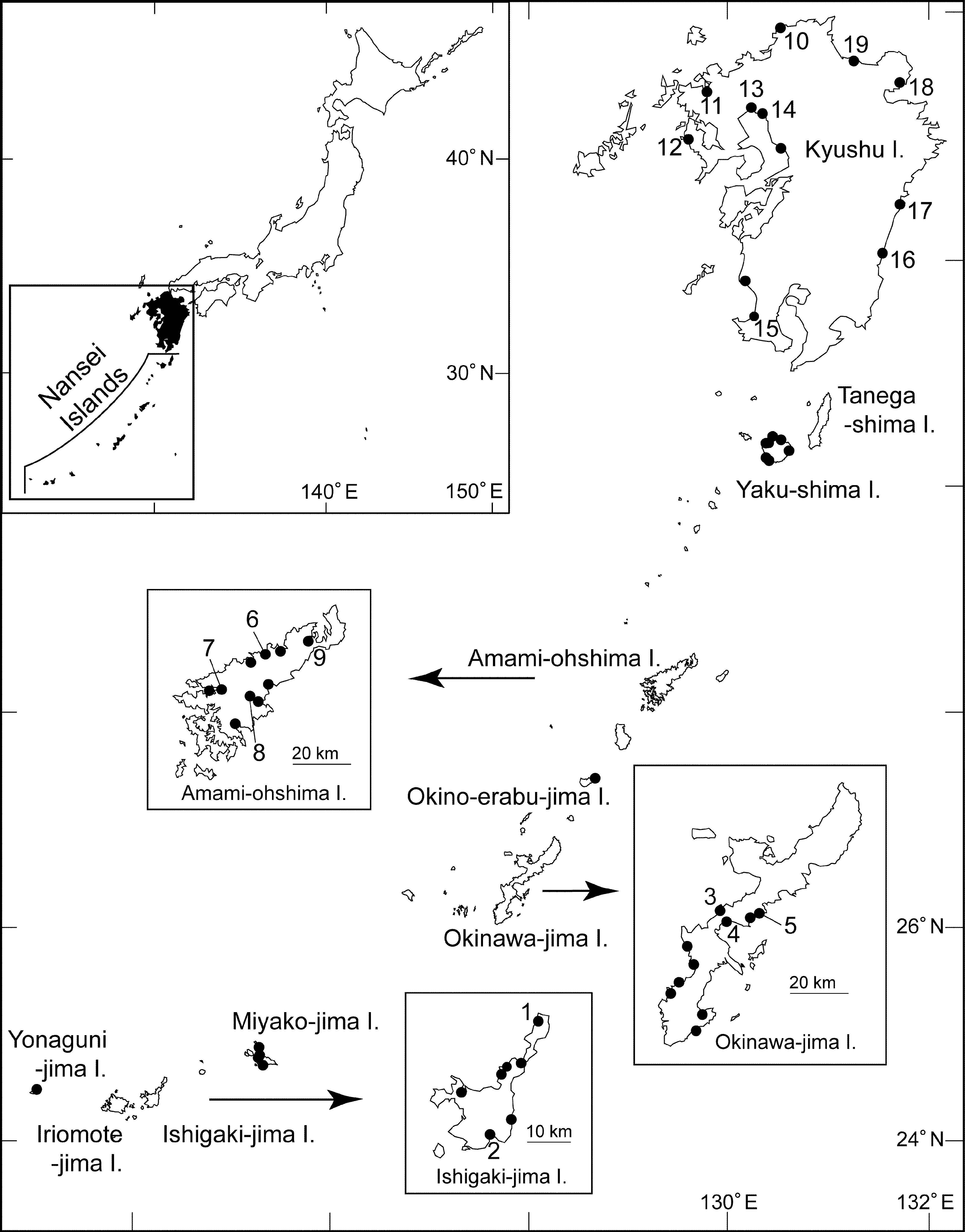
Type material. Female holotype (NSMT-Cr 21259), male allotype (NSMT-Cr 21260) dissected in lactophenol and mounted on 8 glass slides using CMC-10, aqueous mounting medium (Masters Company, Inc., Wood Dale, IL), and undissected 8 female and 10 male paratypes (NSMT-Cr 21261, 21262) in alcohol were deposited in the NSMT. Undissected 10 female and 10 male paratypes in alcohol were deposited in the USNM ( USNM 1145707, 1145708). All type specimens ( 57 females and 48 males including the sequenced specimens) were collected from the mouth of the Hirakubo-gawa R. ( 24°35’39”N, 124°18’48”E, 10 October 2008, colls S.O. Sakaguchi and H. Ueda; # 1 in Fig. 1 View FIGURE 1 ) of Ishigaki-jima Island on 10 October 2008.
Other material examined. Five females and 4 males from the Miyara-gawa R. ( 24°21’31”N, 124°12’42”E, 10 October 2008; # 2 in Fig. 1 View FIGURE 1 ), southern Ishigaki-jima Island; 12 females and 4 males from the Yakashimoguchi-gawa R. ( 26°28’50”N, 127°50’48”E, 12 October 2008; #3) on the west coast, 1 male from the Tokuhina-gawa R. ( 26°27’16”N, 127°51’23”E, 12 October 2008; #4) and 8 males from the Ginozafukuchi-gawa R. ( 26°28’22”N, 127°57’05”E, 12 October 2008; #5) on the east of central Okinawajima Island; 1 female from the Chinaze-gawa R. ( 28°22’46”N, 129°26’47”E, 15 October 2008; #6) and 1 female from the Kawauchi-gawa R. ( 28°16’17”N, 129°18’28”E, 15 October 2008; #7) on the western coast, and 2 females and 8 males from the Yakugachi-gawa R. ( 28°15’17”N, 129°24’14”E, 15 October 2008; #8) and 5 females and 25 males from the Ura-gawa R. ( 28°24’36”N, 129°35’29”E, 15 October 2008; #9) on the eastern coast of Amami-ohshima Island. All the specimens were collected by S.O. Sakaguchi and H. Ueda.
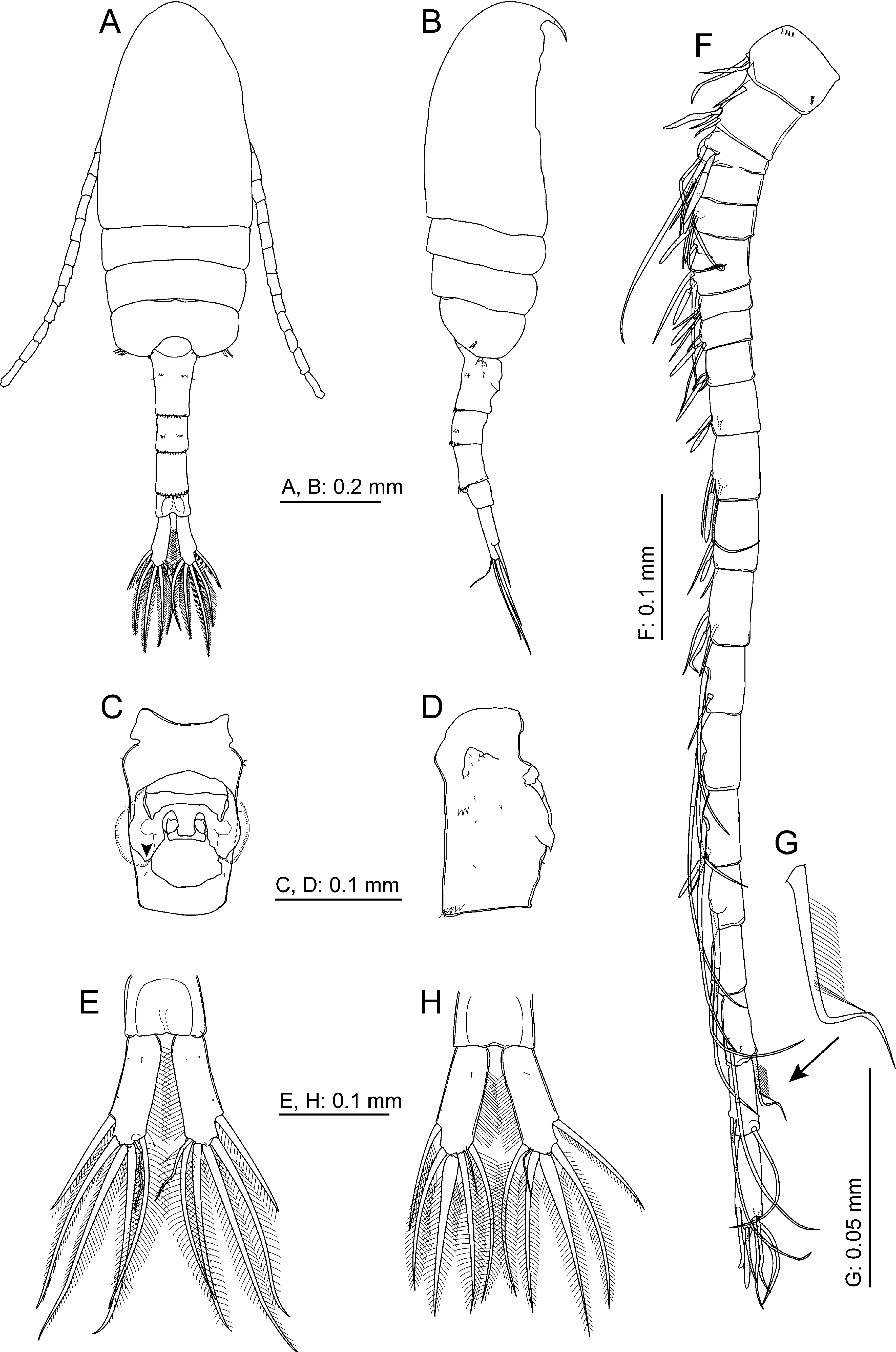
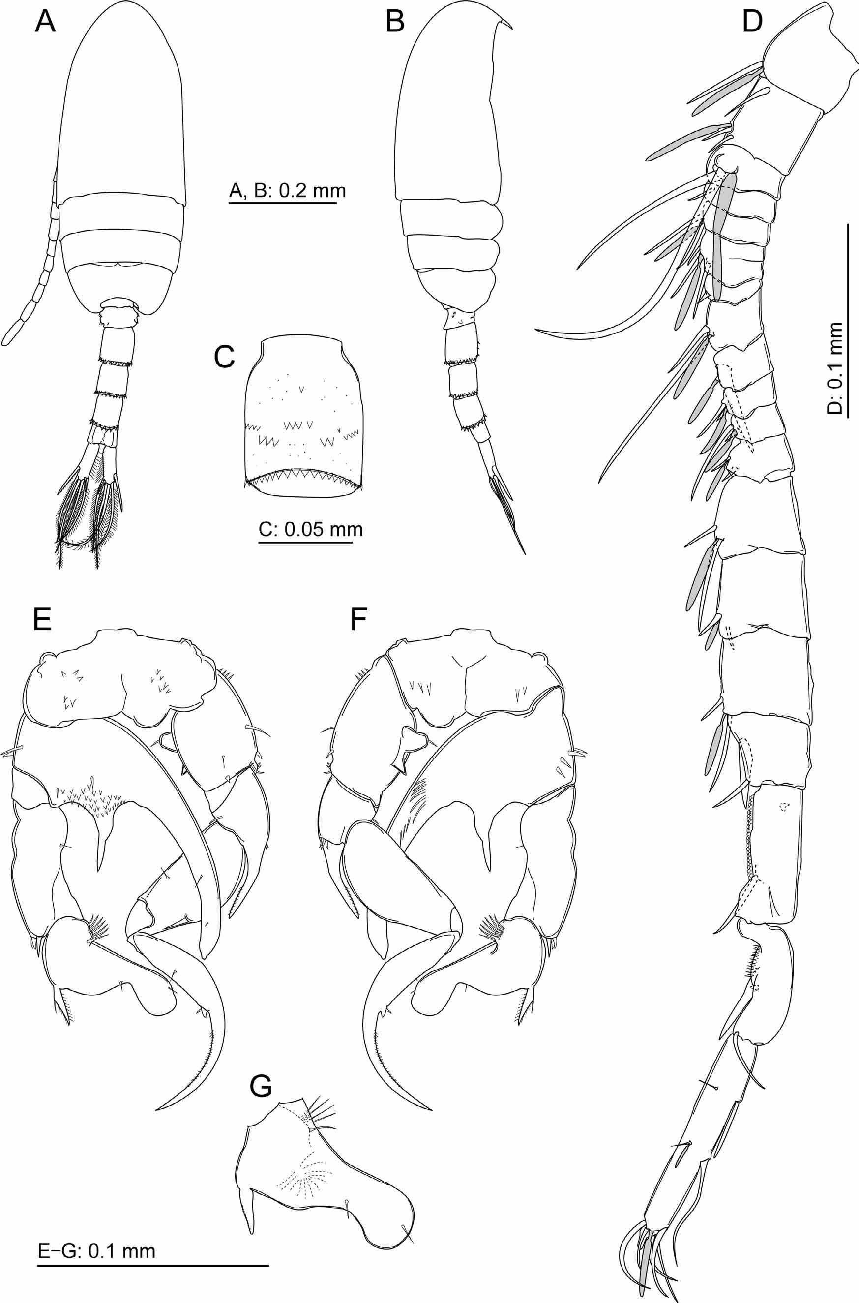
Description. FEMALE ( HOLOTYPE). Body ( Fig. 2 View FIGURE 2 A, B) length 1.13 mm; prosome length 0.65 mm. Forehead rounded in dorsal and lateral views. Cephalosome and first pediger fused; fourth and fifth pedigers fused; dorsal and lateral surfaces of pedigers smooth, except 3–4 spinules on each posterolateral rounded corner of fifth pediger. Urosome symmetrical. Genital double-somite 1.3 times longer than wide, with several spinules on each anterolateral projection, long anterolateral seta and dorsolateral row of spinules at one-third anteriorly on each side; genital operculum ( Figs. 2 View FIGURE 2 C, D, 3A) with broad hyaline frill laterally and rounded posterior process (indicated by arrowhead in Fig. 2 View FIGURE 2 C); second urosomite with dorsal row of spinules at on each side; genital double-somite and second and third urosomites with posterior row of spinules along dorsal margin. Caudal rami ( Fig. 2 View FIGURE 2 E) symmetrical, 3.3 times longer than wide; 1 lateral and 4 terminal caudal setae thin and dorsal seta short and sinuate; lateral medial terminal caudal seta (longest caudal seta) twice longer than ramus.
Antennule ( Fig. 2 View FIGURE 2 F) 22-segmented with incomplete suture between sixth to seventh segments; setal formula as follows: 1=1+ae (aesthetasc), 2 = 3 + ae, 3 = 2 + ae, 4 = 2 + ae, 5 = 3 + ae, 6 = 1 (spiniform), 7 = 2 + ae, 8 = 2 + ae, 9 = 2 + ae, 10 = 2(1 spiniform) + ae, 11 = 2 + ae, 12 = 2 + ae, 13 = 2 + ae, 14 = 2 + ae, 15 = 2, 16 = 2, 17 = 2 + ae, 18 = 1, 19 = 1, 20 = 2, 21 = 2, 22 = 6 + ae; 20th segment with seta bearing unique row of dense hairs ( Fig. 2 View FIGURE 2 G).
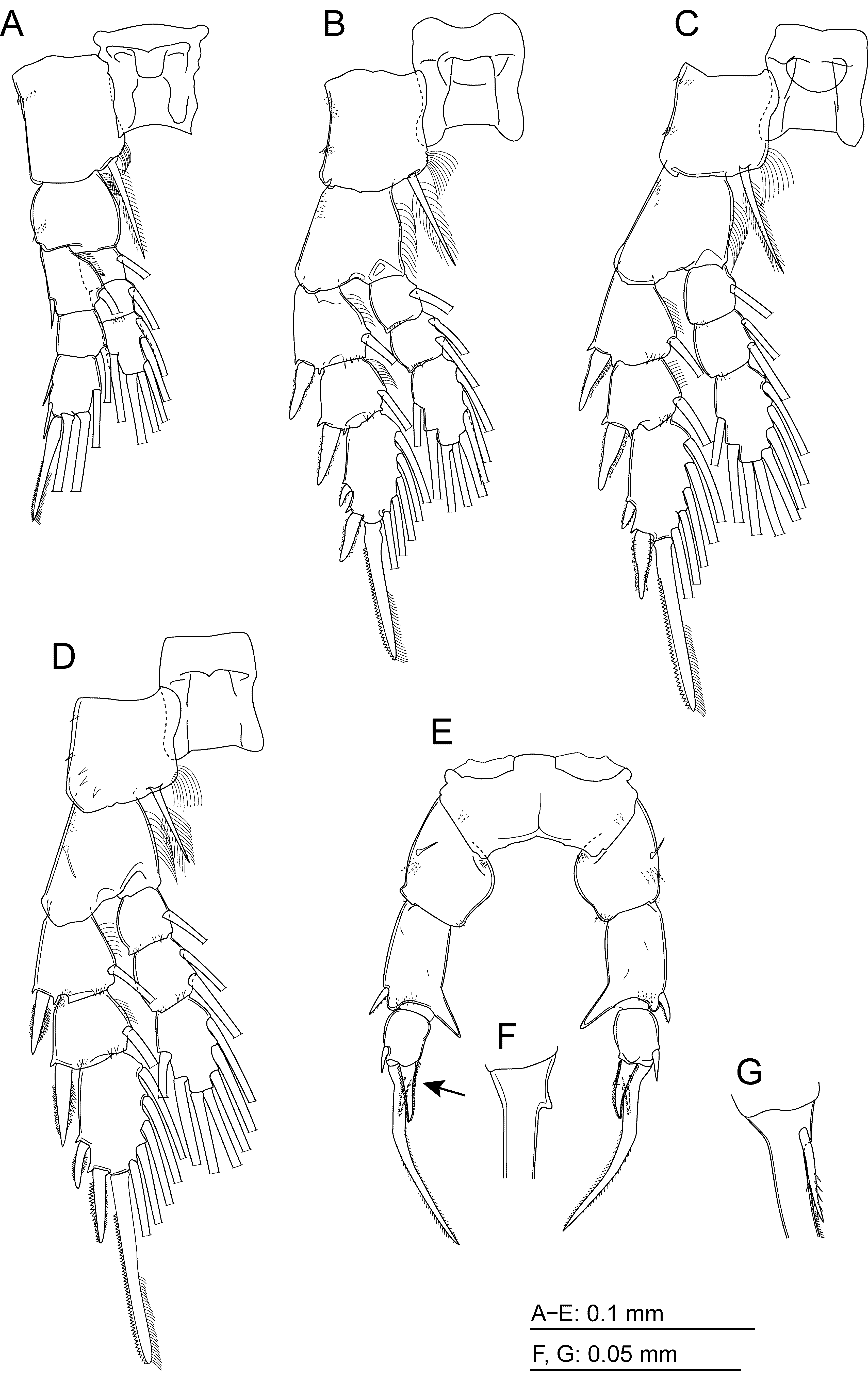
Antenna ( Fig. 4 View FIGURE 4 A) coxa and basis and first endopodal segment completely fused; coxa with seta; basis with 2 setae; first endopodal segment with 2 setae, second segment with 9 subterminal and 7 terminal setae; exopod 5-segmented, with setal formula 1, 5, 1, 2, 3.
Mandible ( Fig. 4 View FIGURE 4 B) basis with 4 setae; exopod 5-segmented, with seta on each first to fourth segments and 2setae on fifth segment; endopod 2-segmented, with 4 setae on first segment and 9 setae.
Maxillule ( Fig. 4 View FIGURE 4 C) praecoxal arthrite with 9 spines and 6 setae; coxal endite with 4 setae, epipodite with 9 setae; basal endites with 3 and 5 setae, exite with seta; exopod with 9 seta; endopod 3-segmented, with setal formula 4, 4, 7
Maxilla ( Fig. 4 View FIGURE 4 D) praecoxal with 4 setae on first endite and 3 setae on second endite; coxal endites with 3 and 3 setae; basal endite with 4 setae; endopod 4-segmented, with setal formula 2, 3, 2, 2.
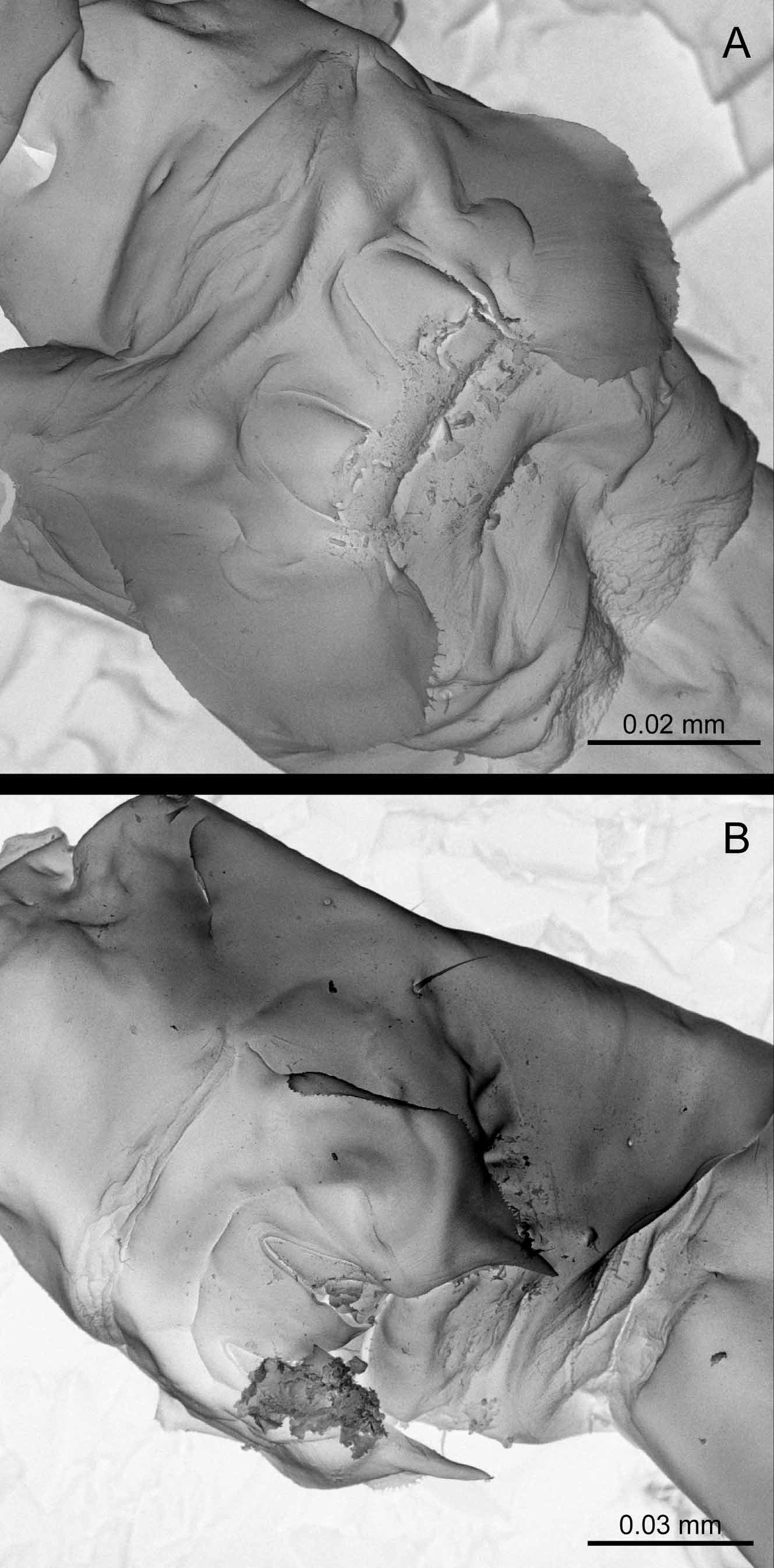
Maxilliped ( Fig. 4 View FIGURE 4 E) coxa with 2, 3, 4 setae on first to third endites; basis with 3 setae; endopod 6- segmented, with setal formula 2, 3, 2, 3, 3, 4; 2 setae on second segment and seta on third segment of endopod modified as shown in Fig. 4 View FIGURE 4 F and seta on fourth segment short and bearing teeth-like spinules.
Legs 1–4 ( Fig. 5 View FIGURE 5 A–D, Table 1). Terminal spines on third exopodal segments of legs 1–3 with row of hairs along distal half of medial margin.
Coxa Basis Exopodal segment Endopodal segment Leg 1 0-1 0-0 I-1; 0-1; II, I, 3 0-1; 0-1; 1, 2, 3 Leg 2 0-1 0-0 I-1; I-1; II, I, 5 0-1; 0-2; 2, 2, 4 Leg 3 0-1 0-0 I-1; I-1; II, I, 5 0-1; 0-2; 2, 2, 4 Leg 4 0-1 1-0 I-1; I-1; II, I, 5 0-1; 0-2; 2, 2, 3 Leg 5 ( Fig. 5 View FIGURE 5 E) symmetrical; coxa with spinules anterolaterally; basis with proximal and distal spinules medially, posterior seta and anterior spinules laterally. Exopod 3-segmented; first segment with pointed distomedial process, distolateral spine, and anterodistal row of spinules; second segment with 1 large medial and 1 small lateral spines; third segment representing long terminal spine with notch ( Fig. 5 View FIGURE 5 F) and short anterior spine at base ( Fig. 5 View FIGURE 5 G).
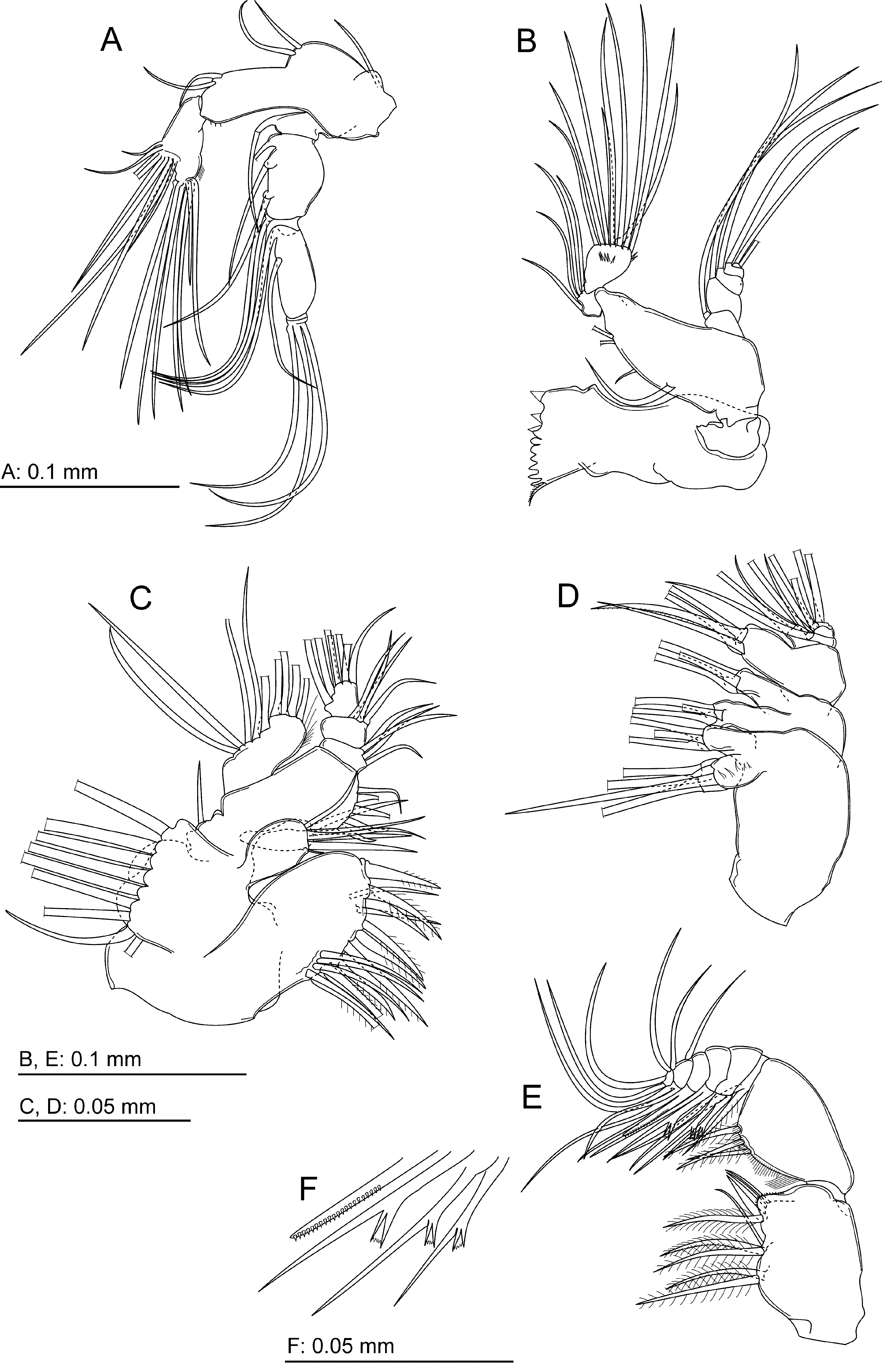
MALE (ALLOTYPE). Body ( Fig. 6 View FIGURE 6 A, B) length 0.90 mm, prosome length 0.54 mm. Cephalothorax as in female. Fifth pediger with no spinules on each posterolateral rounded corners. Genital somite with small posterolateral process on each side; second urosomite ventrally with transverse rows of spinules ( Fig. 6 View FIGURE 6 C); second to fourth urosomites fringed with spinules on whole margins.
Right antennule ( Fig. 6 View FIGURE 6 D) 20-segmented, with sixth to seventh segments incompletely fused; setal formula as follows: 1 = 1 + ae, 2 = 3 + ae, 3 = 2 + ae, 4 = 1, 5 = 2 + ae, 6 = 1, 7 = 2 + ae, 8 = 1 (spine), 9 = 2 + ae, 10 = 1 (spine) + ae, 11 = 2 (1 spine) + ae, 12 = 2 (1 spine) +ae, 13 = 2 (1 spine) + ae, 14 = 2 + ae, 15 = 2 + ae, 16 = 2 + ae, 17 = 2 (1 spine), 18 = 2 (1 pine), 19 = 3 (2 spine), 20 = 9 + 2ae; 18th segment with serrate ridge; 19th segment with hirsute proximal ridge; length ratios of 18–20th segments to 17th segment 1.9, 1.6, 3.2, respectively.
Leg 5 ( Fig. 6 View FIGURE 6 E, F) coxa with spinule patch on both surfaces. Left basis and endopod completely fused, produced into 2 large medial processes, medial one large and laterally curved with anterior row of long spinules at mid length, and distal one smaller, and discontinuously tapering into thin spiniform process with many spinules on posterior surface of base, and with lateral seta and anterolateral row of spinules. Left exopod 2-segmented; first segment rectangular with spine and spinules at distolateral corner, second segment thumb-shaped with lateral notch one-third from tip, proximomedial row of spinules, proximolateral spine, 2 setae each at notch and tip. Right leg basis with 2 medial processes, proximal one rounded, tip bearing hairlike spinule, and distal one triangular, and with proximolateral spinules, lateral seta and distal spinules. Exopod 3-segmented; first segment with fused thick curved terminal spine extending at most to half length of second segment, with proximomedial spinule and anterolateral spinules; third segment shaped falcate long, proximally swollen along lateral margin with maximum width at proximal one-third, bearing medial seta and medial triangular process with seta, terminal third with medial spinules.
Other appendages as in female.
Variability. The body length ranged from 1.10–1.13 mm (n=5) in females and 0.86–0.91 mm (n=5) in males, and the prosome length 0.65–0.67 mm in females and 0.54–0.55 mm in males. One female specimen from the Yakugachi-gawa River had slightly swollen setae on the caudal rami ( Fig. 2 View FIGURE 2 H). The male first urosomite lateral spinules and second urosomite ventral spinule rows varied and in some specimens the rows resembled those of P. inopinus Burckhardt, 1913 described below. Male left leg 5 posterior spinules patch on the fused basis-endopod segment was absent in specimens from the Amami-ohshima Island. Male left leg 5 the second exopodal segment of a specimen from the Ura-gawa River was the intermediate form between thumb- and paddle-types by having a weak lateral depression, and a spinule patch on the anterior surface ( Fig. 6 View FIGURE 6 G), which was absent in the thumb-type specimens.
Etymology. The species name is a noun in apposition derived from the Nansei Islands , the distribution range of the species.
Remarks. This new species was collected from three islands of the Nansei Islands in southern Japan: Ishigaki-jima, Okinawa-jima and Amami-ohshima Islands ( Fig. 1 View FIGURE 1 ), but not found in the samples from Kyushu and the other islands of the Nansei Islands . Salinity and temperature at the sampling sites of the species ranged from 0.3–25.1 and from 21.6–29.7°C, respectively. This species was especially dominant at the site of the Hirakubo-gawa River, Ishigaki-jima Island, where the salinity ranged from 3.1–6.2. Oka et al. ’s (1991, fig. 3) specimens described as P. inopinus from Iriomote-jima, Ishigaki-jima, Okinawa-jima, Amami-ohshima and Tanegashima Islands of the Nansei Islands are here attributed to P. nansei . The authors published illustrations showing the female caudal rami with thin terminal setae and the male right leg 5 with the third segment swollen at the proximal one-third. Oka et al. (1991) and Oka and Saisho (1994) also recorded the dominance of P. nansei in brackish waters of the Nakama-gawa River (salinity 1.5), Iriomote-jima Island, and the Sumiyo-gawa River (salinity 21.8±7.5), Amimi-oshima Island, respectively.
No known copyright restrictions apply. See Agosti, D., Egloff, W., 2009. Taxonomic information exchange and copyright: the Plazi approach. BMC Research Notes 2009, 2:53 for further explanation.
|
Kingdom |
|
|
Phylum |
|
|
Class |
|
|
Order |
|
|
Family |
|
|
Genus |