
Flannerystrongylus abulus, Smales, 2019
|
publication ID |
https://doi.org/ 10.11646/zootaxa.4679.1.7 |
|
publication LSID |
lsid:zoobank.org:pub:49CC819D-538B-4623-A2C6-A947D2AAB18C |
|
DOI |
https://doi.org/10.5281/zenodo.5616781 |
|
persistent identifier |
https://treatment.plazi.org/id/4F3B678F-A947-4EF9-A53B-6658FF654BE9 |
|
taxon LSID |
lsid:zoobank.org:act:4F3B678F-A947-4EF9-A53B-6658FF654BE9 |
|
treatment provided by |
Plazi |
|
scientific name |
Flannerystrongylus abulus |
| status |
sp. nov. |
Flannerystrongylus abulus sp. nov.
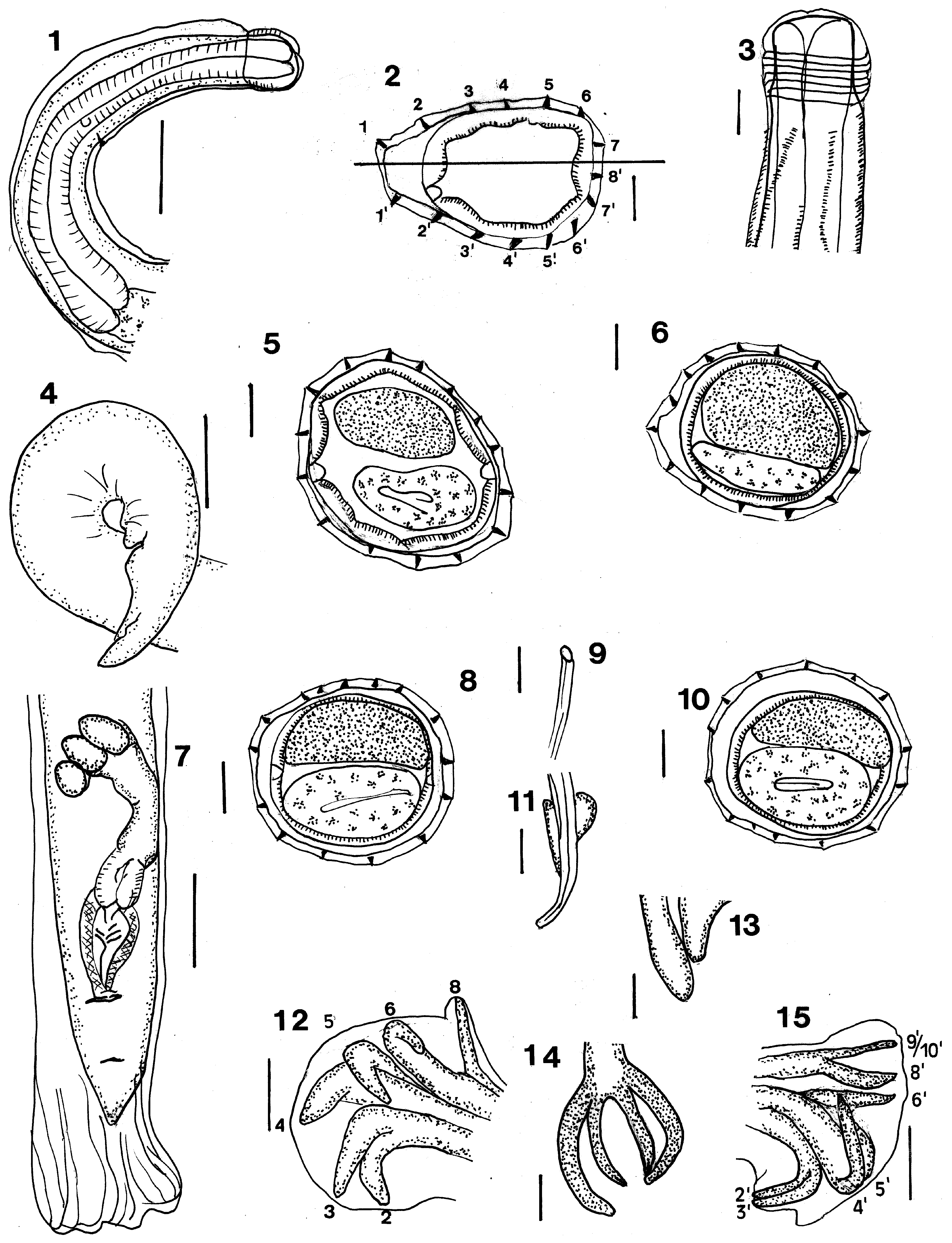
( Figs 1–15 View FIGURES 1–15 )
urn:lsid:zoobank.org:act:4F3B678F-A947-4EF9-A53B-6658FF654BE9
Type host. Paramelomys platyops (Thomas)
Site in host. Small intestine.
Material examined. Holotype male AM W.51743, GoogleMaps allotype female AM W.51744, Kampong Korido , Supiori Island, Papua, Indonesia (00° 50´S 135° 36´E) from Paramelomys platyops coll T. Flannery, A. Szalay 22. ix. 1992. GoogleMaps
Other material examined. From Paramelomys platyops from Papua New Guinea, Madang Province; 3 males, 6 females, 8 male or female pieces Wanuma, BBM NG103708A;
Etymology. The species name is derived from abul, a local name used in Sanduan Province, for P. platyops .
Description. General: Relatively large nematodes, coiled anteriorly. Cephalic vesicle prominent with about 8 transverse annulations, buccal capsule vestigial. Mouth opening triangular, with rudimentary lips; cephalic and labial papillae not seen. Oesophagus claviform, nerve ring, deirids not seen; excretory pore in mid region of oesophagus.
Synlophe: (sections of 10 specimens) Pointed longitudinal ridges extend from posterior margin of cephalic vesicle to anterior to bursa or vulva; 15 anteriorly, 15 mid body, 14–16, usually 15, posteriorly; axis of orientation sub frontal anteriorly, usually 8 dorsal, 7 ventral ridges, no obvious size gradient either left to right or anterior to posterior.
Male: (measurements of 3 specimens) Length 5400–8500 (6740), maximum width 102–148 (133). Cephalic vesicle 51.0–66.0 (59.9) long. Oesophagus not measured; nerve ring, deirids and excretory pore not observed. Bursa dissymmetrical, left lobe larger, pattern of rays 2–3, dorsal lobe shorter than laterals, dorsal ray dissymmetrical, divided at about 1/3 its length, each branch dividing again at tip, terminal divisions rays 9, 10 symmetrical, rays 8 dissymmetrical left ray larger, arising from dorsal trunk at about same level as dorsal ray divides; lateral rays 6 curved dorsally, 4, 5 ventrally, rays 4 largest, rays 2, 3 diverge ventrally, reaching margin of bursa. Genital cone simple, ventral lip longer. Spicules equal, filiform, 440–550 (485) long, spicule to body length ratio 7.2%, tips simple curved. Gubernaculum not symmetrical, dorsal edge shorter and thicker than ventral edge 27.2–29.7 (28.9) long.
Female: (measurements of 7 specimens) Length 7200–10,000 (8400), maximum width 119.0–181.5 (151.7). Cephalic vesicle 53–83 (69) long. Oesophagus 400–710 (539); nerve ring and deirids not seen, excretory pore 390 from anterior end (1 measurement). Vulva opens 139.0–165.0 (148.6) from tail tip; large plug of material covers ventral surface from vulva to tail tip in most specimens, some specimens with praepuce; tail may be flexed ventrally. Monodelphic ovejector with short vagina, sphincter, 60, shortest element, vestibule 100, infundibulum, 150, longest element (single measurements). Tail bluntly conical 41.0–80.0 (62.3) long. Eggs thin-shelled ellipsoidal, numerous eggs in utero, 51.0 long by 37.4 wide.
Remarks. As noted above these specimens were not well fixed and were difficult to handle. Further, the coiling of the anterior end made it difficult to get accurate measurements of the oesophagus and associated features. Nor could the relationship of ridge 1´to the left lateral ridge be determined with any certainty. Sufficient morphological data, however, could be obtained to characterise the new genus.
Following the keys of Beveridge et al. (2014) Flannerystrongylus gen. nov. can be placed in the family Heligmonellidae , subfamily Nippostrongylinae . The genus differs from the 41 genera described in the family ( Beveridge et al. 2014; Digiani & Durette-Desset 2014; Durette-Desset & Digiani 2015; Smales 2014, 2016, 2017, 2018) in the features of the synlophe: usually 15 ridges, 8 dorsal, 7 ventral, with no obvious size gradients and an axis of orientation sub frontal anteriorly. In the key to the 8 genera of nippostrongylins from the Sahul region erected by Durette-Desset & Digiani (2015) the genus Flannerystrongylus is closest to the genus Equilophos Durette-Desset & Digiani, 2015 but differs in having a smaller number of ridges 14–16 compared with 35–36 ridges. Representatives of a further 19 genera from the small intestine have been described from the Sahul and Malaysian regions and can be distinguished by the form of the synlophe as follows: the cosmopolitan Nippostrongylus Lane, 1923 has a careen supported by a single hypertrophied ridge. Heligmonoides, Baylis, 1928 and Maxomystrongylus Hasegawa & Syafruddin, 1997 have type B careens, Orientostrongylus Durette-Desset, 1970 has a primitive arrangement of the synlophe, Hasanuddinia Hasegawa & Syafruddin, 1994 has left ventral and right dorsal ridges larger, Malaistrongylus Ow Yang, Durette-Desset & Obayashi, 1983 has left and right dilatations of the synlophe, Rattustrongylus, Ow Yang, Durette-Desset & Obayashi, 1983 has 18–19 ridges with the left dorsal ridges smallest, Sabanema, Ow Yang, Durette-Desset & Obayashi, 1983 has more than 30 ridges, the left dorsal and right ventral smallest, Bunomystrongylus Hasegawa & Mangali, 1996 has both pointed internally supported and rounded unsupported ridges, Melomystrongylus Smales & Heinrich, 2010 has a ventral hypertrophied ridge anteriorly, Mawsonema Smales & Heinrich, 2010 has 15 sub frontal ridges anteriorly with a size gradient and a type A careen, Montistrongylus Smales & Heinrich, 2010 has 12–15 ridges left ventral largest, oriented at 55° from ventral left to dorsal right in mid body, Parasabanema Smales & Heinrich, 2010 has 45 ridges with a frontal axis of orientation, Pogonomystrongylus Smales, 2014 has 7–10 ridges with the left ventral ridges hypertrophied, Krishnasamyos Digiani & Durett-Desset, 2014 has 19–21 ridges with ridge 1′ forming a comarete, and the left ridge minute, Syafruddinema Digiani & Durette-Desset, 2014 has 14–17 small to minute unevenly sized ridges, Nuigininema Smales, 2016 has 17 ridges mid body with an oblique axis of orientation and the ventral and ventral right ridges hypertrophied, Rodentanema Smales, 2016 has 6–7 ridges mid body, Parvinema Smales, 2017 has 15–17 ridges with an oblique axis of orientation and a careen, the left lateral ridges the largest, Missimstrongylus Smales, 2018 has 14 ridges in the mid body with ventral ridge 5′ the largest (Durette- Desset 1970; Ow Yang et al. 1983; Hasegawa & Syafruddin 1994a, 1997; Hasegawa & Mangali 1996; Smales & Heinrich 2010; Digiani & Durette-Desset 2014; Smales 2014, 2016, 2018).
| AM |
Australian Museum |
| T |
Tavera, Department of Geology and Geophysics |
No known copyright restrictions apply. See Agosti, D., Egloff, W., 2009. Taxonomic information exchange and copyright: the Plazi approach. BMC Research Notes 2009, 2:53 for further explanation.
|
Kingdom |
|
|
Phylum |
|
|
Class |
|
|
Order |
|
|
Family |
|
|
Genus |