
Mastogloia ovulum Hustedt 1933
|
publication ID |
https://doi.org/ 10.11646/phytotaxa.126.1.1 |
|
persistent identifier |
https://treatment.plazi.org/id/F9712264-4D38-FFC5-39B1-FF28D633FE35 |
|
treatment provided by |
Felipe |
|
scientific name |
Mastogloia ovulum Hustedt 1933 |
| status |
|
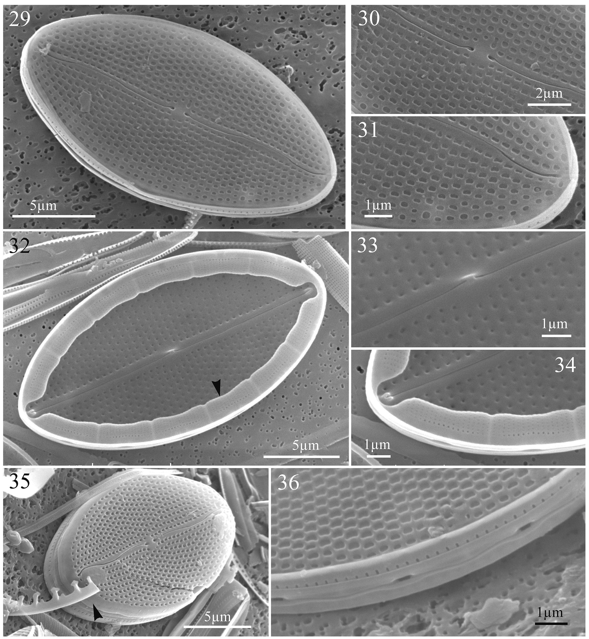
Mastogloia ovulum Hustedt 1933 ( Figs 29–36 View FIGURES 29–36 )
References:— Hustedt 1933, p. 474, fig. 892; non Cholnoky 1968, p. 42, fig. 45; non Montgomery 1978, pl. 141, fig. F (= Mastogloia ovalis ); non Tomàs 1982, figs 18, 19 (= Mastogloia crucicula ); Navarro 1983, p. 121, figs 50, 51; Simonsen 1987, p. 136, pl. 222, figs 1, 2, 6–9, 10?, 11?, non figs 3–5 (= Mastogloia crucicula ); Navarro et al. 1989, p. 352, fig. 70; non Witkowski et al. 2000, p. 255, pl. 75, fig. 14 (= Mastogloia crucicula ); Hein et al. 2008, p. 69, pl. 40, fig. 7.
Material:— Sample from Siladen, Indonesia. SEM stub no. DISVAR-ANS4SP32.
SEM morphology:— The raphe consists externally of two sinuous branches ending centrally in expanded pores and distally in slightly deflected pores toward the same side of the valve ( Figs 29–31, 35 View FIGURES 29–36 ). Internally, the raphe branches run straight and are bordered by ribs, with a rising up central nodule and helictoglossae at poles ( Figs 32–34 View FIGURES 29–36 ). The transapical striae (20–26 in 10 µm) are uniseriate and they vary from parallel at centre to radiate at apices, sometimes forming an irregular quincunx pattern ( Figs 29–31, 35, 36 View FIGURES 29–36 ). Externally, the areolae are apically elongated and more or less rectangular in shape, and they are sunken onto the valve surface ( Figs 29–31, 36 View FIGURES 29–36 ), with none observed on the mantle ( Fig. 35 View FIGURES 29–36 , arrowhead). Internally, the areolae open through small rounded foramen ( Figs 32–34 View FIGURES 29–36 ). Partecta slightly bilobed ( Figs 32 View FIGURES 29–36 , arrowhead, 34), equal in shape and size (1.1–1.5 µm in width), and occupy the entire length of the partectal ring up to the apex ( Figs 32, 34 View FIGURES 29–36 ). Each partectum is ornamented with small pores in parallel rows ( Fig. 34 View FIGURES 29–36 ) and opens externally through an elongate partectal pore positioned opposite to the concave side of the partectal lobes ( Figs 34, 36 View FIGURES 29–36 ). Length: 17.6–23.4 µm; width: 8.9–10.7 µm ( Table 1).
No known copyright restrictions apply. See Agosti, D., Egloff, W., 2009. Taxonomic information exchange and copyright: the Plazi approach. BMC Research Notes 2009, 2:53 for further explanation.
|
Kingdom |
|
|
Phylum |
|
|
Class |
|
|
Order |
|
|
Family |
|
|
Genus |