
Austrognatharia orientis, Achatz, Johannes G. & Sterrer, Wolfgang, 2015
|
publication ID |
https://doi.org/ 10.11646/zootaxa.3955.2.5 |
|
publication LSID |
lsid:zoobank.org:pub:A38F87D7-C55C-4172-831B-D665AFBEF6C6 |
|
DOI |
https://doi.org/10.5281/zenodo.5631770 |
|
persistent identifier |
https://treatment.plazi.org/id/FB11986F-FE77-FF96-FF5B-FF2673D9FE3E |
|
treatment provided by |
Plazi |
|
scientific name |
Austrognatharia orientis |
| status |
sp. nov. |
Austrognatharia orientis View in CoL n. sp.
( Figs. 4–7 View FIGURE 4 View FIGURE 5 View FIGURE 6 View FIGURE 7 ; Table 3 View TABLE 3 )
Diagnosis. Small, fairly slender Austrognatharia (mean length 738 µm, width 96 µm; index 8.40; see Table 3 View TABLE 3 ). Basal plate 6.11 µm long, 20.44 µm wide (index 0.30), with inconspicuous median but pronounced lateral lobes; with at least 8 uneven-sized teeth of which the median and the lateral pair are largest. Jaws 20.78 µm in length, with a strong terminal tooth; dorsal tooth row with two teeth sharing a single root, ventral row with 3–6 unevensized teeth. Conuli small (12.80 µm long by 7.60 µm wide; index 1.76), with a shallow capitulum (length 2.55 µm; index 5.24). Conuli often appear as twins, and may have a flaring cingulum.
Type material. Holotype one adult from Hong Kong (sample #2) in squeeze preparation, USNM 1270622.
Type locality. Sample HK 3 from Hong Kong (see Table 1 View TABLE 1 ).
Other material examined. Seven more specimens (4 adults) from Hong Kong, and five specimens (3 adults) from Shirahama. Semithin and ultrathin sections of one mature specimen from Hong Kong and one mature specimen from Shirahama.
Etymology. From Latin orientis = from the East.
Description. Organization ( Fig. 5 View FIGURE 5 A). Adults measure 600–850 µm in length and 80–120 µm in width (body index 8.40), with a short rostrum (index 0.97).
Body wall. The epidermis is cellular, monociliated, has glandular components and rod-shaped secretory products. The “potatoe-shaped” nuclei of the epidermis lie at the basal side of the cells and possess a dark, homogeneous chromatin. The nuclei are up to 2.5 µm long. The cilia originate from a ciliary pit, which is surrounded by eight microvilli. The ciliary pit has a diameter of 500 nm and is ~ 500 nm deep. The apical cell surface is covered by microvilli, which are 200 nm long, 50 nm wide and occur at a density of ~64/µm2. Many epidermal cells contain vacuoles with glandular content that can nearly fill the entire cell and mitochondria that are ~ 500 nm long and ~ 300 nm in diameter. Specialized cells contain up to 7 rods, which are bluntly pointed at both ends, and measure up to 3.5 µm long and 800–850 µm wide. They are composed of numerous hollow tubes of unknown material, which are arranged parallel to each other in a longitudinal orientation. Occasionally electronlucent vesicles, which have a diameter of ~ 500 nm, are found underneath the apical cell membrane. The epidermal cells are connected with zonula adherentes and apical septate junctions to one another. On the basal side, the epidermis has a distinct basal lamina. Underneath the epidermis lies the body-wall musculature with outer circular muscles and inner longitudinal muscles. However, in the region of the male copulatory organ this pattern is inverted (see below).
Sclerotized parts of the pharynx. The basal plate ( Figs. 4 View FIGURE 4 A, B; 5B, C) is 5–7 µm long and 18–23 µm wide (index 0.30). Its median lobe is shallow whereas its lateral lobes are slightly more defined. Caudally the basal plate is set with up to 9 uneven-sized teeth, of which only three stand out in size. Jaws ( Figs. 4 View FIGURE 4 C; 5B, C) are 19–24 µm long, with a strong terminal tooth and two rows of teeth: a dorsal row of two (more rarely one) teeth of which only the posterior is rooted, and a ventral row in which a larger tooth is followed by up to 5 long but uneven teeth.
Excretory system. The protonephridia ( Fig. 6 View FIGURE 6 A–E) are positioned below the epidermis and are accompanied by two lateral muscles (one muscle shown in Fig. 6 View FIGURE 6 B and C). We found three pairs of protonephridia in the posterior part. They are arranged serially, separated ~50 µm from another, the most posterior pair lying 10 µm in front of the male gonopore. Each protonephridium is composed of three cells: a terminal cell, a canal cell, and a nephroporus cell ( Fig. 6 View FIGURE 6 A–E). The terminal cell encloses the canal cell along its entire apical half or more, its nucleus is situated eccentrically, reaching from the terminal end distally, overlapping with the nucleus of the canal cell for an undetermined distance ( Fig. 6 View FIGURE 6 B, C). At the apical part, the terminal cell lacks cytoplasmic organelles and has lobes with 30-nm small clefts in between, constituting the so-called filtration area. In the central part, the terminal cell, the canal cell and the two adjacent muscles are enclosed by a basal lamina ( Fig. 6 View FIGURE 6 C). The terminal cell has a cilium with the typical 9+2 arrangement and eight microvilli. The cilium of the terminal cell projects into the protonephridial duct of the canal cell, the microvilli of the terminal cell originate from pits and terminate at about half the length of the protonephridial duct ( Fig. 6 View FIGURE 6 A–E). The nucleus of the canal cell is also situated eccentrically and stretches along the distal half of the cell. The cell has many mitochondria and a lacunar system, the tubules of which often appear collapsed in our preparations. The protonephridial duct is surrounded by six clusters of unidentified filamentous material ( Fig. 6 View FIGURE 6 A–E). These clusters form rods, which originate in a position that lies approximately in between the microvilli of the terminal cell ( Fig. 6 View FIGURE 6 A, B) and stretch along the entire length of the canal cell. The nephridial duct opens into the nephroporus, its opening is surrounded either by a stiffened wall or collar or by very long microvilli (at least 250 nm long; Fig. 6 View FIGURE 6 E). The pyriform nephroporus cell encloses the protrusion of the nephridial duct, bears numerous microvilli on its surface, it lacks a cilium but a centriole is present ( Fig. 6 View FIGURE 6 E).
Parenchyma. Wide and flat parenchyma cells occur between the copulatory organs and the body wall. They have more or less rounded nuclei with a diameter of up to 2.5 µm with a small nucleolus and the most electronlucent chromatin of all cell types. The cytoplasm contains only a few organelles. Mitochondria, which can be slightly larger than the ones in the epidermis, are the most numerous ones.
Male system. The single testis is saccular, unpaired and lies dorsally. It contains 6–9 conuli that are usually uniform in size but of various shapes. Conuli of the Hong Kong specimens measured 12–15 µm in length and 7–10 µm in width (index 1.60), with a very shallow capitulum (index 5.40) sitting atop a narrow, rarely flaring cingulum. The larger conuli usually appear as more or less distinct ‘twin conuli’. Conuli of the three mature Shirahama specimens were quite similar in size and proportions but showed the ‘twin’ aspect only once, in a conulus wedged in the penis. The overall structure of the testis as well as the development of the gametes is in concordance with the description in Austrognathia sp. by Lanfranchi and Falleni (1998). Spermatogenesis starts from primary spermatogonia, with a homogeneously condensed nucleus with pronounced nucleolus ( Fig. 7 View FIGURE 7 A) and proceeds to spermatogonia, primary spermatocytes and secondary spermatocytes, which no longer show a nuclear envelop, to spermatids. Late spermatids are completely enclosed by a nurse cell and the chromatin starts to change from an ovoid to a mushroom-like shape with the chromatin showing three grades of condensation ( Fig. 7 View FIGURE 7 B). The cytoplasm of spermatids and sperm is rich in mitochondria, and the more mature the sperm are the more restricted the mitochondria are to the area lateral and below the cingulum ( Fig. 7 View FIGURE 7 B, C, D). In mature sperm, a layer of flocculent electron-dense material appears 150 nm apart from the top of the cingulum ( Fig. 7 View FIGURE 7 E, F, G). The usually single conulus within the bursal tissue goes through a disintegration whereby the restricted distribution of the mitochondria is lost and the tubular system disappears ( Fig. 7 View FIGURE 7 F, G). Like Lanfranchi and Falleni (1998), we found single basal bodies below the cingulum in disintegrating conuli ( Fig. 7 View FIGURE 7 F, G, inserts). The anterior, glandular part of the male copulatory organ ( Fig. 6 View FIGURE 6 G) comprises 24 gland cells, which have nuclei with dark chromatin and a distinct nucleolus, lack microvilli on the apical surface, are rich in cytoplasmic organelles, and produce electron-dense, potato-shaped vesicles with a diameter of ~ 500 nm. More distally a ring of 6 cells, which produce unshaped and chiseled secretion and spherical, electron-dense vesicles with a diameter of ~1 µm joins the penis, which is ~10 µm long, has a diameter of ~9 µm, and consists of ~24 cells. The male genital duct lies in the center of the tube. Distally the penis tube is composed of 16 cells with peripherally positioned nuclei with dark, homogeneous heterochromatin. These cells contain a large number of cytoplasmic organelles, mitochondria measuring ~ 500 nm in length and ~ 300 nm in diameter, and electron-lucent vesicles with a diameter of ~ 800 nm ( Fig. 6 View FIGURE 6 F). These cells lack cilia but bear microvilli with a length of up to 700 nm and are connected with zonulae adherentes and septate junctions to adjacent cells; the basal part is delineated from other tissues by a basal lamina ( Fig. 6 View FIGURE 6 F). The zonulae adherentes stretch 300 nm from apical to basal, the septate junctions up to 1 µm further. The male gonopore lies ~20 µm in front of the posterior end. All ventrally situated longitudinal body-wall muscles bend around the gonopore caudally. In the region from 6 µm in front of the gonopore to 6 µm posterior to the gonopore the circular muscles are positioned inside the longitudinal muscles. About six muscle fibers run along the male copulatory organ and connect it to the testis. At the distal part a thin sphincter encloses the penis tube.
Female system. In a Hong Kong specimen, the ovary (including the single mature, 220 µm long egg) extends from U51.8 to U82.4. No distinct bursa is present but various conuli were found in a state of disintegration close to the most posteriorly lying oocyte ( Fig. 7 View FIGURE 7 FL, G). The conuli were partially enclosed either by gut cells or cells that closely resemble parenchymal cells, except that their nucleus is darker, their cytoplasm richer in organelles, and their cell surface highly interdigitated with gut cells. In one case a laterally lying parenchymal cell extended towards the center of the body and partially covered a conulus.
Remarks. The species is similar to A. homunculus Sterrer, 1991 , and A. mooreensis Sterrer, 1991 , in having one or two teeth in the dorsal row of its jaws, two teeth being more common than one. Both of these species, however, differ in having much larger jaws and basal plates and vastly larger conuli (mean conulus length in A. homunculus is 43.47 µm and in A. mooreensis 39.78 µm). A. orientis has the second smallest conuli in the genus, and A. barbadensis Sterrer, 2011 , has the smallest (length 6.00 µm). The heterogeneity of conuli in A. orientis (within specimens and populations as much as between populations) is puzzling and leaves room for gaining better knowledge of this species.

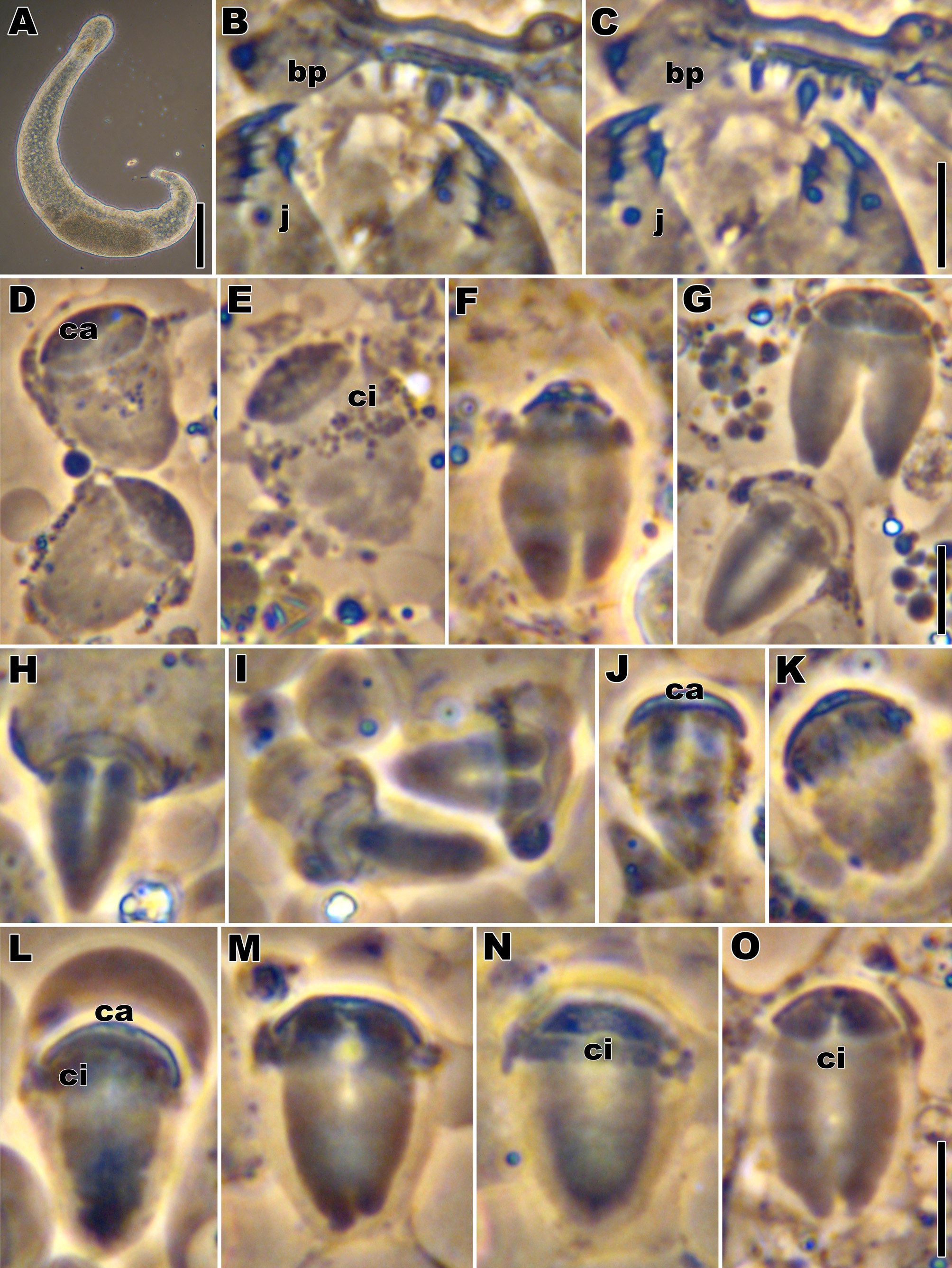


TABLE 3. Morphometric data (in µm) for Austrognatharia orientis n. sp.
| Hong Kong | Mean | StDev | Max | Min | n |
|---|---|---|---|---|---|
| Body length of adults | 760.00 | 95.39 | 850 | 660 | 3 |
| Body width of adults | 92.50 | 10.61 | 100 | 85 | 2 |
| Body index of adults | 8.78 | 0.40 | 9.06 | 8.50 | 2 |
| Rostrum index of adults | 0.97 | 1 | |||
| Jaw length | 19.75 | 0.96 | 21 | 19 | 4 |
| Basal plate length | 6.25 | 0.50 | 7 | 6 | 4 |
| Basal plate width | 20.25 | 2.22 | 23 | 18 | 4 |
| Basal plate index | 0.31 | 0.02 | 0.33 | 0.29 | 4 |
| Sperm length | 13.50 | 1.38 | 15 | 12 | 6 |
| Sperm width | 8.50 | 1.05 | 10 | 7 | 6 |
| Sperm index (sp l/w) | 1.60 | 0.15 | 1.80 | 1.40 | 6 |
| Capitulum length | 2.60 | 0.55 | 3 | 2 | 5 |
| Capit. index (sp l/cap l) | 5.40 | 0.55 | 6 | 5 | 5 |
| USNM |
Smithsonian Institution, National Museum of Natural History |
No known copyright restrictions apply. See Agosti, D., Egloff, W., 2009. Taxonomic information exchange and copyright: the Plazi approach. BMC Research Notes 2009, 2:53 for further explanation.