
Enchodelus ameliae, Guerrero & Liébanas & Santiago, 2008
|
publication ID |
https://doi.org/ 10.1163/156854108785787208 |
|
DOI |
https://doi.org/10.5281/zenodo.8114884 |
|
persistent identifier |
https://treatment.plazi.org/id/FC5FD70C-FFB8-FFBF-4C6E-FB2AFE78FE55 |
|
treatment provided by |
Carolina |
|
scientific name |
Enchodelus ameliae |
| status |
sp. nov. |
Enchodelus ameliae * sp. n.
( Figs 4B View Fig ; 5 View Fig , 6 View Fig )
MATERIAL EXAMINED
Three females and eight males from only one sample in Sierra Mágina, province of Jaén.
MEASUREMENTS
See Table 1. View Table 1
DESCRIPTION
Female
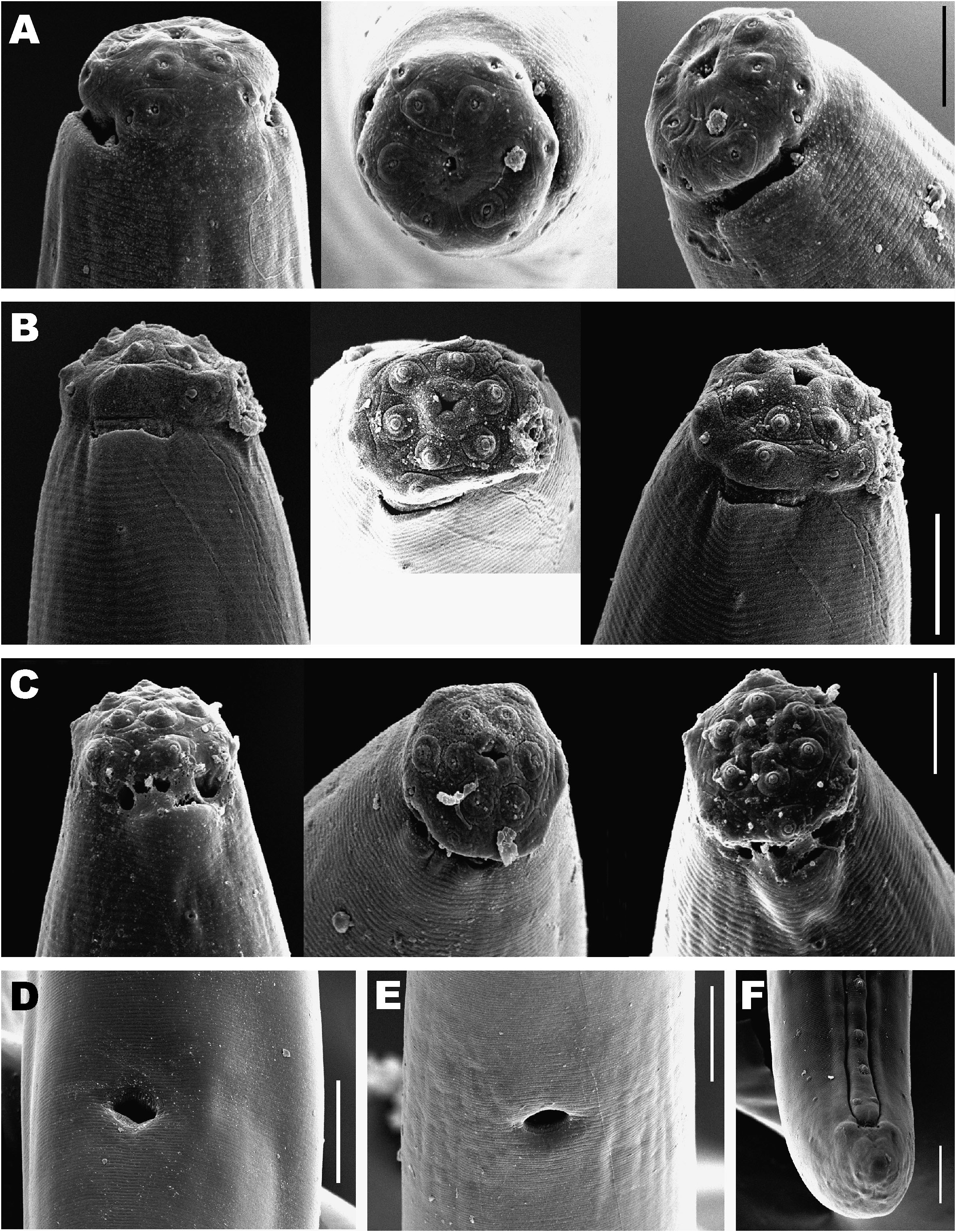
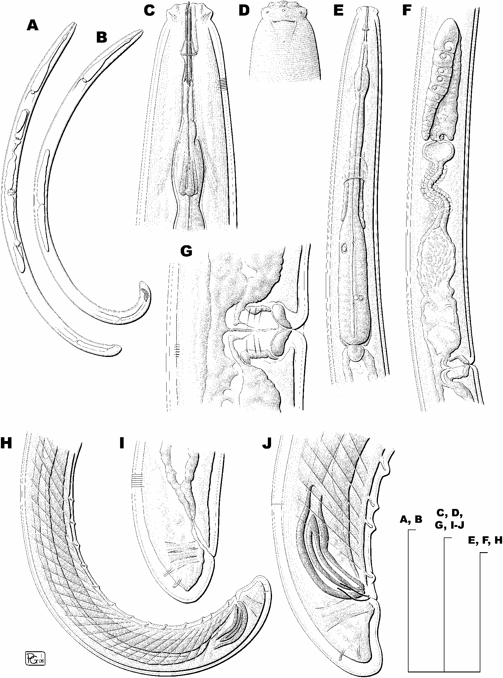
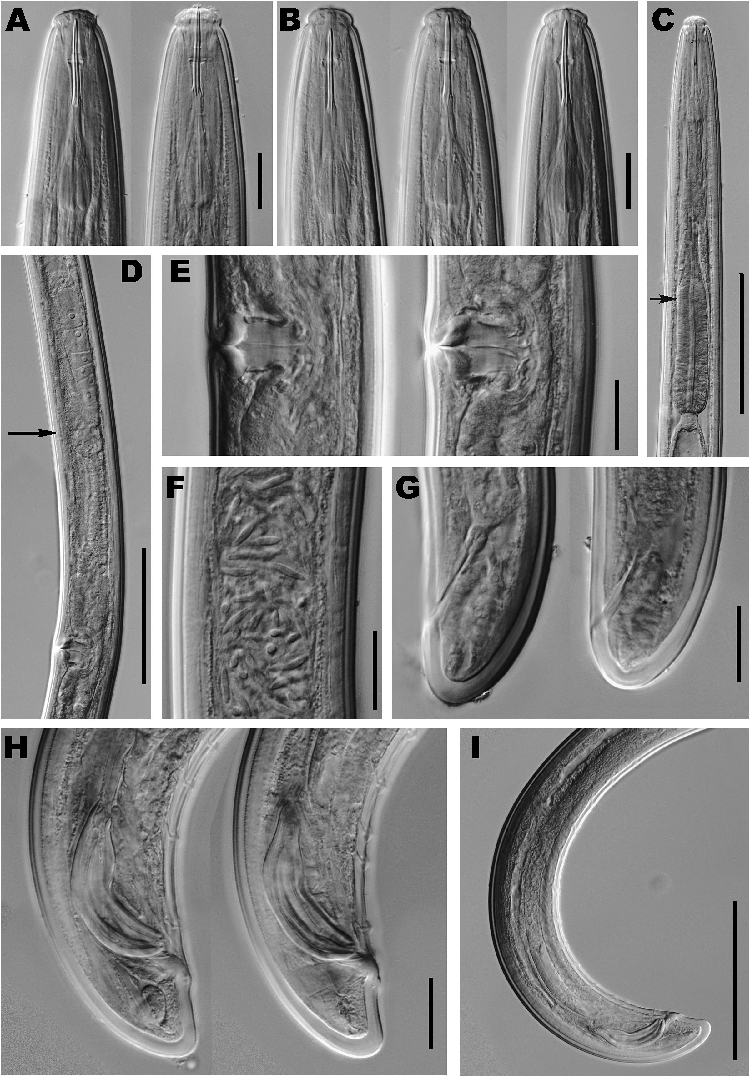
Moderately slender, medium-sized nematodes, 1.37- 1.55 mm long. Habitus after fixation curved ventrad, adopting a rather open C-shape. Body cylindrical, tapering towards both ends but more so anteriorly. Cuticle 2.5- 3.0 µ m in anterior region, 3.0 µ m at mid-body and 5.0-6.5 µ m on tail; its outer layer marked with very fine transverse striations, and thinner than inner one, especially at tail level. Lateral chord very narrow, ca 5 µ m wide or occupying 9% of mid-body diam., lacking any particular differentiation. Lateral pores obscure. Lip region moderately angular, offset by more or less marked constriction, 2.6-2.7 times as broad as high and ca one-third to two-fifths of body diam. at neck base. Lips amalgamated; labial and cephalic papillae distinct, visibly protruding above cephalic contour. SEM pictures showing a rhomboid oral aperture surrounded by elevated perioral area; inner labial papillae offset from adjacent surface by a circular striation, outer labial and cephalic papillae lacking this differentiation. Amphid fovea cup-shaped, opening at level of cephalic constriction, occupying ca one-half of corresponding body diam; under SEM, one pore visible a little posterior to amphid fovea surface. Cheilostom almost cylindrical but with slight discontinuity at anterior third. Odontostyle long and slender, 13- 17 times as long as wide or 1.8-1.9 times longer than lip region diam.; aperture small, occupying 8-9% of total length. Odontophore 1.2-1.4 times longer than odontostyle, provided with distinct basal flanges along posterior half, consisting of six sclerotised pieces with distinct basal knobs. Guiding ring double, situated at 18- 19 µ m or 1.1 times lip region diam. from anterior end. Pharynx consisting of slender muscular anterior part expanding gradually into basal expansion at 61-62% of total neck length, reaching full diam. at 68-70%; pharyngeal expansion occupying 34-35% of total neck length, ca one-half to two-thirds of corresponding body diam. Pharyngeal gland nuclei located as follows: DN = 68-71; S 1 N 1 and S 1 N 2 not observed; S 2 N 1 = 51-53; S 2 N 2 = 52- 57. Delicate membrane-like structure surrounding base of pharyngeal expansion. Cardia rounded conoid, practically as wide as long, 11-13 × 12-14 µ m. No greenish material inside intestine. Genital system didelphic-amphidelphic, both branches equally and well developed. Ovaries 92- 139 µ m long, reaching sphincter level; oocytes first in two or more rows, then one. Oviduct 82-123 µ m or 1.6- 2.3 corresponding body diam. long, consisting of slen- der portion with prismatic cells and well developed pars dilatata with distinct lumen and containing sperm. Distinct sphincter located between oviduct and uterus. Uterus long, 159-188 µ m or 2.9-3.6 times corresponding body diam., occasionally twisted; tripartite, i.e., consisting of: i) wide proximal region with distinct lumen; ii) followed by a narrower intermediate section with very narrow lumen and surrounded by long cluster of hyaline cells; and iii) ending with well developed spheroid pars dilatata distalis. No eggs observed. Two females with sperm inside proximal region of uterus and pars dilatata uteri. Vagina extending inwards for 48-62% of body diam.; pars proximalis longer than broad, 16-21 × 12-14 µ m, with sigmoid walls and surrounded by weak circular musculature; pars refringens consisting of (in lateral view) two arcuate drop-shaped sclerotisations with combined width of 17-19 µ m; pars distalis 3.0-5.0 µ m long. Vulva a transverse oval slit preceded by slight depression in body contour. Prerectum 3-4 and rectum 1.0-1.1 anal body diam. long. Tail rounded conoid; terminal cuticle 4.0-6.5 µ mthick or 28-31% of total tail length, its inner layer with barely visible radial striation, neither irregularities nor saccate bodies observed. Two pairs of subterminal caudal pores in posterior half of tail, one lateral, another subdorsal.
Male
General morphology similar to female, albeit posterior region more curved, adopting a J-shape. Lateral chord very narrow, ca 2 µ m wide. Pharyngeal gland nuclei located as follows (n = 4): DN = 70-72; S 1 N 1 and S 1 N 2 not observed; S 2 N 1 = 51-57; S 2 N 2 = 55-61. Genital system diorchic, testes opposed. Sperm spindle shaped, 8.0-10.0 µ m long. In addition to acloacal pair, a series of 11-14 irregularly spaced ventromedian supplements present, posteriormost two being within spicule range; in two cases only posteriormost supplement located inside spicule range. Posterior supplement located at 18-25 µ m from adcloacal pair with latter at 9-11 µ m from cloaca aperture. Spicules dorylaimoid, 3.9-4.8 times as long as wide or 1.5-1.9 times longer than anal body diam. Lateral guiding pieces 11.5-14.5 × 3.0-3.5 µ m, with bifurcate tip. Prerectum longer than in female, 4-6 times anal body diam. Tail more conoid than in female, with straight ventral contour, terminal cuticle 4.0-6.0 µ m thick or 16- 26% of total tail length; cuticle lacking differentiations as in female.
TYPE HABITAT AND LOCALITY
Hedgehog heath at 1950 m a.s.l., in Sierra Mágina (province of Jaén, Spain).
TYPE MATERIAL
Two females (holotype and paratype) and six paratype males on slides 308-310, deposited in the Departamento de Biología Animal, Biología Vegetal y Ecología, Universidad de Jaén, Spain. One paratype female and two paratype males deposited in the Nematode Collection of the
United States Department of Agriculture, Beltsville, MD, USA.
DIAGNOSIS AND RELATIONSHIPS
Enchodelus ameliae sp. n. is characterised by its body length of 1.37-1.55 mm, verynarrow lateral chord, slightly angular lip region offset by a constriction and 16-17 µ m broad, odontostyle 30-32 µ m long or 1.8-1.9 times lip region diam., odontophore with distinct basal flanges and 1.2-1.4 times longer than odontostyle, neck 282-307 µ m long, pharyngeal expansion 100-107 µ m long or 34-35% of total neck length, female genital system amphidelphic, uterus long and tripartite, pars refringens vaginae with two arcuate drop-shaped sclerotisations, vulva a transverse slit and almost equatorial (V = 47-50), tail rounded conoid (15-21 µ m long, c = 71-91, c ļ = 0.6- 0.7 in females and 23-30 µ m long, c = 46-68, c ļ = 0.7-1.0 in males), spicules 49-60 µ m long and presence of 11-14 irregularly spaced ventromedian supplements, the one or two posteriormost ones being located within the range of the spicules.
Because of its short rounded conoid tail and medium sized odontostyle (ca 30 µ m) this species resembles E. altherri, E. analatus, E. arcticus, E. georgiensis , E. hopedoroides, E. hopedorus and E. ponorensis. However, it can be distinguished from them by the following characteristics: from E. altherri by its slightly longer body (1.37-1.55 vs 1.18-1.38 mm) and longer odontostyle (30- 32 vs 25-27 µ m), lip region offset by constriction vs slight depression, absence of saccate bodies on tail vs presence of small bodies and a greater number of ventromedian supplements (11-14 vs 7-9); from E. analatus by its lip region offset by constriction vs slight depression, shorter odontostyle, odontophore and total stylet (30- 31, 37-44 and 67-76, respectively, vs 34, 47 and 81 µ m, as estimated from original drawing), guiding ring located further posterior (18-19 µ m from anterior end vs 15 µ m, as estimated from original drawing) and males more frequent than females vs males not known; from E. arcticus by its broader lip region (16-17 vs 12 µ m, as estimated from original drawing) offset by constriction vs scarcely differentiated by a shallow depression, longer odontostyle, odontophore and total stylet (30-31, 37-44 and 67-76, respectively, vs 24-29, 29-33 and 56-64 µ m, as maximum range estimates from data on stylet length and original drawing) and presence of 11-14 ventromedian supplements vs absence [sic] of any supplements apart from the adanal pair; from E. georgiensis by its longer body (1.37-1.55 vs 0.88-1.10 mm), broader lip region (16- 17 vs ca 12 µ m), and longer spicules (49-60 vs 36-44 µ m); from E. hopedoroides by its narrower lip region (16-17 vs 18-20 µ m) offset by a constriction vs a slight depression, shorter neck (282-307 vs 317-442 µ m), shape of pars refringens vaginae (drop-shaped vs semicircular), shorter prerectum (95-111 vs 140-369 µ m) and males more frequent than females vs males not known; from E. hopedorus by its slightly broader lip region (16-17 vs 15-16 µ m) offset by a constriction vs depression, slightly shorter neck (282-307 vs 305-374 µ m), shorter tail (15-21 vs 27-39 µ m) lacking saccate bodies (vs present) and males frequent vs not known; and, finally, from E. ponorensis by its shorter body (1.37-1.55 vs 1.65-2.07 mm), longer odontostyle, odontophore and total stylet (30-32, 37-44 and 67-76, respectively, vs 23-25, 33- 38 and 55-63 µ m), shorter pharyngeal expansion (100- 107 vs 150-245 µ m), uterus tripartite vs a simple tube, as illustrated in the original description, although this feature must be interpreted with caution), shorter prerectum (95- 111 vs 107-281 µ m), shorter tail (15-21 µ m long and c ļ = 0.6-0.7 vs 35-45 µ m long and c ļ = 0.9-1.3) and frequent males vs absence.
No known copyright restrictions apply. See Agosti, D., Egloff, W., 2009. Taxonomic information exchange and copyright: the Plazi approach. BMC Research Notes 2009, 2:53 for further explanation.