
Enchodelus, Thorne, 1939
|
publication ID |
https://doi.org/ 10.1163/156854108785787208 |
|
DOI |
https://doi.org/10.5281/zenodo.8114882 |
|
persistent identifier |
https://treatment.plazi.org/id/FC5FD70C-FFBE-FFB4-4D92-FE63FE02FEE3 |
|
treatment provided by |
Carolina |
|
scientific name |
Enchodelus |
| status |
|
Enchodelus hopedoroides Altherr, 1963
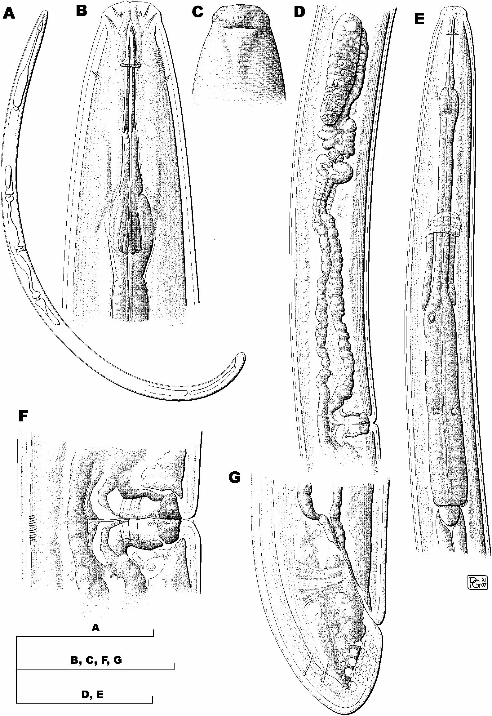
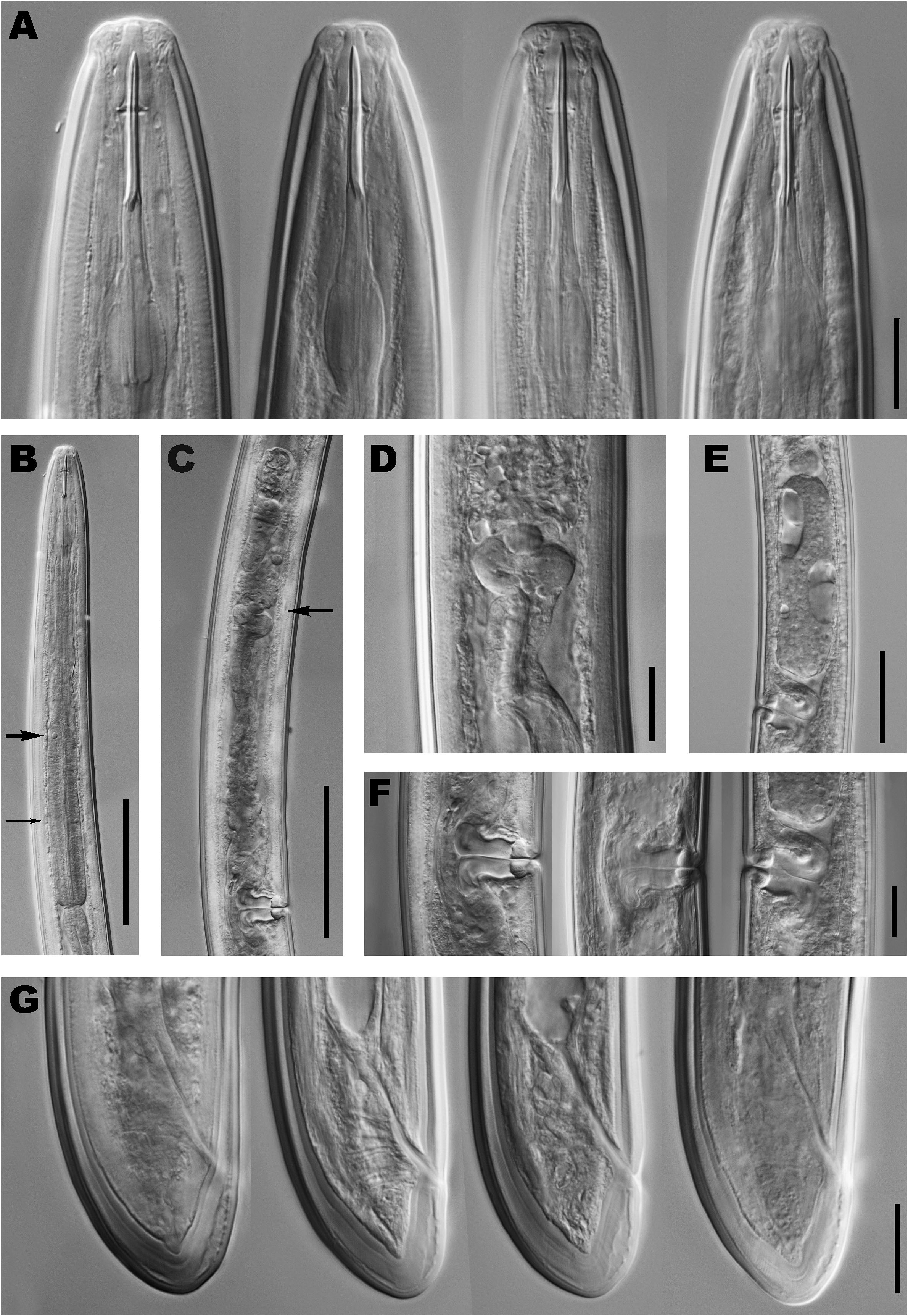
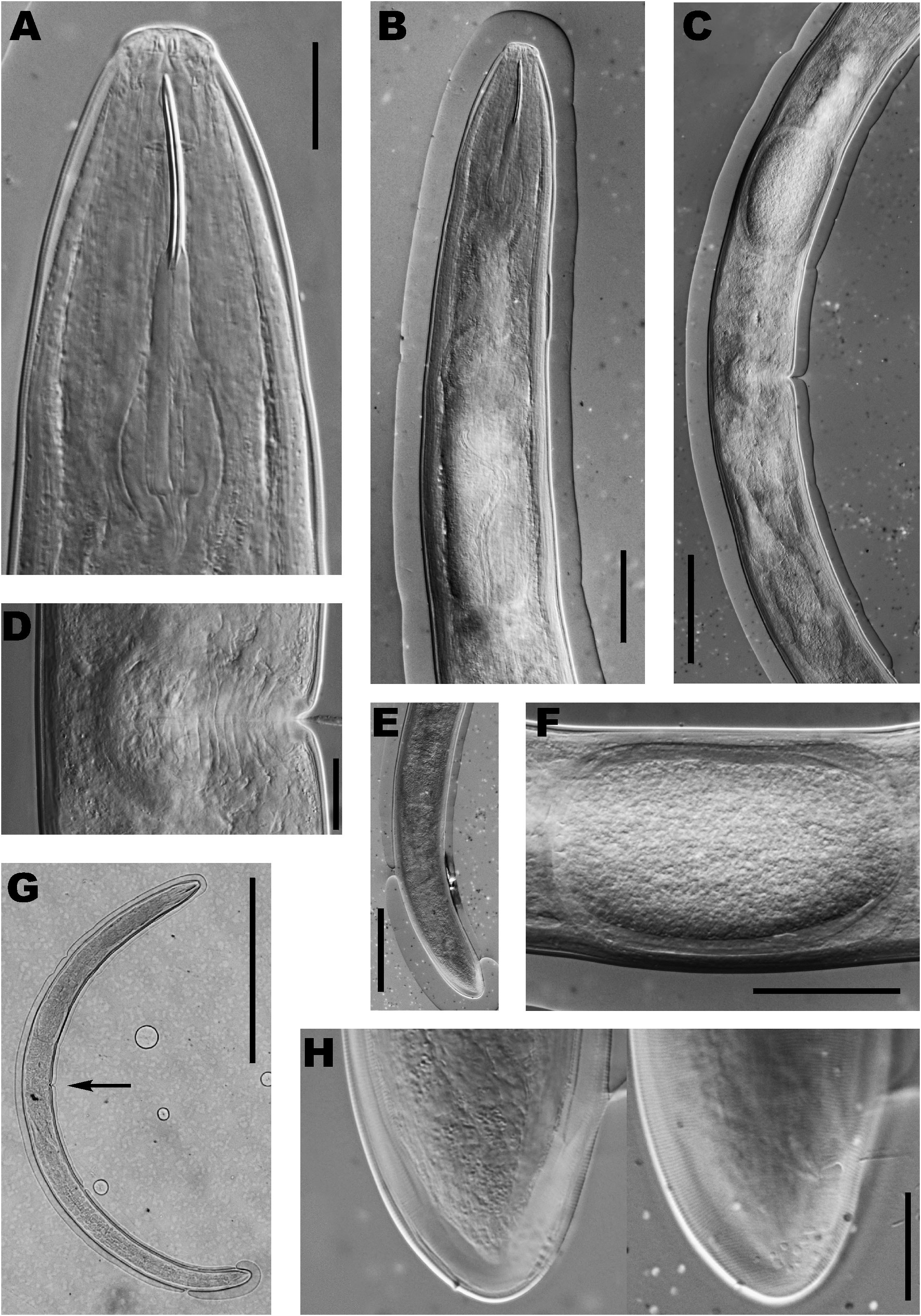
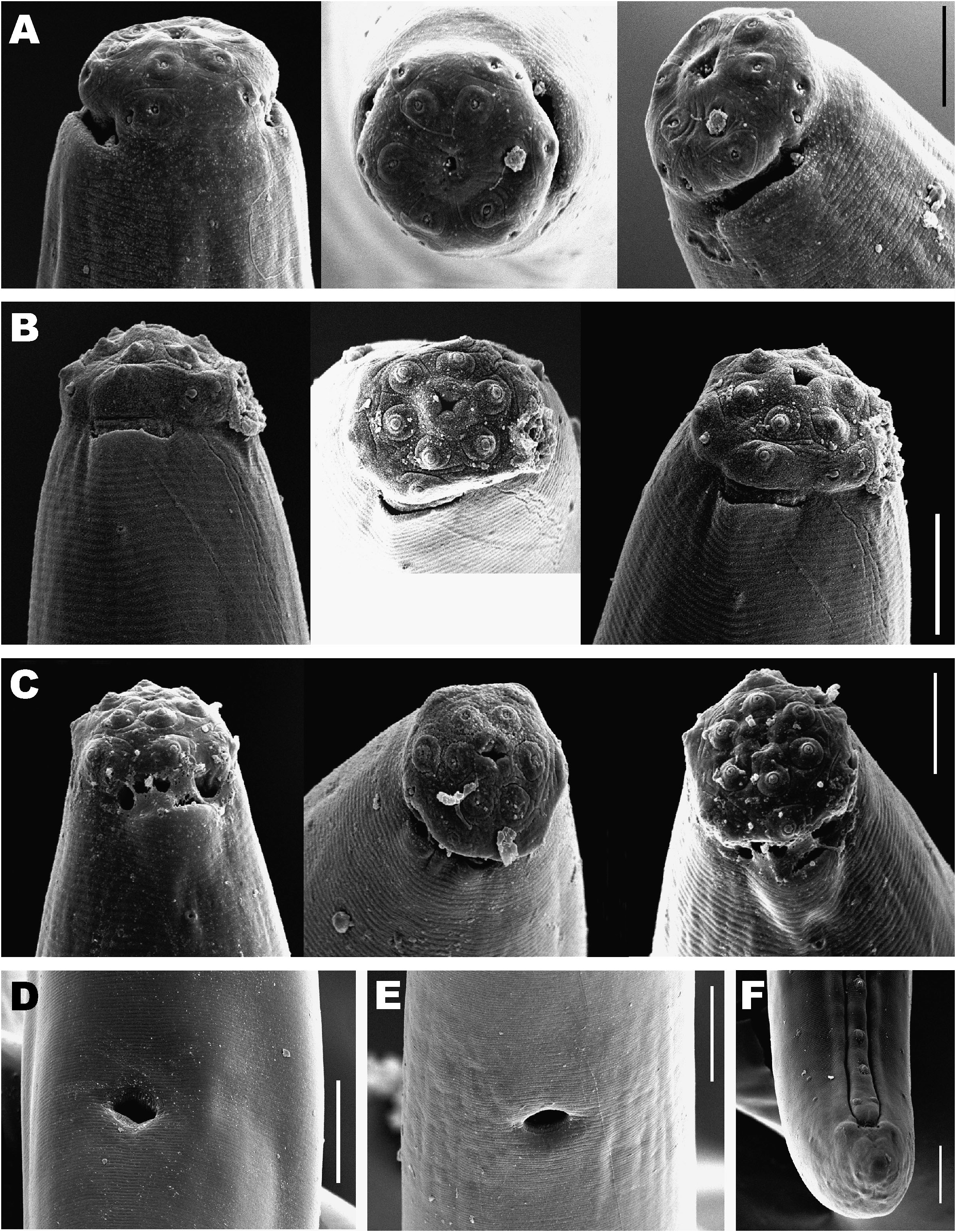
( Figs 1-3 View Fig View Fig View Fig ; 4A, D View Fig )
MATERIAL EXAMINED
Twenty-four females collected from the province of Granada. In addition, the holotype female deposited at the Museo Cantonale di Storia Naturale di Lugano ( MCSN, Switzerland) was made available for study by courtesy of Ms Lucia Pollini.
MEASUREMENTS
See Table 1. View Table 1
DESCRIPTION
Female
Based on Andalusian individuals. Moderately slender to slender nematodes of medium size, 1.69-1.99 mm long. Habitus after fixation curved ventrad, adopting a C-shape. Body cylindrical, tapering towards both ends but more so anteriorly. Cuticle 4.0-6.0 µ m in anterior region, 3.0-5.0 µ m at mid-body and 7.0-9.5 µ m on tail; its outer layer with very fine transverse striations and much thinner than inner one, especially at tail level. Lateral chord 8-16 µ m wide or occupying 11-27% of mid-body diam., lacking any particular differentiation. Lateral pores obscure. Lip region more or less rounded, offset by shallow depression, 2.2-3.5 times as broad as high and ca one-third to two-fifths of body diam. at neck base. Lips amalgamated; labial and cephalic papillae visible, scarcely protruding above cephalic contour. Under SEM, oral aperture appearing as circular to square opening surrounded by a slight elevation of perioral area; inner labial papillae located on ring-like protuberances offset from adjacent surface by circular striation; outer labial papillae on similar but less differentiated areas; cephalic papillae lacking such differentiation. Amphid fovea cup-shaped, opening at level of cephalic depression, occupying two- to three-fifths of corresponding body diam; under SEM, one pore visible on amphid fovea surface. Cheilostom curly bracket-shaped. Odontostyle long and slender, 12-17 times as long as wide or 1.6-1.9 times longer than lip region diam.; aperture small, occupying 7-15% of total length. Odontophore 1.1- 1.4 times as long as odontostyle, bearing distinct basal flanges extending over posterior half of its total length and consisting of three sets of two sclerotised pieces with basal knobs. Guiding ring double, located at 19-23 µ m or 1.0-1.3 times lip region diam. from anterior end. Pharynx consisting of slender muscular anterior portion expanding gradually into basal expansion at 54-64% of total neck length, reaching full diam. at 61-70%; pharyngeal expansion occupying 34-43% of total neck length, ca twoto three-fifths of corresponding body diam. Pharyngeal gland nuclei located as follows (n = 6): DN = 61-67; S 1 N 1 = 32 (n = 1); S 1 N 2 = 43 (n = 1); S 2 N 1 = 50-57; S 2 N 2 = 50-58. Weak membrane-like structure surrounding base of pharyngeal expansion. Cardia rounded conoid, wider than long, 12-18 × 8-16 µ m. Some individuals with a cell mass located on dorsal surface, ca one body diam. posterior to cardia. Intestine not containing any greenish material. Genital system didelphic-amphidelphic, both branches equally and well developed. Ovaries 86-128 µ m long, normally not reaching sphincter level; oocytes first in two or more rows, then one. Oviduct 104-190 µ m or 1.6-2.7 corresponding body diam. long, consisting of slen- der portion with prismatic cells and well developed pars dilatata with distinct lumen. Sphincter prominent, located between oviduct and uterus. Uterus long, 136-255 µ m or 2.3-4.1 corresponding body diam., sometimes twisted, tripartite, i.e., consisting of: i) a wider proximal region with distinct lumen; ii) followed by a narrower and shorter intermediate portion with narrow lumen and surrounded by a cluster of hyaline cells; and iii) ending with a well developed, spheroid, pars dilatata distalis. Two females bearing one egg each, eggs measuring 113-116 × 34-36 µ m. Sperm not observed in genital tract. Vagina extending inwards for 42-67% of body diam.; pars proximalis longer than broad, 15-27 × 13-21 µ m, with sigmoid to straight walls and enveloped by weak circular musculature; pars refringens with (in lateral view) two massive trapezoidal sclerotisations with combined width of 15-21 µ m and an apparently granular texture; pars distalis 4.0-6.5 µ m long. Vulva a transverse oval slit, preceded by shallow depression in body surface. Prerectum 4-12 and rectum 0.8-1.7 anal body diam. long. Tail rounded conoid, with abundant saccate bodies, especially on ventral surface but sometimes extending to subdorsal region; terminal cuticle 8.0- 11.5 µ m thick or 25-54% of total tail length, its inner layer marked by radial striation and divided into two by a discontinuity along its middle; ventral margin of inner layer rather irregular, somewhat separated from outer. Two pairs of subterminal caudal pores, one subdorsal, another practically lateral.
Male
Unknown.
Based on the original description and data of Andalusian individuals, E. hopedoroides is distinguished by its 1.42-1.99 mm long body, lip region offset by a shallow depression and 18-20 µ m diam., odontostyle 31-34 µ m long or 1.6-1.9 lip region diam. long, odontophore with distinct basal flanges and 1.1-1.4 times longer than odontostyle, neck length 317-442 µ m long, pharyngeal expansion 114- 161 µ m long or 34-43% of total neck length, female genital system amphidelphic, uterus long and tripartite, pars refringens vaginae with two trapezoidal sclerotisations, vulva a transverse pre-equatorial slit (V = 40-48), tail rounded conoid (16-33 µ m long, c = 47-117, c ļ = 0.5- 0.9) with ventral margin of inner cuticle layer distinctly irregular and separated from outer layer and males unknown.
Because of its short rounded conoid tail and medium sized odontostyle (ca 30 µ m), E. hopedoroides resembles E. altherri Vinciguerra & De Francisci, 1973, E. analatus (Ditlevsen, 1927) Thorne, 1939, E. arcticus Nesterov, 1976, E. distinctus Ahmad & Jairajpuri, 1980, E. georgiensis Eliava, Tskitishvili & Bagathuria, 2006 , E. hopedorus (Thorne, 1929) Thorne, 1939 and E. ponorensis Popovici, 1995. However, it can be distinguished from these species by the following characters: from E. altherri by its longer body (1.42-1.99 vs 1.18-1.38 mm) and longer odontostyle (31-34 vs 25-27 µ m), broader lip region (18- 20 vs 14.5 µ m as calculated from original drawing), vulva located more anterior (V = 40-48 vs 50-52), longer prerectum (3.8-12.3 vs 1.5-2.5 times longer than anal body diam.) and males absent vs present; from E. analatus, a little known species described on the basis of only two females, by its guiding ring located further posterior (19- 23 vs 15 µ m) from the anterior end (as estimated from the original drawing), presence of flanges in odontophore vs absence – a striking feature which should be viewed with caution, vulva pre-equatorial (V = 40-48 vs 51), and longer prerectum (4-12 vs 1.5 times anal body diam., as calculated from original drawing); from E. arcticus by its longer body (1.42-1.99 vs 1.2-1.5 mm), longer odontostyle, odontophore and total stylet (31-34, 38-45 and 70- 79, respectively, vs 27-31, 29-33 and 56-64 µ m as maximum range estimates from data on stylet length and original drawing), vulva pre-equatorial (V = 40-48 vs 49-51), and absence of males vs presence; from E. distinctus, a species described on the basis of only a single female, by its lip region offset by a shallow depression (vs a clear constriction as depicted in the original drawing), slightly shorter odontostyle, odontophore and total stylet (31-34, 38-45 and 70-79, respectively, vs 36, 46 and 82 µ m), and vulva pre-equatorial (V = 40-48 vs 53); from E. georgiensis by its rather longer body (1.42-1.99 vs 0.88-1.10 mm), broader lip region (18-20 vs ca 12 µ m), longer odontostyle (31-34 vs 27-29 µ m) and absence of males vs presence; from E. hopedorus, the closest species and one recently redescribed by Guerrero and Peña-Santiago (2007), by its more rounded (vs moderately angular) and broader lip region (18-20 vs 15-16 µ m), odontostyle slightly but consistently longer (33 (31-34) vs 31 (29-33) µ m), guiding ring located posterior (20 (19-23) vs 18 (17-21) µ m), vulva located anteriorly (V = 40-48 vs 48-55), pars refringens vaginae differently shaped (trapezoidal and with granular texture vs drop-shaped and smooth), longer eggs (113-120 vs <90 µ m long), longer prerectum (140-369 µ m long or 4-12 times anal diam. vs 62-162 µ m or 1.5- 4.5 times anal diam.) and tail more rounded and relatively shorter (c ļ = 0.5-0.9 vs 0.7-1.0 and more conoid); and, finally, from E. ponorensis by its broader lip region (18- 20 vs 15-16 µ m) offset by a depression vs a constriction, rather longer odontostyle, odontophore and total stylet (31-34, 38-43 and 70-77, respectively, vs 23-25, 33-38 and 55-63 µ m), guiding ring located further posterior (19-23 vs 15-17 µ m from anterior end), uterus tripartite (vs a simple tube as illustrated in original description, although this feature should be considered with caution) and shorter tail (16-33 µ m long and c ļ = 0.5-0.9 vs 35-45 µ m long and c ļ = 0.9-1.3).
DISTRIBUTION
This species was collected in only one sampling point, a wet meadow on a glacial valley in Sierra Nevada , province of Granada, at 2800 ma.s.l.
REMARKS
To date, the only available information concerning E. hopedoroides was the original description by Altherr (1963) which was based on three females from Dischma valley, Switzerland, and a detailed drawing of the anterior region of a type specimen by Siddiqi (1969). After the death of Dr Altherr in 1976, his nematode collection was inherited by the Station Fédérale de Recherches Agronomiques de Changins, Nyon ( Switzerland). It is now deposited in the Museo Cantonale di Storia Naturale di Lugano ( Switzerland), under the charge of Ms Lucia Pollini, who kindly provided the holotype of E. hopedoroides for study. The original material is still in acceptable condition, although the pharynx is twisted, an obvious flattening of the body distorts some of the measurements and the low contrast prevents accurate observations on the genital system. General measurements and morphology, especially those relative to lip region, odontostyle, odontophore and guiding ring, fit well with the individuals collected in Sierra Nevada. Only minor differences have been observed, i.e., the body seems to be slightly longer in the Andalusian individuals (1.69-1.99 vs 1.42-1.85 mm) whereas the odontophore is somewhat shorter (38-43 vs 43-45 µ m).
No known copyright restrictions apply. See Agosti, D., Egloff, W., 2009. Taxonomic information exchange and copyright: the Plazi approach. BMC Research Notes 2009, 2:53 for further explanation.