
Sinopoda lebar, Grall & Jäger, 2020
|
publication ID |
https://doi.org/ 10.11646/zootaxa.4797.1.1 |
|
publication LSID |
lsid:zoobank.org:pub:6219676C-8533-4D6F-AEFC-7276C70554D9 |
|
persistent identifier |
https://treatment.plazi.org/id/FD388D4B-FFD3-FFD4-FF23-FC91FD7DFE6B |
|
treatment provided by |
Plazi |
|
scientific name |
Sinopoda lebar |
| status |
sp. nov. |
Sinopoda lebar View in CoL spec. nov.
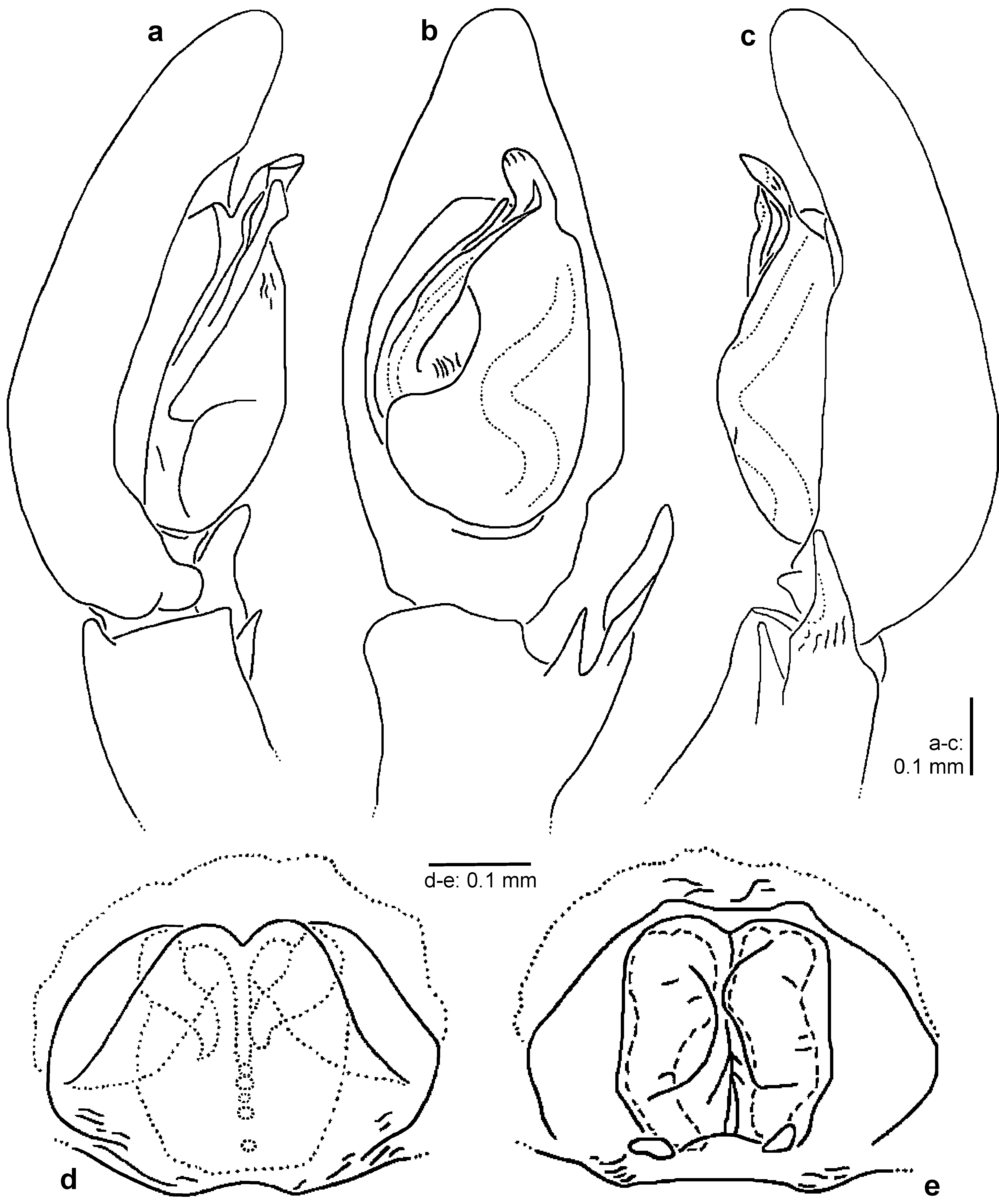
Figs 25 View FIGURES 25 a–b, 59 m–n
Type material: Holotype: female, INDONESIA: Northern Sulawesi: Tomohon, Rurukan , Gunung Mahawu , 1200 m, sifted, 30 November 1999, A. Riedel leg. ( MHNG).
Paratype: 1 female (SD 1618) with same data as for holotype ( MHNG) .
Additional material examined: 1 juvenile (SD 1562) with same data as for holotype ( MHNG) .
Etymology: The species name is derived from the Indonesian word “lebar” meaning “broad/wide” and referring to the thickened glandular appendages of the females; term in apposition.
Diagnosis: Females of S. lebar spec. nov. are similar to those of S. cornuta spec. nov. ( Figs. 7 View FIGURES 7 d–e) and S. sulawesia spec. nov. ( Figs. 41 View FIGURES 41 d–e) in having the glandular appendages running ventrally of spermathecae. They can be distinguished from S. cornuta spec. nov. by 1. glandular appendages thickened, distal part rounded and only slightly curved (thinner, distal part tapering and strongly curved in S. cornuta spec. nov.), 2. spermathecae fused along median line (posteriorly not completely fused at median line in S. cornuta spec. nov.) and 3. fertilization ducts long, curved and arising medially at posterior part of spermathecae (short and arising posteriorly at posterior part of spermathecae in S. cornuta spec. nov.); from S. sulawesia spec. nov. by 1. glandular appendages thickened and slightly curved (divided into two pairs of appendages and strongly curved in S. sulawesia spec. nov.) and 2. fertilization ducts curved and posterior part nearly as wide as distal part (nearly straight and posterior part wider than anterior part in S. sulawesia spec. nov.).
Description: Female (holotype): Total length 4.70; prosoma 2.20 long, 2.10 wide, anterior width of prosoma 1.30; opisthosoma 2.50 long, 1.60 wide. Eye measurements: AME 0.12; ALE 0.20; PME 0.12; PLE 0.24; AME– AME 0.13; AME–ALE 0.05; PME–PME 0.19; PME–PLE 0.22; AME–PME 0.23; ALE–PLE 0.24; clypeus AME 0.17; clypeus ALE 0.16. Leg formula: 2431; measurements of palp and legs: palp: 3.00 (0.90, 0.50, 0.70, 0.90); I: 7.25 (1.90, 0.95, 1.90, 1.80, 0.70); II: 8.60 (2.40, 1.00, 2.40, 2.10, 0.70); III: 7.60 (2.10, 0.95, 1.95, 1.90, 0.70); IV: 7.95 (2.20, 0.85, 2.00, 2.20, 0.70). Spination: palp: 131, 101, 2121, 1004; legs: Fe I–III 323, IV 321; Pa I–IV 000; Ti I–II 1016, III 2026, IV 2226; Mt I–II 0006, III–IV 3036. Cheliceral furrow with 3 anterior and 4 posterior teeth.
Colouration in ethanol: Prosoma yellow, anteriorly with brown spot around PLE, laterally brown with white stripes, postero-laterally with yellow U-shaped pattern. Sternum yellowish-white with six brown spots laterally. Opisthosoma brown with yellowish-white spots, medially with white longitudinal stripe, posterior part with four yellow spots in front of spinnerets, ventrally yellowish-grey, posteriorly brown. Chelicerae yellow with two brown stripes. Palps yellowish-brown; Fe, Pa & Ti distal-laterally brown. Legs yellow; Fe prolaterally, medially and distally with brown band, Pa laterally brown, Ti proximally and distally brown, Mt distally brown.
Copulatory organ as in diagnosis. Epigynal field wider than long without anterior bands or slit sensilla. Epigynal pockets running from latero-posterior to medio-anterior. Lateral lobes fused. Lobal septum wide, anteriorly with median indentation. Internal duct system roughly as long as wide. Glandular appendages only visible in ventral view and extending not in posterior half of internal duct system. Fertilization ducts arising medio-laterally in posterior half of internal duct system.
Variation: Female (SD 1618): Total length 5.20; prosoma 2.30 long; opisthosoma 2.90 long, 1.95 wide. measurements leg I: 7.6 (2.00, 1.00, 2.00, 1.90, 0.70). Spination: legs: Ti I–II 0016.
Male: Unknown.
Distribution: Known only from the type locality.

| MHNG |
Museum d'Histoire Naturelle |
No known copyright restrictions apply. See Agosti, D., Egloff, W., 2009. Taxonomic information exchange and copyright: the Plazi approach. BMC Research Notes 2009, 2:53 for further explanation.