
Encarsia hayati Li & Geng
|
publication ID |
https://doi.org/ 10.11646/zootaxa.4162.3.7 |
|
publication LSID |
lsid:zoobank.org:pub:27697FCD-BDAE-4256-85E6-8B22C1DD0E67 |
|
DOI |
https://doi.org/10.5281/zenodo.6079516 |
|
persistent identifier |
https://treatment.plazi.org/id/FE17F410-FFB8-FFDD-FF03-FF6E2398FF5E |
|
treatment provided by |
Plazi |
|
scientific name |
Encarsia hayati Li & Geng |
| status |
sp. nov. |
Encarsia hayati Li & Geng , sp. nov.
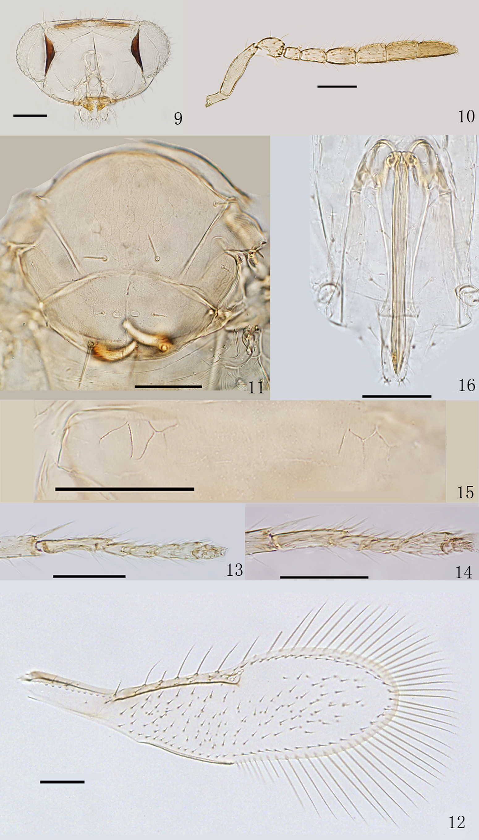
Figs 9–16 View FIGURES 9 – 16
Type material. Holotype. ♀ [on slide], ( NEFU), CHINA, Jiangxi Province, Shangrao City, Wuyishan Protection Station, 780m, 2.VII. 2013, Chao Zhang , sweeping.
Paratype. 1♀ [on slide], CHINA, Hubei Province, Suizhou City, Santan, 14. VIII. 2015, Hui Geng , Yan Gao , Zhi-Guang Wu , sweeping. ( NEFU).
Diagnosis. Female. Length, mesosoma plus metasoma, 0.49–0.51 mm. Body pale yellow except clypeus, malar sulcus and postocellar bars brown. Antennal formula 1:1:3:3. F1 and F2 without longitudinal sensilla. Mid lobe of mesoscutum with 4 setae; each side lobe and axilla with 1 seta; placoid sensilla on scutellum narrowly separated; distance between anterior pair of scutellar setae distinctly less than that between posterior pair. Fore wing 3.35–3.49× as long as wide, sparsely setose, with a large asetose area around stigmal vein; marginal fringe 0.88–0.89× as long as wing width. Tarsal formula 5:4:5. Mid tibial spur 0.70–0.76× as long as corresponding basitarsus. Petiole with sculpture laterally. Ovipositor slightly exerted, 1.15–1.19× as long as mid tibia; third valvula 0.35–0.36× as long as second valvifer.
Description. Female. Holotype. Length, mesosoma plus metasoma, 0.51 mm. Head and body including ovipositor and legs pale yellow except clypeus, malar sulcus and postocellar bars brown. Antennae yellow except last two segments slightly brown. Wings hyaline, venation pale brown.
Head ( Fig. 9 View FIGURES 9 – 16 ), in frontal view, 1.77× as wide as high, and about as wide as mesosoma. Ocelli forming about an obtuse triangle, POL distinctly less than OOL. Maxillary and labial palpi 1-segmented. Mandibles with two teeth and a truncation. Eyes with fine and transparent setae. Frontovertex with short setae. Antennal formula, 1:1:3:3 ( Fig. 10 View FIGURES 9 – 16 ); radicle (R), scape (S), pedicel (P), 3 funicle segments (F1–F3) and 3 club segments (F4–F6) with the following ratios of length to width: R: 2.00, S: 4.05, P: 1.47, F1: 1.40, F2: 2.00, F3: 1.89, F4: 1.65, F5: 1.70 and F6: 2.33; relative lengths of segments R–F6 to length of F1: R: 1.15, S: 3.68, P: 1.61, F1: 1.00, F2: 1.61, F3: 1.95, F4: 1.90, F5: 1.95 and F6: 2.41; flagellum with the following numbers of longitudinal sensilla: F1: 0, F2: 0, F3: 2, F4: 3, F5: 3, F6: 3.
Mesosoma ( Fig. 11 View FIGURES 9 – 16 ) 0.61× as long as metasoma. Mid lobe of mesoscutum and axillae weakly reticulate. Mid lobe of mesoscutum with 4 setae, each side lobe of mesoscutum with 1 seta. Axilla with 1 short seta. Scutellum 1.94× as wide as long, and 0.75× as long as mid lobe of mesoscutum. Distance between placoid sensilla on scutellum about own maximum width. Anterior pair of scutellar setae clearly shorter than posterior pair, and distance between anterior pair 0.7× that between posterior pair. Endophragma long and rounded at apex, extending to posterior margin of T1. Fore wing ( Fig. 12 View FIGURES 9 – 16 ) 3.49× as long as wide, sparsely setose, with a large asetose area around stigmal vein; costal cell with 4 short setae in basal half; basal cell with 2 setae, with proximal one distinctly shorter; submarginal vein with 2 setae; marginal vein 1.38× as long as submarginal vein, with 5 setae along anterior margin; marginal fringe 0.89× as long as wing width. Hind wing 8× as long as wide, marginal fringe 1.84× as long as wing width. Tarsal formula 5:4:5, mid leg with last two tarsal segments fused but indicated by a transverse suture ( Fig. 13 View FIGURES 9 – 16 ). Mid tibial spur 0.7× as long as corresponding basitarsus ( Fig. 13 View FIGURES 9 – 16 ), and the latter 0.34× as long as mid tibia. Hind tibia 0.99× as long as mid tibia.
Metasoma with petiole ( Fig. 15 View FIGURES 9 – 16 ) sculptured laterally. T1–T5 with scale-like reticulation laterally. T2–T7 with 1+1, 1+1, 1+1, 2+2, 1+2+1 and 4 setae, respectively. T7 1.56× as wide as long. Ovipositor ( Fig. 16 View FIGURES 9 – 16 ) not exerted, apparently originating from middle of T3, 1.19× as long as mid tibia, and 0.89× as long as mid tibia and basitarsus combined. Third valvula 0.36× as long as second valvifer.
Male. Unknown.
Host. Unknown.
Variation. Sole paratype with antennal F2 1.41× as long as wide, F3 1.63× as long as wide and with 2 longitudinal sensilla; fore wing with 4 setae along anterior margin; hind tibia 1.04× as long as mid tibia. Other characters the same as holotype.
Etymology. This species is named in honor of Prof. Mohammad Hayat (Department of Zoology, Aligarh Muslim University, Uttar Pradesh, India) for his contributions to the study of Hymenoptera, Chalcidoidea.
Comments. Mid tarsal structure of E. hayati is similar to that of the foregoing new species (see also comments under E. dianensis ). Placement to species-group of this new species is uncertain. Among the species with a 5:4:5 tarsal formula and fore wing with a clear asetose area around the stigmal vein (meghalayana - and cubensis -group), E. hayati is unique by having a completely yellow body, and narrowly separated placoid sensilla on the scutellum.
This species is closely related to E. dianensis based on similar structures of the mid tarsi, body color, and shape and characters of the fore wing, but can be separated from the latter by the differences given in the key and the comments under E. dianensis .
No known copyright restrictions apply. See Agosti, D., Egloff, W., 2009. Taxonomic information exchange and copyright: the Plazi approach. BMC Research Notes 2009, 2:53 for further explanation.