
Eurytoma infracta Mayr, 1904
|
publication ID |
https://doi.org/10.5252/z2011n3a3 |
|
persistent identifier |
https://treatment.plazi.org/id/03E487A7-AF5E-276B-05F6-F9F36B58FDE8 |
|
treatment provided by |
Felipe (2021-02-17 23:16:28, last updated by Plazi 2023-11-01 21:12:10) |
|
scientific name |
Eurytoma infracta Mayr, 1904 |
| status |
|
Eurytoma infracta Mayr, 1904 View in CoL
MATERIAL EXAMINED. — Ex Aylax minor : Spain. Madrid, Rivas Vaciamadrid, 14. V.2003, J. L. Nieves leg (n = 2).
Ex Neaylax verbenacus : Spain. Madrid, Dehesa de Arganda, 1. VI.2003, J. L. Nieves leg (n = 4). — Same locality, 6. VI.2004, J. L. Nieves leg (n = 8, of which 2 specimens MNHN-EY 6408).
DESCRIPTION
n = 14; body length 1.67 mm (range 1.3-2.0); body width 0.73 mm (0.7-0.8) ( Figs 5D View FIG ; 7D View FIG ); body more or less fusiform, ratio L/W = 2; ventral margin of body segments clearly convex; anterodorsal protuberances present from second thoracic segment to the third abdominal and not protruding beyond the dorsal margin of body segments ( Fig. 7D View FIG ).
Head with upper margin of vertex straight; anteromedial setae of the antennal area situated at the same level as the antennae; ratios SA/LAA = 0.38 and SA/DAV = 1.1; antero-medial setae of vertex situated relatively high on the upper face ( Fig. 9C View FIG ); dorsal-labral setae as long as clypeal setae; ventral margin of clypeus indistinct; labrum with slight divisions restricted to its apical part; flaps or lobes not well differentiated; maxillary palps conspicuous ( Fig. 11D View FIG ).
Mandibles with two teeth and exposed in part, with the tip of the first tooth visible ( Fig. 11D View FIG ); first tooth relatively long and slender; ratio between the length at its base and the width 2.38; outer margin of first tooth strongly convex, tip moderately recurved; apex of the second tooth straight, directed upwards in the same direction as the first tooth and more or less acute; inner margin of mandible from the base of second tooth strongly convex ( Fig. 13D View FIG ).
BIOLOGY
This species is a primary oligophagous ectoparasitoid, reared from galls of Aylacini cynipids in fruits of species of Salvia (Lamiaceae) and Papaver (Papaveraceae) ( Askew et al. 2006). It is univoltine; fully-grown larvae usually overwinter and adults emerge in summer when the new host galls are developing.
REMARKS
The straight upper margin of the vertex and the strongly convex inner margin of the base of the second mandible tooth allow separation of the larva of this species from the others in this study.
GOMEZ J. F., HERNANDEZ NIEVES M., GARRIDO TORRES A. M., ASKEW R. R. & NIEVES- ALDREY J. L. 2006. - Los Chalcidoidea (Hymenoptera) asociados con agallas de cinipidos (Hymenoptera, Cynipidae) en la Comunidad de Madrid. Graellsia 62: 293 - 331.
WALKER F. 1832. - Monographia Chalciditum. The Entomological Magazine 1 (1): 12 - 29.

FIG. 5.— Ventral views ofterminal-instar eurytomidlarvae:A, Eurytomaaspila (Walker,1836) ex Phanacis caulicola; B, E.brunniventris Ratzeburg, 1852 ex Cynips quercus (A); C, E. cynipsea Boheman, 1836 ex Phanacis hypochoeridis; D, E. infracta Mayr, 1904; E, E. mayri Ashmead, 1887; F, E. pediaspisi Pujade i Villar,1994; G, E. robusta Mayr, 1878; H, E. rosae Nees, 1834 ex Diplolepis rosae. Scale bars:1 mm.
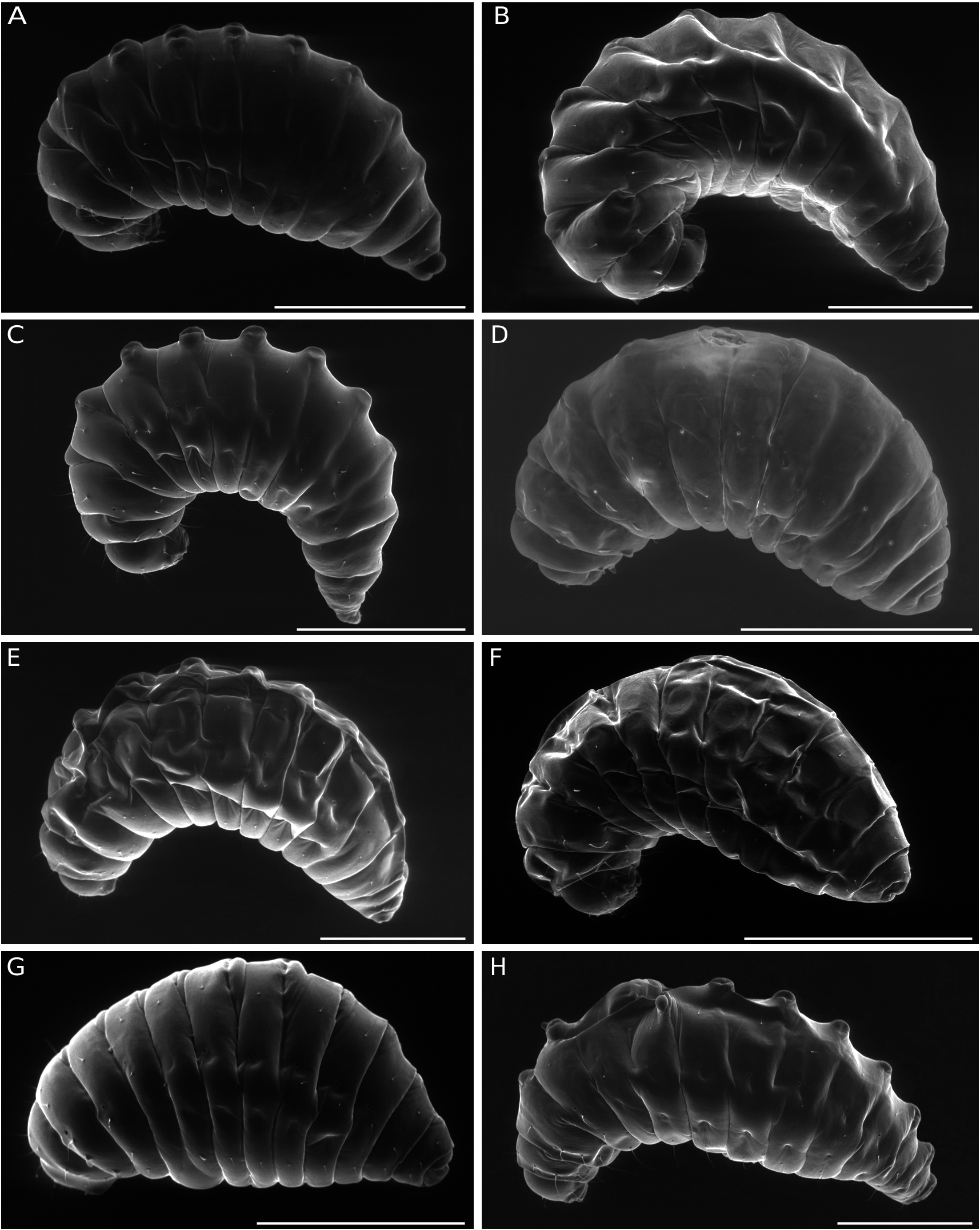
FIG. 7.— Lateralviews ofterminal-instar eurytomidlarvae:A,Eurytoma aspila (Walker,1836) ex Phanacis caulicola; B, E.brunniventris Ratzeburg, 1852 ex Cynips quercus (A); C, E. cynipsea Boheman, 1836 ex Phanacis hypochoeridis; D, E. infracta Mayr, 1904; E, E. mayri Ashmead, 1887; F, E. pediaspisi Pujade i Villar,1994; G, E. robusta Mayr, 1878; H, E. rosae Nees, 1834 ex Diplolepis rosae. Scale bars:1 mm.
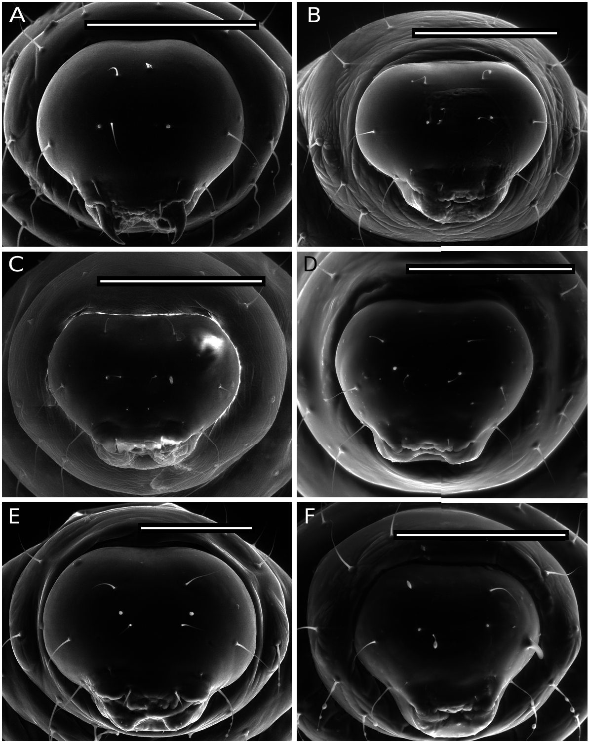
FIG. 9. — Anterior views of the head of terminal-instar eurytomid larvae: A, Eurytoma aspila (Walker, 1836) ex Phanacis caulicola; B, E. brunniventris Ratzeburg, 1852 ex Cynips quercus (A); C, Eurytoma infracta Mayr, 1904; D, Eurytoma mayri Ashmead, 1887; E, Eurytoma pediaspisi Pujade i Villar, 1994; F, Eurytoma robusta Mayr, 1878. Scale bars: A, B, D, F, 400 μm; C, 300 μm; E, 200 μm.
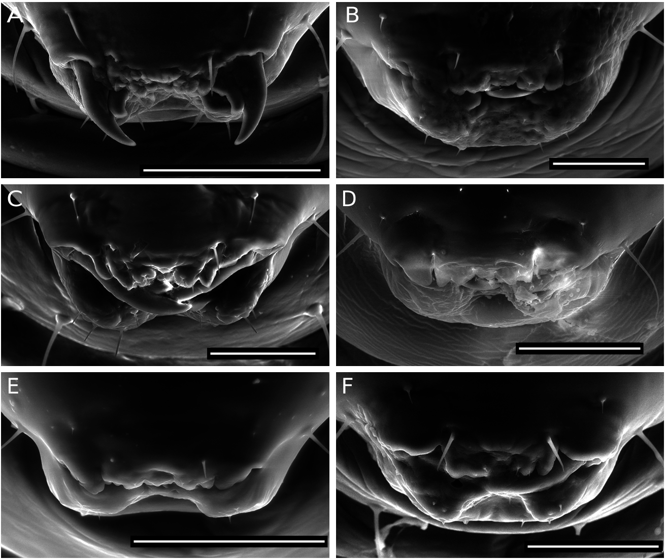
FIG. 11. — Anterior views of mouthparts of terminal-instar eurytomidae larvae:A, Eurytoma aspila (Walker,1836) ex Phanacis caulicola; B, E. brunniventris Ratzeburg, 1852 ex Cynips quercus (A); C, E. cynipsea Boheman, 1836 ex Phanacis hypochoeridis; D, E. infracta Mayr, 1904; E, E. mayri Ashmead, 1887; F, E. pediaspisi Pujade i Villar, 1994. Scale bars: A, E, 200 μm; B-D, F, 100 μm.

FIG. 13.— Anterior views of the right mandible of terminal-instar eurytomid larvae:A, Eurytoma aspila (Walker,1836) ex Phanacis caulicola; B, E. brunniventris Ratzeburg, 1852 ex Cynips quercus (A); C, E. cynipsea Boheman, 1836 ex Phanacis hypochoeridis; D, E. infracta Mayr, 1904; E, E. mayri Ashmead, 1887; F, E. pediaspisi Pujade i Villar, 1994; G, E. robusta Mayr, 1878; H, E. rosae Nees, 1834 ex Diplolepis rosae; I, E. rufipes Walker, 1832; J, E. sp. cf. aspila; K, E. sp. cf. jaceae; L, E. strigifrons Thomson, 1876 (right mandible); M, E. strigifrons (left mandible); N, E. timaspidis (Mayr, 1904). Scales bars: A, D-G, I-N, 50 μm; B, 20 μm; C, H, 100 μm.
| V |
Royal British Columbia Museum - Herbarium |
| VI |
Mykotektet, National Veterinary Institute |
No known copyright restrictions apply. See Agosti, D., Egloff, W., 2009. Taxonomic information exchange and copyright: the Plazi approach. BMC Research Notes 2009, 2:53 for further explanation.
|
Kingdom |
|
|
Phylum |
|
|
Class |
|
|
Order |
|
|
Family |
|
|
Genus |