
Agathodesmus steeli Silvestri, 1910
|
publication ID |
https://doi.org/ 10.3897/zookeys.12.206 |
|
publication LSID |
lsid:zoobank.org:pub:702BCAE3-5B0A-49EA-8603-D60826241F2C |
|
DOI |
https://doi.org/10.5281/zenodo.3792148 |
|
persistent identifier |
https://treatment.plazi.org/id/9B0D9E62-FF9C-CC31-FF5E-5E9D854FB194 |
|
treatment provided by |
Plazi |
|
scientific name |
Agathodesmus steeli Silvestri, 1910 |
| status |
|
Agathodesmus steeli Silvestri, 1910 View in CoL
Figs 1 View Figure 1 , 3 View Figure 3 A-D, 4 View Figure 4 A-D, 5 View Figure 5 A, 5 View Figure 5 B, 6 View Figure 6 A-C, 7 View Figure 7 A
Agathodesmus steeli View in CoL . Silvestri 1910:362. Attems 1914:283, 1940:488. Chamberlin 1920:137. Jeekel 1971:310, 1985:50. Golovatch et al. 2009:3.
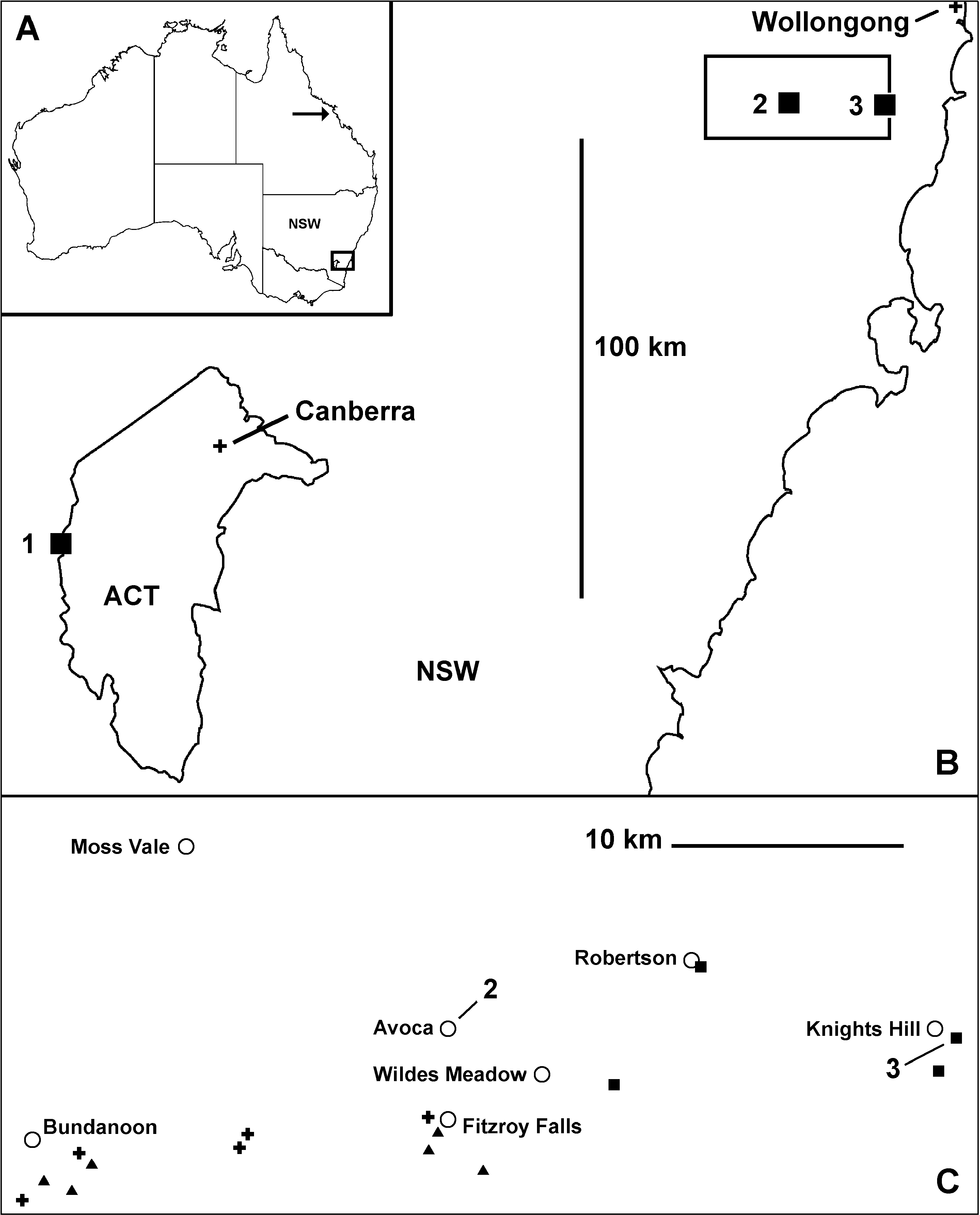
Holotype. Female , permanently mounted on microscope slide in two pieces, with a break between rings 9 and 10 ( Figs 1 View Figure 1 , 3A). Avoca , NSW, Australia, Thomas Steel, date not known. In MCSN.
Paratypes. None designated.
Other material examined. 7 males, 4 females, 1 stadium VI female, 2 stadium V females, 1 stadium IV female, Knights Hill, NSW, 34°37’07”S 150°42’38”E ± 25 m, 720 m, 14 May 2009, R. Mesibov and T. Moule, wet eucalypt forest, AM KS107964 (three males, two females dissected).
Diagnosis. Head + 19 rings; gonopod telopodite with distal portion directed basally and slightly laterally near origin and with broad lateral branch apically expanded and divided into three anterobasally curving lobes.
Description. The original description ( Silvestri 1910) is quoted in the Appendix. What follows is based on my examination of both the holotype and the Knights Hill material.
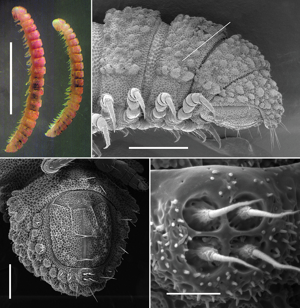
Adult with head + 19 rings ( Fig. 4A View Figure 4 ). Live and freshly preserved adults pale with faint reddish pigmentation dorsally; in some individuals, pigment concentrated in transverse band at rear of metazonite. Male/female approximate dimensions: length 4.5/5.5 mm, maximum width with paranota 0.5/0.6 mm, midbody vertical diameter 0.4/0.4 mm. Head ( Figs 3C, 3D View Figure 3 ) about as wide as collum, overall body width almost uniform, tapering only slightly posteriorly from collum. Head facing downwards ( Fig. 3C View Figure 3 ), with clypeus, frons and ventral part of vertex almost parallel to substrate and only slightly convex. Antenna ( Fig. 3D View Figure 3 ) short, stout, clavate, held close to head, antennomeres 2 and 3 lying in broad, shallow excavation on head; antennomere 6 widest and longest; antennomeres 2-5 about equal in length, decreasing slightly in diameter from 5 to 2. Collum with slightly convex anterior margin and broadly convex posterior margin; corners rounded ( Fig. 3C View Figure 3 ). Ring 2 tergite largest, extending basally, later- ally and anteriorly well below collum corner ( Figs 3A, 3B, 3C View Figure 3 ). Ring 2 and 3 tergites edged with 5-6 and 4 large tubercles, respectively ( Figs 3A, 3B, 3C View Figure 3 ); posterior rings, including apodous ring 18, with row of 4 large tubercles just above leg bases forming all or part of lateral extension of metatergite, the anteriormost tubercle smaller than the posterior 3. Prozonites sharply demarcated from metazonites ( Figs 3C View Figure 3 , 4B View Figure 4 , 6C View Figure 6 ). Ozopore ( Fig. 4B View Figure 4 ) very small, not raised, in small, non-tuberculated area just above middle of group of 4 larger tubercles forming lateral metatergal extension; pore formula 5, 7, 9, 10, 12, 13, 15-18. Sternites on diplosegments ( Fig. 5A View Figure 5 ) longer than wide, not setose, with distinct longitudinal and transverse impressions. Legs short, stout; relative podomere lengths tarsus>(prefemur, femur)>(postfemur, tibia); claw large, about twothirds tarsus length. Spiracles not evident. Telson facing downwards ( Fig. 4B View Figure 4 ), anal valves parallel to substrate and almost flat. Hypoproct trapezoidal ( Fig. 4C View Figure 4 ); spinnerets in square array ( Figs 4C, 4D View Figure 4 ); spinneret setae with single, low sheath, each seta in deep, walled depression.
Integument richly and densely sculptured ( Figs 3D View Figure 3 , 4C View Figure 4 , 5A, 5C View Figure 5 , 6 View Figure 6 A-C, 7A, 7B). Most of body covered with cuticle raised in cellular mesh of narrow folds, often with minute bumps (adorned with even smaller bumps) at or near fold junctions. Integument raised further as tubercles ( Fig. 6A View Figure 6 ) of varying sizes on head, collum, tergites, metatergites and telson, the largest tubercles forming paranotum-like extensions on posterior rings; tubercles and some other parts of integument with minute, finger-like projections ( Figs 4D View Figure 4 , 6A, 6B View Figure 6 ), often arising along ‘mesh-cell’ boundaries. Cell boundaries at rear of metazonite extended as lappets, forming secondary limbus above primary limbus of uniform, triangular elements ( Fig. 6C View Figure 6 ). Setae of normal type on legs and some other surfaces; a bisegmented seta with flattened, expanded tip ( Fig. 6B View Figure 6 ) on each ‘paranotum’ tubercle and in association with some dorsal tubercles.
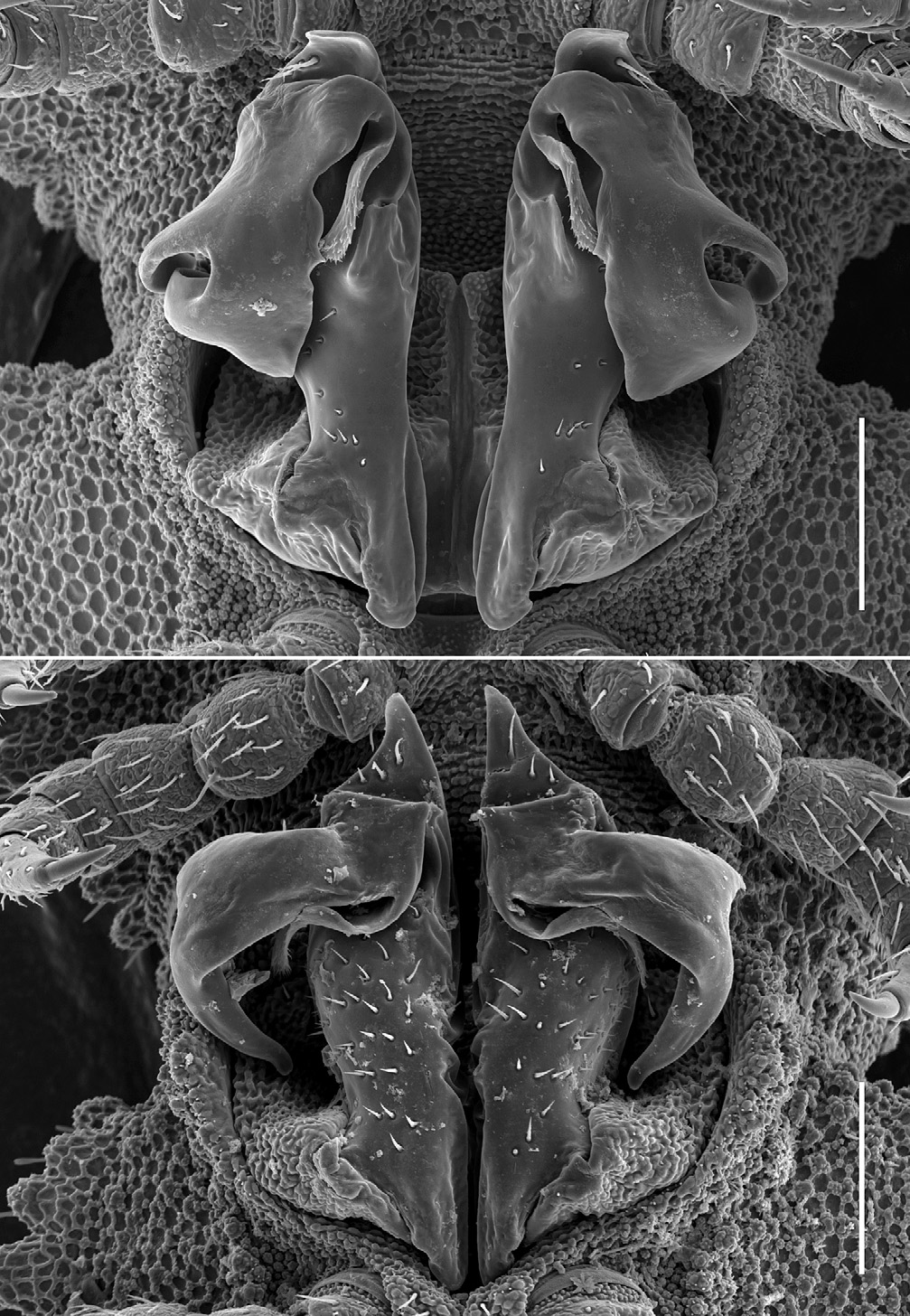
Male with gonopore opening at tip of cylindrical projection about 1/3 the length of leg 2 coxa, arising distomedially on the coxa. First legs somewhat swollen ( Fig. 3C View Figure 3 ), no other anterior legs enlarged; neither sphaerotrichomes nor brush setae on any legs. Leg 7 bases well separated; leg 6 bases slightly separated, with a pair of short, rounded projections between coxae. Gonopod aperture oval ( Fig. 7A View Figure 7 ), rim a little raised laterally. Gonocoxae ( Fig. 7A View Figure 7 ) occupying full width of aperture; tapering a little distally; with mesh-like integumental sculpture and without setae; firmly joined medially near distal end. Telopodite ( Fig. 7A View Figure 7 ) short, compact, when retracted reaching leg 7 base; broadly joined to gonocoxa ( Figs 5B View Figure 5 , 7A View Figure 7 ); no trace of cannula or prostatic groove; no integumental sculpturing; divided into more or less cylindrical basal portion and flattened distal portion. Basal portion of telopodite with blunt, basally directed projection arising posteromedial to junction with gonocoxa; portion terminating in flat, rounded tab bending posteriorly; with a few short setae on basal half of posterior surface of portion and three large setae in a row on lateral edge of posterior surface of terminal tab. Distal portion of telopodite a large, flattened structure arising on posterior surface of basal portion of telopodite just below terminal tab; curving basally and slightly laterally; divided near base into narrow medial branch, flattened apically with minute, spine-like protrusions on posterior and medial surfaces, and much larger lateral branch, the latter much expanded, curving anterobasally and divided by two deep notches into three broad, rounded lobes ( Figs 5B View Figure 5 , 7A View Figure 7 ).
Female longer and more robust than male ( Fig. 4A View Figure 4 ); epigynum inconspicuous, posterior margin barely raised; cyphopods not examined.
Distribution and habitat. Known so far from eucalypt forest (historically in the case of Avoca) at two localities ca. 20 km apart in southeastern New South Wales ( Figs 2B, 2C View Figure 2 ). Both sites are above 700 m with annual rainfall probably> 900 mm, in a temperate climate with cool winters. At the Knights Hill site, the 15 A. steeli specimens were found in narrow spaces in part of a large, moist, well-rotted log, either a Eucalyptus species or Acacia melanoxylon . Also in that part of the log were Siphonophorida, Symphyla, Cryptops sp. centipedes, fly and beetle larvae and terrestrial isopod crustaceans.
Remarks. Live A. steeli are very slow-moving and do not curl up, even when disturbed. Unlike adults of the morphologically, ecologically and behaviourally similar species of Asphalidesmus Silvestri, 1910 ( Mesibov 2002, 2009), A. steeli adults are not heavily encrusted with soil particles.
The apparent absence of well-defined spiracles in A. steeli is remarkable. I have so far been unable to detect spiracles either with light microscopy (cleared specimens) or scanning electron microscopy (see also Fig. 5C View Figure 5 ). A histological study is needed to determine whether the tracheal system is also modified from the norm in Polydesmida .


| MCSN |
Museo Civico di Storia Naturale, Verona |
| AM |
Australian Museum |
No known copyright restrictions apply. See Agosti, D., Egloff, W., 2009. Taxonomic information exchange and copyright: the Plazi approach. BMC Research Notes 2009, 2:53 for further explanation.
|
Kingdom |
|
|
Phylum |
|
|
Class |
|
|
Order |
|
|
Family |
|
|
Genus |
Agathodesmus steeli Silvestri, 1910
| Mesibov, Robert 2009 |
Agathodesmus steeli
| Silvestri 1910:362 |
| Attems 1914:283 |
| 1940:488 |
| Chamberlin 1920:137 |
| Jeekel 1971:310 |
| 1985:50 |
| Golovatch et al. 2009:3 |