
Amphicyclus japonicus Bell, 1884
|
publication ID |
https://doi.org/ 10.11646/zootaxa.4455.3.2 |
|
publication LSID |
lsid:zoobank.org:pub:A01F20BE-F0C4-4B45-97C5-E996A2C84EE1 |
|
DOI |
https://doi.org/10.5281/zenodo.5951756 |
|
persistent identifier |
https://treatment.plazi.org/id/6D72879E-FFA1-0C72-D7A2-F9D3E2B38BB5 |
|
treatment provided by |
Plazi |
|
scientific name |
Amphicyclus japonicus Bell, 1884 |
| status |
|
Amphicyclus japonicus Bell, 1884 View in CoL
[Common Japanese name: Okina-gumimodoki]
( Figs 1A View FIGURE 1 , 2A View FIGURE 2 , 3A–E View FIGURE 3 )
Amphicyclus japonicus Bell, 1884: 253 View in CoL ; Lampert 1885: 181; Théel 1886: 126; Ohshima 1912: 71, Taf. I, figs 5–6, Textfigs 2–3; Heding & Panning 1954: 87 –88, Abb. 28a–f.
Pseudocucumis japonicus ( Bell, 1884) : Ludwig 1887: 1239; Augstin 1908: 29.
Material examined. 4 specimens, WMNH-INV-2015-13–16 (St. 1, 26 August 2014, length 29–37 mm, width 6–8 mm); 11 specimens, WMNH-INV-2015-310–320 (St. 2, 25 June 2015, length 34–51 mm, width 6–13 mm); 1 specimen, WMNH-INV-2015-347 (St. 3, 24 June 2015, length 22 mm, width 6 mm).
Description. Body fusiform, curved, with both ends tapered and turned slightly upwards ( Fig. 1A View FIGURE 1 ); body wall soft, and often wrapped in brownish membrane. Body color not seriously affected by fixation/preservation. Some specimens extremely swollen, with thin transparent skin, white to pale brownish. 25 tentacles arranged in double circle (15 + 10), comprising five pairs of large interradial tentacles and five single medium radial tentacles in outer circle and five pairs of small radial tentacles in inner circle. Numerous thin short villi-like projections surrounding oral opening. Color of tentacles, oral periphery, and introvert white. Pedicels forming two longitudinal rows along each radius, apparently non-retractile, lacking on introvert, each row with approximately 25–33 pedicels. Color of pedicels same as body wall, however, pedicel tip white. Five anal papillae and five anal teeth in radii.
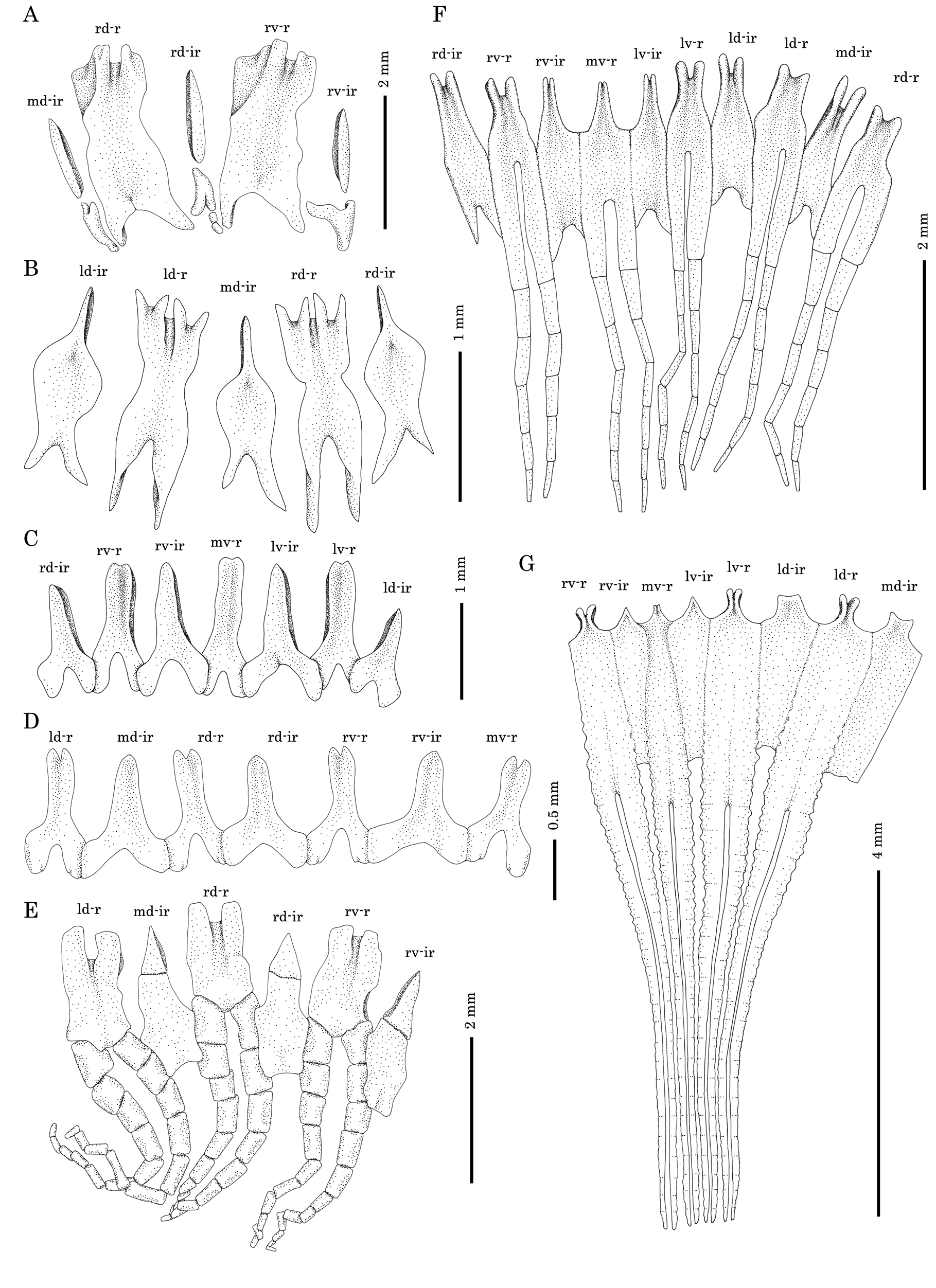
Calcareous ring simple, no posterior bifurcation of the radial plates; radial elements unfragmented, slightly constricted in the middle and with deep posterior depression ( Fig. 2A View FIGURE 2 ) and shallow anterior bifurcations; interradial elements of calcareous ring fragmented, composed of several slender pieces, proximal/posterior pieces wider. Polian vesicles two, stone canal single. with a large disc-shaped madreporite (reaching 1.4 mm in width). Gonad situated in mid-body, in two clumps, one on each side of dorsal mesentery, most tubules unbranched.
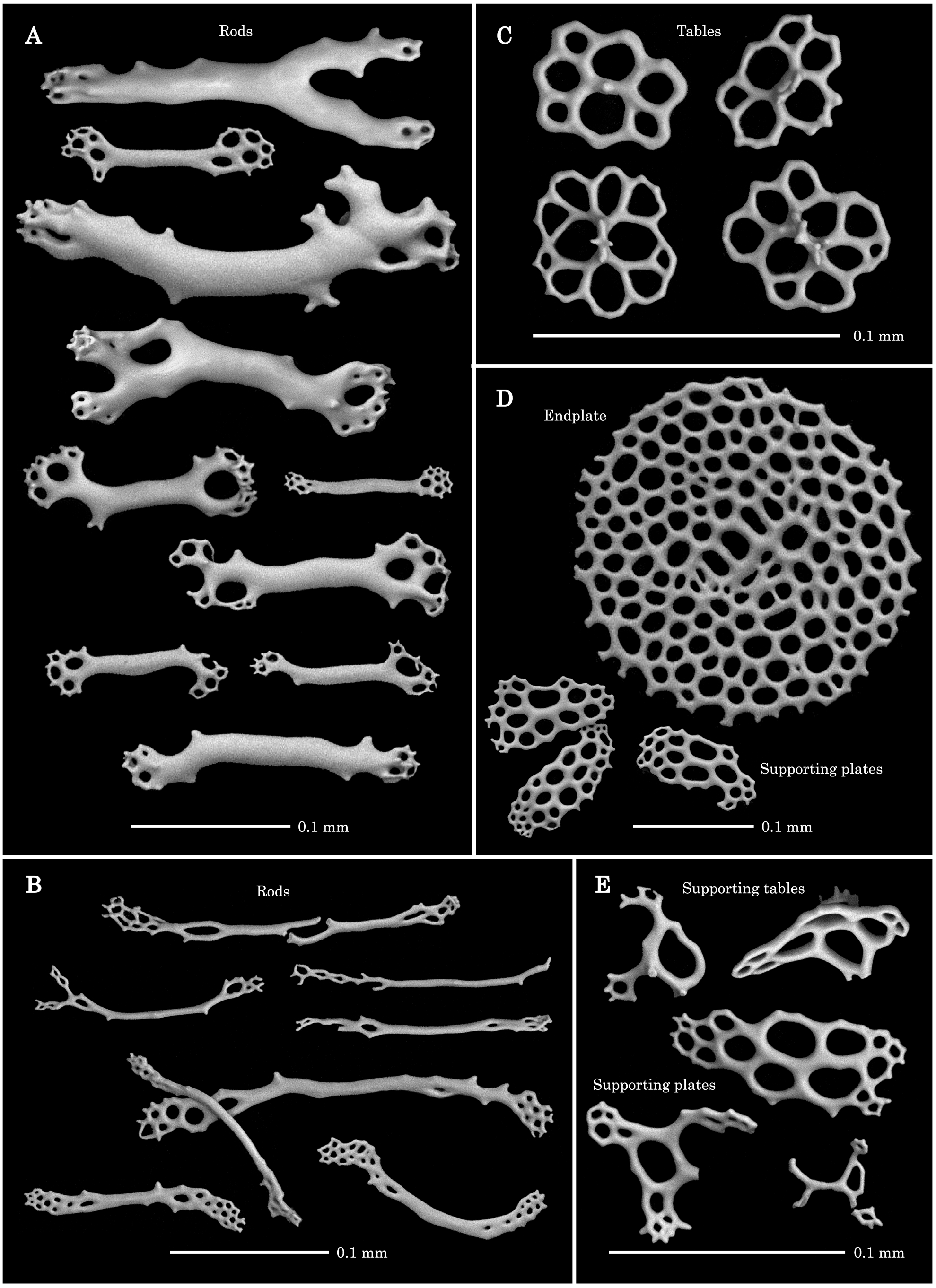
Body wall lacking ossicles. Tentacle ossicles in most specimens comprising thick rods, variously-sized ( Fig. 3A View FIGURE 3 , Table 1), most with distal perforations, rarely with central perforations, sometimes rods with one or two large branches, resulting in X- or Y-shaped bodies. Peri-oral skin lacking ossicles, while pharyngeal villi have many thin long rods, with many distal perforations ( Fig. 3B View FIGURE 3 , Table 1); sometimes rods possess one or two branches, also resulting in X- or Y-shaped structures. Introvert with tables with broad disc and low spire with two (or rarely four) pillars and one cross-beam ( Fig. 3C View FIGURE 3 , Table 1), disc with angular perforations. Such tables sometimes occur also in basal part of tentacles.
Pedicels with large circular endplate and comparatively small supporting plates ( Fig. 3D View FIGURE 3 , Table 1). Endplate with approximately uniform-sized perforations. Perforations of supporting plates mostly arranged in double rows in centre; but three or more rows sometimes present. Anal papilla exhibit mostly supporting plates, rarely supporting table with low spire also present, both small and distorted ( Fig. 3E View FIGURE 3 , Table 1). Skin around anus devoid of ossicles. Gonad lacking ossicles.
Remarks. The description and sketches of ossicles given by Ohshima (1912) are so precise that most of our observations yielded no further variation. However, the existence of the thin long rod ossicles in the pharynx ( Fig. 3B View FIGURE 3 ), is here reported for the first time; these may be useful in future studies of the systematics of Amphicyclus . Furthermore, we did not observe any ossicles in the body wall reported by Ohshima (1912) to be present. Ohshima (1912) reported that imperfect tables were found very sparsely from the body wall in only one young specimen (body size was not reported). Those tables had two rudimentary pillars with four central and some peripheral holes. No ossicles were observed in the body wall of WMNH-INV-2015-311 ( Fig. 1A View FIGURE 1 , Table 1), a difference which is likely attributed to the specimen’s small size.

No known copyright restrictions apply. See Agosti, D., Egloff, W., 2009. Taxonomic information exchange and copyright: the Plazi approach. BMC Research Notes 2009, 2:53 for further explanation.
|
Kingdom |
|
|
Phylum |
|
|
Class |
|
|
Order |
|
|
Family |
|
|
Genus |
Amphicyclus japonicus Bell, 1884
| Yamana, Yusuke & Kohtsuka, Hisanori 2018 |
Amphicyclus japonicus
| Bell, 1884 : 253 |
| Lampert 1885 : 181 |
| Théel 1886 : 126 |
| Ohshima 1912 : 71 |
| Heding & Panning 1954 : 87 |
Pseudocucumis japonicus (
| Ludwig 1887 : 1239 |
| Augstin 1908 : 29 |