
Asterocheres scutatus Stock, 1966
|
publication ID |
https://doi.org/ 10.1080/00222933.2012.742588 |
|
publication LSID |
lsid:zoobank.org:pub:1507EC09-372A-4C75-9DD3-6AE64A90DF70 |
|
persistent identifier |
https://treatment.plazi.org/id/03C27E20-FFC2-657A-FE50-04F9FDABFEA0 |
|
treatment provided by |
Felipe |
|
scientific name |
Asterocheres scutatus Stock, 1966 |
| status |
|
Asterocheres scutatus Stock, 1966
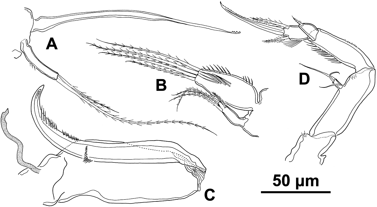
( Figure 7 View Figure 7 )
Material examined
Holotype female ( ZMA Co. 100.974a) and one paratype female ( ZMA Co. 100.9746) from near Hotel Coral Beach , Eilat ( Israel); associated with the anemone
Rhodactis rhodostoma (Ehrenberg, 1834) and collected at 1 m depth by J.H. Stock, April 1962.
Description
Female. Body cyclopiform, almost circular in outline and slightly dorsoventrally flattened (fig. 1A; Stock 1966). Total length of holotype 540 µm and maximum width 430 µm. Prosome comprising cephalothorax (fully incorporating first pedigerous somite) and three free pedigerous somites. Cephalothorax with posterolateral angles slightly pointed, somite bearing leg 2 with posterolateral angles very pointed, somite bearing leg 3 with posterolateral angles rounded and somite bearing leg 4 partly covered by previous somite. Urosome four-segmented, comprising leg 5-bearing somite, genital double-somite, and two free abdominal somites, which are wider than long. Leg 5-bearing somite wider than long, not visible in lateral view as completely concealed by last abdominal somite. Genital double-somite laterally expanded, 1.5 times wider than long, paired genital apertures bipartite, each comprising lateroventral copulatory pore and dorsolateral gonopore; lateral margins with rows of long spinules in distal half. Each genital area with one smooth seta (fig. 1B; Stock 1966).
Caudal rami slightly wider than long, with six terminal setae, two longest plumose (fig. 1B; Stock 1966).
Antennule 19-segmented, about 260 µm long. All setae plumose. Described and illustrated by Stock (fig. 1C; Stock 1966).
Antenna biramous ( Figure 7D View Figure 7 ), about 200 µm long; coxa small with tuft of spinules; basis elongate. Exopod one-segmented, small, twice as long as wide; with one lateral and two terminal setae, all of them smooth. Endopod three-segmented; first segment elongate with row of long spinules; second segment produced distally on medial side but articulating with distal segment proximally on lateral side, triangular, with one plumose seta; third segment with row of setules on inner margin and two plumose subterminal setae plus terminal claw.
Siphon short, about 160 µm long, conical, reaching to maxilliped insertion.
Mandible ( Figure 7A View Figure 7 ) consisting of stylet-like gnathobase with four distal teeth and small one-segmented palp. Palp with few spinules on lateral margin and two plumose terminal setae, unequal in length.
Maxillule bilobed ( Figure 7B View Figure 7 ); inner lobe oval and twice as long as outer one, with tuft of long spinules medially, row of shorter spinules laterally, and four plumose distal setae. Outer lobe with two terminal and two subterminal setae, one of them shorter and barbed.
Maxilla ( Figure 7C View Figure 7 ) two-segmented but with partial transverse surface suture on syncoxa (proximal segment) possibly marking plane of praecoxa–coxa fusion; praecoxal portion bearing flaccid aesthetasc-like element medially, representing tubular extension of external opening of maxillary gland. Coxal portion unarmed. Basis claw-like with rows of spinules on distal half.
Maxilliped (fig. 1I; Stock 1966) five-segmented with armature formula (1,0,2,1,1+claw) as described and illustrated by Stock in 1966.
Swimming legs 1–4 biramous (fig. 2A–E; Stock 1966) with three-segmented rami and intercoxal sclerite present in all of them. Swimming legs as described by Stock (1966).
Fifth and sixth legs as described by Stock (fig. 1B; Stock 1966).
Colour light yellowish.
Male. Unknown.
Remarks
This species was described by Stock from two females collected in Eilat ( Israel) in 1962. Asterocheres scutatus lives associated with the sea anemone Rhodactis rhodostoma (Ehrenberg, 1834) . Some oral appendages of this species are slightly different from those described by Stock: (1) the endopod of the antenna has three well-defined segments and the terminal seta of the exopod is approximately twice as long as the seta illustrated by Stock; (2) the stylet of the mandible is illustrated for the first time; (3) the setae of the inner and the outer lobes of the maxillule are plumose and the inner lobe has a patch of long spinules; (4) the maxilla bears a flaccid element medially, representing tubular extension of external opening of maxillary gland.
This species belongs to the group of species with 18- or 19-segmented antennules in females that comprises 13 species. These species are: A. bahamensis Kim, 2010 , A. brevisurculus Kim, 2005 , A. canui Giesbrecht, 1897 , A. dysideae Humes, 1996 , A. enewetakensis Humes, 1997 , A. fastigatus Kim, 2010 , A. hongkongensis Malt, 1991 , A. pilosus Kim, 2004 , A. plumosus Kim, 2010 , A. rotundus Malt, 1991 , A. serrulatus (Humes, 1996) , A. unioviger Kim, 2010 and A. walteri Kim, 2004 .
Asterocheres scutatus differs from 11 of these 13 species ( A. bahamensis , A. canui , A. dysideae , A. enewetakensis , A. fastigatus , A. pilosus , A. plumosus , A. rotundus , A. serrulatus , A. unioviger and A. walteri ) in the possession of a two-segmented mandibular palp, in contrast to the one-segmented mandibular palp shown by the present species ( Giesbrecht 1899; Humes 1996b, 1997; Kim 2004a, 2010; Bandera and Conradi 2009b).
Asterocheres hongkongensis differs from A. scutatus in the extremely short caudal setae (described and illustrated by Malt 1991 as about as long as the caudal rami). In contrast, A. scutatus has long caudal setae, as usual for the genus.
As for the shape of the urosome, A. scutatus has a genital double-somite that is much wider than long, whereas A. brevisurculus has a genital double somite that is longer than wide ( Kim 2005).
| ZMA |
Universiteit van Amsterdam, Zoologisch Museum |
No known copyright restrictions apply. See Agosti, D., Egloff, W., 2009. Taxonomic information exchange and copyright: the Plazi approach. BMC Research Notes 2009, 2:53 for further explanation.
|
Kingdom |
|
|
Phylum |
|
|
Class |
|
|
Order |
|
|
Family |
|
|
Genus |