
Cacopsylla (Thamnopsylla) burckhardti, Luo, Xinyu, Li, Fasheng, Ma, Yanfang & Cai, Wanzhi, 2012
|
publication ID |
https://doi.org/ 10.5281/zenodo.213975 |
|
publication LSID |
lsid:zoobank.org:pub:2C43EA7B-94F7-4133-9070-21AC4A8AB734 |
|
DOI |
https://doi.org/10.5281/zenodo.6178502 |
|
persistent identifier |
https://treatment.plazi.org/id/03B1723D-FFE8-FF8E-FF60-FF2756547469 |
|
treatment provided by |
Plazi |
|
scientific name |
Cacopsylla (Thamnopsylla) burckhardti |
| status |
sp. nov. |
Cacopsylla (Thamnopsylla) burckhardti View in CoL sp. n.
( Figs 9–19 View FIGURES 9 – 16 View FIGURES 17 – 19 )
Adult. Coloration: Body yellowish green. Vertex yellowish brown; discal foveae dark brown; areas along median suture, fore margin and middle of hind margin yellowish green. Genal processes yellowish brown. Ocelli orange, compound eyes brown to black. Antenna brown, with black apices on segments IV–VIII and segments IX–X entirely black. Thoracic terga yellowish green, with brown stripes. Legs yellow, dorsal surface of femora with irregular brown pattern, apical tarsal segment brown. Fore wing transparent; veins light brown, vein A1 darkened near anal break, not forming a marking. Abdomen yellowish green, middle of terga black, connected together into a wide stripe; sterna black or yellowish green. Male and female terminalia yellowish green.
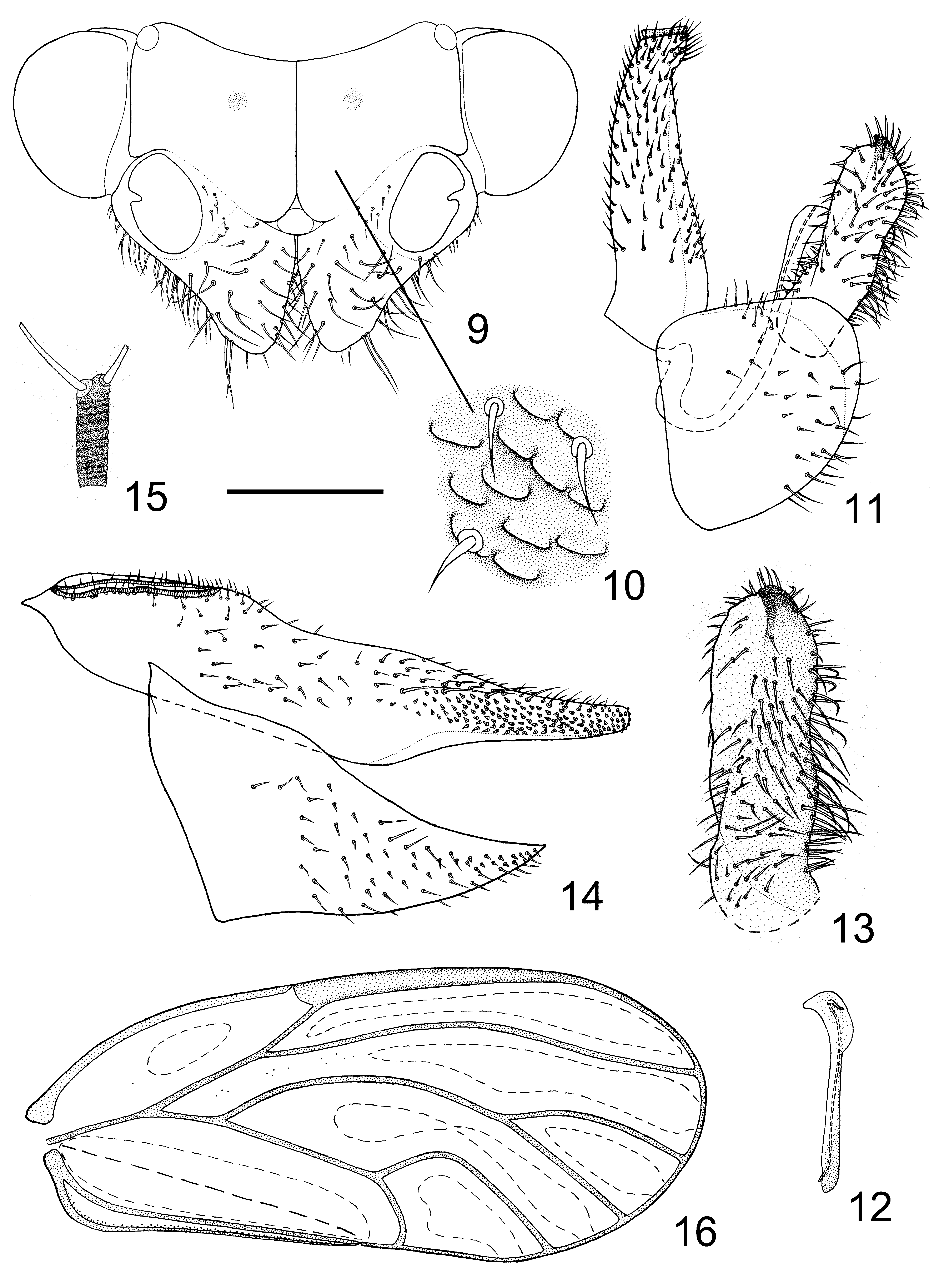
Structure: Body glabrous and robust. Head strongly inclined from longitudinal body axis, slightly wider than mesoscutum and relatively transverse. Vertex ( Fig. 10 View FIGURES 9 – 16 ) finely sculptured with microscopic setae and scaly micro structures that are relatively small, prominent and sparse. Genal processes ( Fig. 9 View FIGURES 9 – 16 ) cone-shaped, robust and relatively blunt apically, almost as long as vertex along median suture, covered with long setae. Ocelli relatively large. Compound eyes moderately protruding. Antenna relatively short and slender, terminal setae ( Fig. 15 View FIGURES 9 – 16 ) of very different lengths, the longer one about twice as long as the shorter one, and about as long as antennal segment X. Metatibia with short basal spine, apical spurs arranged as 1+3+1. Fore wing ( Fig. 16 View FIGURES 9 – 16 ) oblong oval, widest in apical third; pterostigma relatively long, ending in the apical third of cell r1; cell cu1 nearly quadrate, curvature of vein Cu1a almost forming right angle; surface spinules present in all cells except for c+sc, leaving wide spinule-free stripes along the veins, fields narrowing along wing margin in cells r2, m1, m2 and cu1; 4 sets of radular spinules present in cells r2, m1, m2 and cu1, in r2 less developed. For texture of surface spinules: normal (as is in most known Cacopsylla spp. represented by C. chinensis ) basally, gradually turning into singularly contrasting (black and slightly larger in individual) apically, with radular spinules also contrasting.
Male terminalia: Proctiger ( Fig. 11 View FIGURES 9 – 16 ) slender, densely covered with short setae. Paramere ( Figs 11, 13 View FIGURES 9 – 16 ) lamellar and broad, apical tooth subacute and inflexed; anterior margin expanding into narrow elongate extension, posterior margin strongly sinuate; setae present on both inner and outer surface, sparser and shorter on anterior margin, denser and longer in basal 2/3 of inner surface and posterior margin, several slightly thicker short setae present near apical tooth. Apex of basal aedeagus segment ( Fig. 11 View FIGURES 9 – 16 ) strongly projected caudad; apical dilatation ( Fig. 12 View FIGURES 9 – 16 ) of aedaegus relatively elongate and lens-shaped; sclerotised end tube of ductus ejaculatorius weakly sinuate. Subgenital plate ( Fig. 11 View FIGURES 9 – 16 ) nearly triangular, partly covered with setae that vary in length near dorsal margin and sparse short setae ventrally.
Female terminalia ( Fig. 14 View FIGURES 9 – 16 ) elongate. Proctiger sinuate dorsally, covered with rather short setae; apical part densely covered with peg setae, with the involved field not completely surrounded by fields of setae. Ventral surface of subgenital plate covered with short setae and peg setae.
5th instar nymph. Coloration: For specimens preserved in absolute ethanol and not dissected. General colour yellow, mature individuals darker. Dorsal sclerites ochreous, ventral ones lighter or light brown. Wing pads light brown except for the base. Compound eyes brown. Apical 2/3 of antennal segment 7 black.
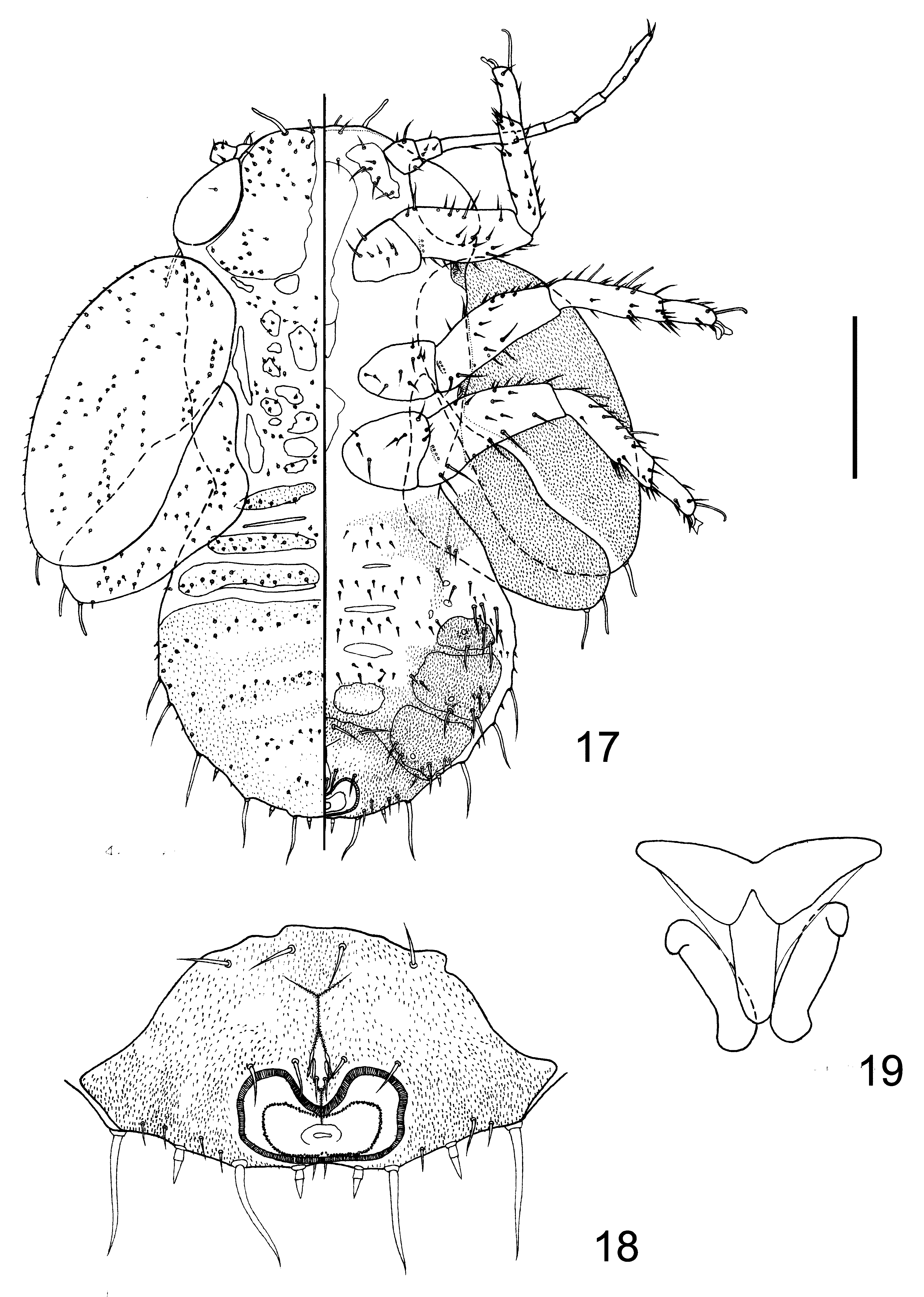
Structure: Body oblong oval. Dorsal surface ( Fig. 17 View FIGURES 17 – 19 ) unevenly covered with minute capitate setae, on abdomen arranged in transverse stripes, indicating the division of abdominal segments. Ventral surface of abdomen ( Fig. 17 View FIGURES 17 – 19 ) covered with simple setae, longer laterally. Micro spinules present on both dorsal and ventral surfaces, fields as shown in Fig. 17 View FIGURES 17 – 19 , on dorsal surface short, lamellar and multicuspid, on ventral long, spinous and unicuspid. Ocular seta ( Fig. 17 View FIGURES 17 – 19 ) very short, capitate. A long capitate seta ( Fig. 17 View FIGURES 17 – 19 ) present posterior to compound eye at body margin. Antenna ( Fig. 17 View FIGURES 17 – 19 ) slender, 7-segmented, with a single rhinarium on apices of segments 3 and 5, two rhinaria on segment 7. A sclerite with a spiracle present in ventral surface between praecoxa and mesocoxa, and a sclerite with a spiracle present on ventral surface between mesocoxa and metacoxa ( Fig. 17 View FIGURES 17 – 19 ). A pair of long capitate setae present in anterior margin of head, with a pair of short capitate setae between them ( Fig. 17 View FIGURES 17 – 19 ). Fore wing pad with one, and hind wing pad with two short capitate setae on distal angle ( Fig. 17 View FIGURES 17 – 19 ). Dorsal surface of mesotibia and metatibia with 2 long capitate setae ( Fig. 17 View FIGURES 17 – 19 ). Tarsal arolium ( Fig. 19 View FIGURES 17 – 19 ) petiolate, fan-shaped. Abdomen ventrally with 3+3 lateral free sclerites ( Fig. 17 View FIGURES 17 – 19 ) each bearing a spiracle. Outer circum-anal ring ( Fig. 18 View FIGURES 17 – 19 ) oval, with anterior margin sharply depressed, posterior margin near straight and lateral margins weakly indented. Inner circum-anal ring ( Fig. 18 View FIGURES 17 – 19 ) of similar shape as outer one but lateral margins not indented. Ventral surface of caudal plate ( Fig. 18 View FIGURES 17 – 19 ) with 2+2 simple setae near anterior margin, 2+2 simple setae right in front of outer circumanal ring, 2 simple setae within the suture and a series of simple setae near posterior margin. Abdominal margin ( Fig. 17 View FIGURES 17 – 19 ) bearing 5 pairs of long and pointed simple setae and 3 pairs of sectasetae.
Material examined. Holotype: male, dry mounted, China, Gansu, Sanshilipu, Hezheng, on Pyrus ussuriensis 15.vi.2011, Ma Yanfang.
Paratypes: 7 male, 26 female with same data as holotype, together with numerous nymphs. Non-paratypic specimens: China, Gansu: Sanshilipu, Hezheng, 16 male, 23 female on Pyrus ussuriensis , 9.vi.2010, Ma Yanfang, dry mounted. Additional materials (from the same series with the type materials) are preserved in absolute ethanol.
Etymology. This species is named after Dr. Daniel Burckhardt for his great contribution to study of psyllids and his help in our research.
Remarks. This species resembles Cacopsylla pyrisuga (Foerster) in the fore wing shape, the strongly contrasting surface spinules, setation of female proctiger and broad parameres. It differs from the latter in shape of distal segment of aedeagus, the more produced anterior margin and the more sinuate posterior margin of the paramere and more reduced areas of surface spinules of the fore wing. C. pyrisuga is recorded from the whole Palaearctic Region ( Klimaszewski, 1973; Kwon, 1983; Ossiannilsson, 1992), though never formally from China by Chinese authors. Kwon’s (1983) record of C. pyrisuga from Korea may concern C. burckhardti rather than C. pyrisuga judging from his illustrations, e.g. distal segment of aedeagus, sinuate hind margin of paramere and shape of female proctiger. The fore wing illustration of Kwon does not show the areas of surface spinules, but he states in his redescription that they are “completely covering cell c+sc”, which is not the same with C. burckhardti . Kwon’s record of Cacopsylla pyricola may also concern other species.
No known copyright restrictions apply. See Agosti, D., Egloff, W., 2009. Taxonomic information exchange and copyright: the Plazi approach. BMC Research Notes 2009, 2:53 for further explanation.