
Chilibathynella kotumsarensis Ranga Reddy
|
publication ID |
https://doi.org/ 10.5281/zenodo.174899 |
|
DOI |
https://doi.org/10.5281/zenodo.5678634 |
|
persistent identifier |
https://treatment.plazi.org/id/03BC1C72-FFA5-D738-FEE8-FC24D44A38D8 |
|
treatment provided by |
Plazi |
|
scientific name |
Chilibathynella kotumsarensis Ranga Reddy |
| status |
sp. nov. |
Chilibathynella kotumsarensis Ranga Reddy n. sp.
( Figs 2–6 View FIGURE 2 View FIGURE 3 View FIGURE 4 View FIGURE 5 View FIGURE 6 )
Etymology
The specific epithet alludes to the type locality of the new species.
Type locality
The Kotumsar Cave, a limestone cave, is one of the largest caves in India. It lies on the bank of the River Kanger, flowing through the Kanger Valley National Park (18º52'09" N; 81º56'05" E), at an altitude of 560 m near Jagdalpur town, Bastar District, Chhattisgarh State, India ( Fig. 1 View FIGURE 1 ).
The cave entrance is a vertical fissure in the wall of a hill. It has a narrow, twisted opening, measuring about 15 m in length. The cave is honeycombed in its structure, consisting of several irregular chambers. The main tunnel of the cave is nearly 500 m long and has several lateral and downward passages. The roofs and walls of the different chambers are lined with colorful dripstone formation, resulting from the precipitation of calcite-dissolved carbonate lime. The chambers are floored with either rocks or pebbles of varying dimensions or surface-derived soil/clay deposits.
Some of the abiotic parameters of the cave, as determined by Pati & Agrawal (2002), between May 1987 and March 1988, were as follows: air and water temperatures remained relatively stable, at an annual average of 28.25 ± 1.23 ºC and 26.33 ± 0.96 ºC respectively (range = 25.0–32.7 ºC for air; 22.9–29.3 ºC for water). The water pools were alkaline, with an annual average pH of 8.04 ± 0.36. Conductivity peaked during December, with an annual average of 0.27 ± 0.03 m Mhos. The annual mean for dissolved oxygen and percentage saturation for oxygen in the cave water were 6.42 ± 0.52 mg /l and 74.83 ± 5.91%, respectively. The cave is subject to frequent flooding during monsoon activity, which generally begins in the middle of June.
.
Material examined
Holotype, adult male, dissected on four slides. Paratypes, adult male, dissected on four slides, and four juveniles, one of them dissected on two slides, three mounted as whole specimens on a single slide. Type material has been deposited in the National Museum of Natural History, Paris; registration numbers: holotype MNHN-Sy 16 (male); paratypes: MNHN-Sy 17 (male), MNHN-Sy 18 (1 juvenile), MNHN-Sy 19 (3 juveniles). Leg. Y. Ranga Reddy, 0 1 December 2004.
Diagnosis
Parabathynellid of small size (1.25 mm). Antennal organ small, represented by two contiguous dentate structures. Fifth antennular segment with two aesthetascs. Antenna sixsegmented. Thoracopod I with well-developed epipodite. Pleopod I with two setae. Uropodal sympod with inhomonomous row of spines. Uropodal exopodite with three setae, two apical and one lateral, and endopodite without spines. Anal operculum convex.
Description of male (holotype)
Total length 1.25 mm (male paratype 1.28 mm). Body elongate, maximum width at first abdominal segment. Abdominal segments wider than thoracic ones. Head 25% longer than wide and slightly longer than first three thoracic segments combined.
Antennule ( Fig. 2 View FIGURE 2 d) consisting of seven, somewhat elongate, slender segments, 29% longer than head; length of first three segments only slightly exceeding that of last four segments. First segment longest, 1.7 times longer than wide, with two dorso-medial, simple setae near distal margin; two dorsal plumose setae at about distal outer corner, one tiny seta at distal inner corner, and one plumose seta on sub-distal outer margin. Second segment with two dorsal and one ventral simple setae near distal inner angle; antennal organ much reduced, represented by two conical, dentate and nearly contiguous hyaline structures; one plumose and one simple seta on sub-distal outer margin, one dorsal, plumose seta near distal outer corner, and one ventral plumose seta close to mid-outer margin. Third segment with one seta at sub-distal outer margin and one ventral and one dorsal seta near distal inner corner. Inner flagellum of third segment slightly longer than wide, with three simple setae. Fourth segment with three plumose setae, two unequal setae on the tip of apophysis and one at its base on outer side; apophysis reaching about proximal third of fifth segment. Fifth segment with two aesthetascs and two simple setae dorsally. Sixth segment longer than seventh one and with three aesthetascs and four setae dorsally. Seventh segment with three aesthetascs and four setae.
Antenna ( Fig. 3 View FIGURE 3 a) six-segmented (basal additional segment, if any, is not discernible with the optics used); right one curved backwards, bending between third and fourth segments; left one nearly straight, antero-laterally directed, 0.7 as long as antennule; percentage lengths of segments 1–6 as follows: 5:13:20:16:19:27; segments 1 and 4 without seta; segments 2, 3, 5, and 6 with 1, 2, 1, and 4 (3 apical and 1 lateral) setae, respectively.
Labrum ( Fig. 3 View FIGURE 3 b,c) flat, symmetrical, free margin straight, bearing eight main, nearly uniform teeth and 1 smaller marginal tooth on each side (N. B. Unfortunately, the labrum was folded in permanent preparation of the holotype as in Fig. 2 View FIGURE 2 b).
Mandible ( Fig. 3 View FIGURE 3 d,e): distal part of pars incisiva with 4 teeth, tooth of the ventral edge small and pointed. Pars molaris (”Borstenlobus”) with 8 claws, of which the distal two smooth, relatively large, forming a separate group; other claws with fine denticles on proximal margins; proximal outer corner of pars molaris with a row of spinules. Palp onesegmented, about three times as long as wide, bearing a terminal seta, slightly exceeding pars incisiva in length.
Maxillule ( Fig. 3 View FIGURE 3 f) with two endites; proximal endite small, somewhat oval in outline, with four apical claw-like spines, distalmost one longest. Distal endite bending inward, gradually tapering posteriad and with two terminal and four inner marginal claws. Outer distal margin with three simple setae.
Maxilla ( Fig. 3 View FIGURE 3 g) consisting of four segments. First segment with an elongately oval endite, carrying two elongate plumose and two short simple setae. Second segment with six (four inner-marginal, two medial) and third segment with 13 setae. Fourth segment tiny, with one claw and three setae.
Thoracopods I–VII ( Figs 4 View FIGURE 4 a–d, 5a–c) well developed; length gradually increasing from pairs I–III. Thoracopods III–VII almost similar in size. Thoracopods I–VII each bearing one-segmented epipodite on coxa and one inner marginal seta on basis. Exopodite one-segmented, with two unequal terminal setae; an additional subterminal seta present on dorsal side of thoracopod V alone (see Variation). Endopodite four-segmented. First and second endopodal segments of thoracopods I–VII with a rudimentary seta each at distal outer corner (not considered for setal formulae). Setal formulae:
Thoracopod I: 2 + 0/2 + 0/2 + 0/3(1)
Thoracopod II: 1 + 0/ 1 + 0/1 + 0/3(1)
Thoracopods III–IV: 1 + 0/ 1 + 0/0 + 0/3(1) Thoracopods V–VII: 0 + 0/ 1 + 0/0 + 0/3(1)
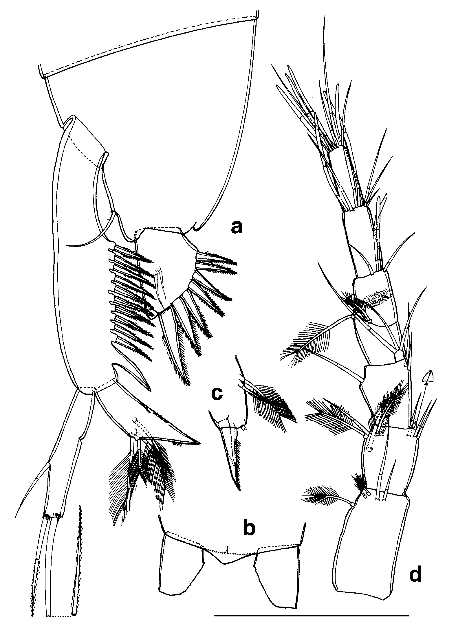
Thoracopod VIII ( Fig. 5 View FIGURE 5 d, e) massive, characteristic in shape. Basal segment of protopodite large, oblong, juxtaposing basis and originating at the same level as the basis. Dentate lobe shorter than the rounded inner lobe and with straight or somewhat convex free margin, carrying five or six teeth in a row. Basis roughly conical, extending well beyond the level of dentate lobe and with one subapical seta. Epipodite (external lobe) conical and arising from the basis. Exopodite curved, sharply bending backward and with a row of apical denticles. Endopodite rectangular, with one subapical and two unequal apical setae.
Pleopod I ( Fig. 5 View FIGURE 5 f) one-segmented, nearly four times as long as wide, with one long apical and one short subapical seta.
Uropod ( Fig. 2 View FIGURE 2 a). Sympod more than twice the length of endopodite and 4.6 times longer than its own maximum width, with eleven spines, distalmost spine largest and smooth; all other spines acutely pointed, almost similar in size and with serrulate lateral margins. Exopodite almost cylindrical, 5.4 times as long as wide, measuring 52% of sympodite length and bearing two apical setae, each with a row of spinules at base, inner seta twice as long as outer seta, and one short seta at about distal third of outer margin. Endopodite somewhat dagger-shaped, reaching 41% of sympodite length; bearing two unequal plumose setae at about the middle of outer margin and two equal plumose setae medially; spinules occurring on distal outer margin and on inner margin, as illustrated.
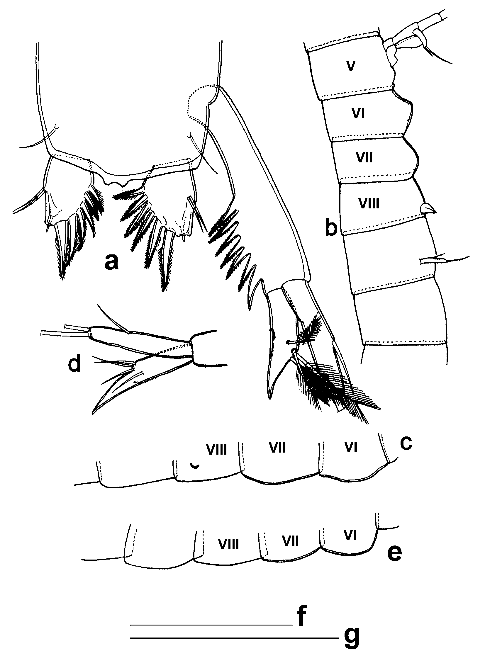
Pleotelson ( Fig. 2 View FIGURE 2 a) with one seta on either side near the base of furcal ramus; seta bare, shorter than furcal ramus. Anal operculum broadly triangular in outline, with rounded tip ( Fig. 2 View FIGURE 2 b).
Furcal rami ( Fig. 2 View FIGURE 2 a) 36.5% longer than maximum width, maximum width occurring at proximal third, outer margin nearly straight and ending in large, blunt ventral projection (furcal organ) ( Fig.2 View FIGURE 2 c), distal two-thirds of inner margin expanded, with two terminal and five inner, pointed, serrulate spines and two unequal dorsolateral setae; terminal spine largest and with a row of dorsal spinules at its base, other spines gradually decreasing in size, as illustrated.
Female: Not known.
Description of juveniles ( Fig. 6 View FIGURE 6 a–e)
Four sexually undifferentiated juveniles were recorded.
Juvenile 1 ( Fig. 6 View FIGURE 6 a, b): Total length 0.96 mm. Body eight times longer than wide. Head 24% longer than wide. Antennule 29% longer than head. Habitus and the various structural details of the cephalic appendages, thoracopods I–V and pleopod I are as in the adult. Sixth and seventh thoracic segments with rounded sternum in lateral view ( Fig. 6 View FIGURE 6 b), but without any trace of thoracopods. Thoracopod VIII represented by an undifferentiated, triangular lobe. Anal operculum slightly projecting backwards, somewhat rectangular in outline and depressed at the middle. Furcal rami with only six spines, the largest spine of the adult rudimentary, in the form a short, filamentous structure. Uropod as in the adult except for the sympod carrying seven spines, distal most spine largest.
Juveniles 2 and 3 ( Fig. 6 View FIGURE 6 c, d): Total length 0.83 mm. Both are identical to each other and differ from the juvenile 1 in two respects: thoracopod VIII is a very small crescentic lobe, and pleopod 1 is absent.
Juvenile 4 ( Fig. 6 View FIGURE 6 e): Total length 0.78 mm, similar to juveniles 2 and 3 except for the absence of any trace of thoracopod VIII.
Va r i a t i o n
The number of spines borne by the uropodal sympod varies between 9 and 11 in the adults and, 5 and 8 in the juveniles. The exopodite of thoracopod V has one dorsal seta in the holotype whereas it is absent in the male paratype as well as juveniles. The anal operculum is different between the adults and juveniles. No variation has been noticed in the armature of caudal furca.


No known copyright restrictions apply. See Agosti, D., Egloff, W., 2009. Taxonomic information exchange and copyright: the Plazi approach. BMC Research Notes 2009, 2:53 for further explanation.