
Chitonaster felli ( H.E.S. Clark, 1971 )
|
publication ID |
https://doi.org/ 10.5281/zenodo.276783 |
|
DOI |
https://doi.org/10.5281/zenodo.6184331 |
|
persistent identifier |
https://treatment.plazi.org/id/03D28792-FFC8-FF93-84E4-11BC686D83F3 |
|
treatment provided by |
Plazi |
|
scientific name |
Chitonaster felli ( H.E.S. Clark, 1971 ) |
| status |
|
Chitonaster felli ( H.E.S. Clark, 1971) View in CoL
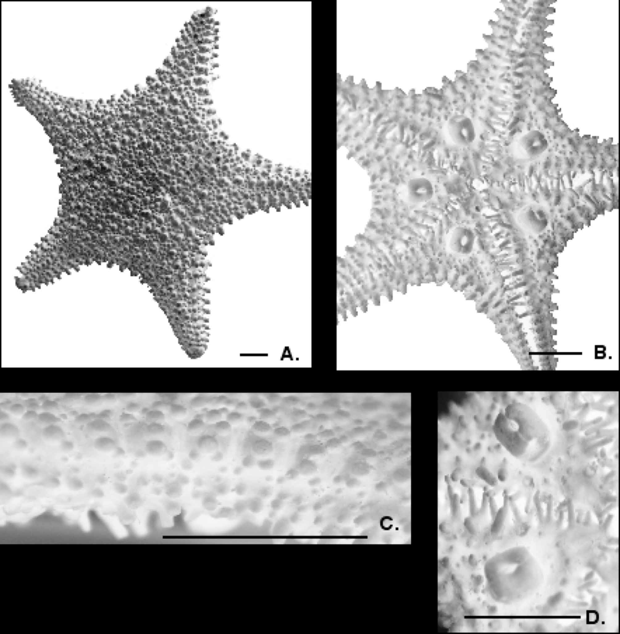
( Figure 5 View FIGURE 5 A–D)
H.E.S. Clark, 1971: 546; A.M. Clark, 1993: 275 (as Pentoplia)
Distribution. South Atlantic-Scotia Sea region near South Georgia /South Orkney Islands. 2355–3714 m.
Comments. The type specimen for this species, as designated by H.E.S. Clark (1971) was apparently never deposited in the USNM collections. An unsuccessful search for the holotype of Pentoplia felli was undertaken at the TePapa museum and in the collections of the National Institute of Water and Atmosphere in Wellington, New Zealand and the holotype could not be located. However, several original paratype specimens have recently been discovered, suggesting the possibility that the holotype may still be extant. Thus, designation of a neotype is considered premature until a complete search can be performed.
Distinctions from other species. Chitonaster felli shares several characters with C. johannae , including clustered spines on superomarginal and abactinal plates, the presence of transverse arm plates, and a similar triangular arm and large disk body shape. Chitonaster felli is distinguished by the autapomorphic large and distinctively shaped pedicellariae present in each actinal interradial region. However, other then this large actinal pedicellariae, C. felli lacks pedicellariae on other body surfaces. Chitonaster johannae possess enlarged and valvate pedicellariae on the actinal interradial and abactinal body surfaces that are similar but not identical to those pedicellariae on C. felli . Chitonaster felli and C. johannae are maintained herein as separate, but given that these two species are distinguished by only one or two characters but share at least four characters. These shared characters suggest that additional specimens and understanding of further morphological variation, could show C. felli is a synonym of C. johannae . However for now, the characters show distinctiveness and support discontinuity between species.
Material examined. USNM 1018660 PARATYPE. Southwest of South Georgia Island, Scotia Sea. 55°06’S, 44°22’W, 3623–3714 m. Coll. R/V Eltanin, Sta. 469, cruise 7. 12 Feb. 1963. (1 dry spec. R=1.3, r=0.5). USNM 1018861, Northeast of South Shetland Islands, Scotia Sea , South Atlantic Ocean. 59˚01’S, 52˚00’W to 59˚10’S, 51˚45’W, 3010–3510.0 m. Coll R/V Eltanin, Sta. 1511, Cruise 22. 26 Jan. 1966. (5 dry specs. R=2.4, r=1.1; R=1.7, r=1.1; R=1.4, r=0.9; R=1.8, r=1.1; R=1.6, r=1.1); USNM 1018862, Off west tip of South Georgia Island, South Atlantic Ocean. 55˚01’S, 39˚55’W to 55˚10’S, 39˚46’W. 2886–3040.0 m. coll. R/V Eltanin Sta. 1537, cruise 22 8 Feb. 1966 (1 dry spec. R=1.3, r=0.6); USNM 1018863, east of South Orkney Islands, Scotia Ridge. 61°4’S, 39°55’W to 61°07’S, 39°42’W. 2355–2897 m. Coll. R/V Eltanin 11 Feb. 1966. (1 dry spec? fragments only).
Description. Body is stellate (R/r=1.0 to 2.2), weakly swollen, convex with triangular arms, large disk ( Fig. 5 View FIGURE 5 A).
Abactinal plates flattened, scalar, forming thin parchment-like body wall. Abactinal plates polygonal in outline, with length and width relatively equidistant. Plate abutting or weakly overlapping. Fasciolar grooves weakly present. Abactinal plates relatively small with six to seven extending from superomarginal to superomarginal at the base of each arm. Each plate with one to three (usually one or two) low, blunt, spines with one to 12 small, coarse tubercular granules. These latter secondary granules are similar in overall shape to the spines but occur irregularly on each plate surface, sometimes around the edge of the plate. On the arms, the primary spines present on the carinal series in conjunction with the adjacent lateral plates form distinctly transverse rows plates. These transverse rows are present on the six to eight distalmost series on the arm plates ( Fig. 5 View FIGURE 5 A). Other then the larger primary spines and the granules, no other accessories are present and the plate surfaces are otherwise bare. Papulae absent. (These are listed as “indistinct” in the original description).
Superomarginals and inferomarginals are most commonly 1:1 with little to no offset ( Fig. 5 View FIGURE 5 C). Each plate is swollen and quadrate in outline, especially interradially. Two to three larger, primary blunt spines are present on the superomarginal and the inferomarginal plates ( Fig. 5 View FIGURE 5 C). Each spine series is arranged in a linear series, forming a distinct border. Smaller secondary spines, which are conical and pointed, are approximately 10–15% of the size of the primary spines are present between the superomarginal and inferomarginal spine series on the lateral facing of each plate series ( Fig. 5 View FIGURE 5 C). Otherwise, accessories, such as granules, spinelets, etc. are absent from the surface of the marginal plates with no peripheral granules, etc. In larger specimens (R=2.2) superomarginal plates number 14–24 in each interradius (armtip to armtip). Inferomarginals number 14–26 from armtip to armtip. Terminal plates are large and often possess two to six blunt spines on the surface of each.
Actinal intermediate plates are restricted to the disk. Each very large pedicellaria present ( Fig. 5 View FIGURE 5 B) is surrounded by, fewer than ten, small blunt, spine-bearing plates. These pedicellariae are composed of two large quadrate valves, each with two prominent blunt projections ( Fig. 5 View FIGURE 5 B). A distinct space is present between the two valves, forming an open area between the projections on the pedicellariae ( Fig. 5 View FIGURE 5 D). The pedicellariae each occupy a distinct concavity in each actinal interradial region, which can be observed within the coelomic cavity of the animal. Aside, from the large, individual conspicuous pedicellariae on the actinal surface pedicellariae are otherwise absent from the abactinal, marginal, and actinal plate surfaces. Each actinal plate with short, sharp, conical spinelets, approximately one to three per plate irregularly distributed over actinal surface ( Fig. 5 View FIGURE 5 B). Actinal plate boundaries obscured by tissue.
Adambulacral plates rectangular to quadrate with two (a third may be present) blunt, pointed furrow spines. A single, shorter subambulacral spine is present, separated by a discrete space from the two furrow spines. Smaller spinelets present, flanking larger furrow spines. Oral plates with three to four (usually three) furrow spines. One prominent blunt, oral spine present on each plate (two per interradius), projecting into the mouth.
H.E.S. Clark (1971) further adds several details on internal anatomy and variation.
| USNM |
Smithsonian Institution, National Museum of Natural History |
No known copyright restrictions apply. See Agosti, D., Egloff, W., 2009. Taxonomic information exchange and copyright: the Plazi approach. BMC Research Notes 2009, 2:53 for further explanation.