
Claudiella jeaneae, Benetti, Cesar João & Hamada, Neusa, 2016
|
publication ID |
https://doi.org/ 10.11646/zootaxa.4205.2.4 |
|
publication LSID |
lsid:zoobank.org:pub:9DE6E9D3-8EEE-4AEB-A7AF-257A516C768E |
|
DOI |
https://doi.org/10.5281/zenodo.6057624 |
|
persistent identifier |
https://treatment.plazi.org/id/03AC6C48-C16D-FFB4-F79B-FB3CFA0FF807 |
|
treatment provided by |
Plazi |
|
scientific name |
Claudiella jeaneae |
| status |
sp. nov. |
Claudiella jeaneae sp. n.
( Figs. 3–4, 8 View FIGURES 7 – 9 , 11 View FIGURES 10 – 12 , 15–16 View FIGURES 13 – 18 , 21–22 View FIGURES 19 – 24 , 26 View FIGURES 25 – 27 )
Type locality. Brazil: Minas Gerais state, Carrancas county , “ Cachoeira da Esmeralda ” (21°28'17.8"S 44°42'15.1"W). GoogleMaps
Type material. Holotype male ( INPA): Brazil: Minas Gerais state, Carrancas county , “ Cachoeira da Esmeralda ” (21°28'17.8"S 44°42'15.1"W), 11.iv.2014, leg. N. Hamada, J.M.C. Nascimento and L.M. Fusari GoogleMaps . Condition of holotype: stored in ethanol 99% with the dissected male genitalia stored in microvials. Paratypes (18): same data as holotype [one male and one female stored in ethanol, deposited at MZUSP; one male mounted on slides and 15 females stored in ethanol, deposited at INPA]. GoogleMaps
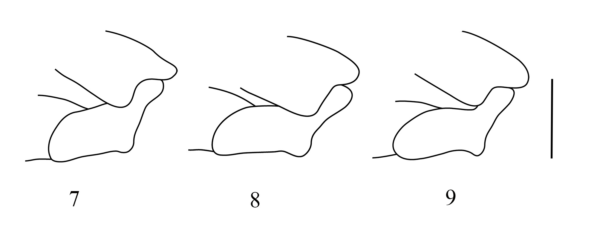
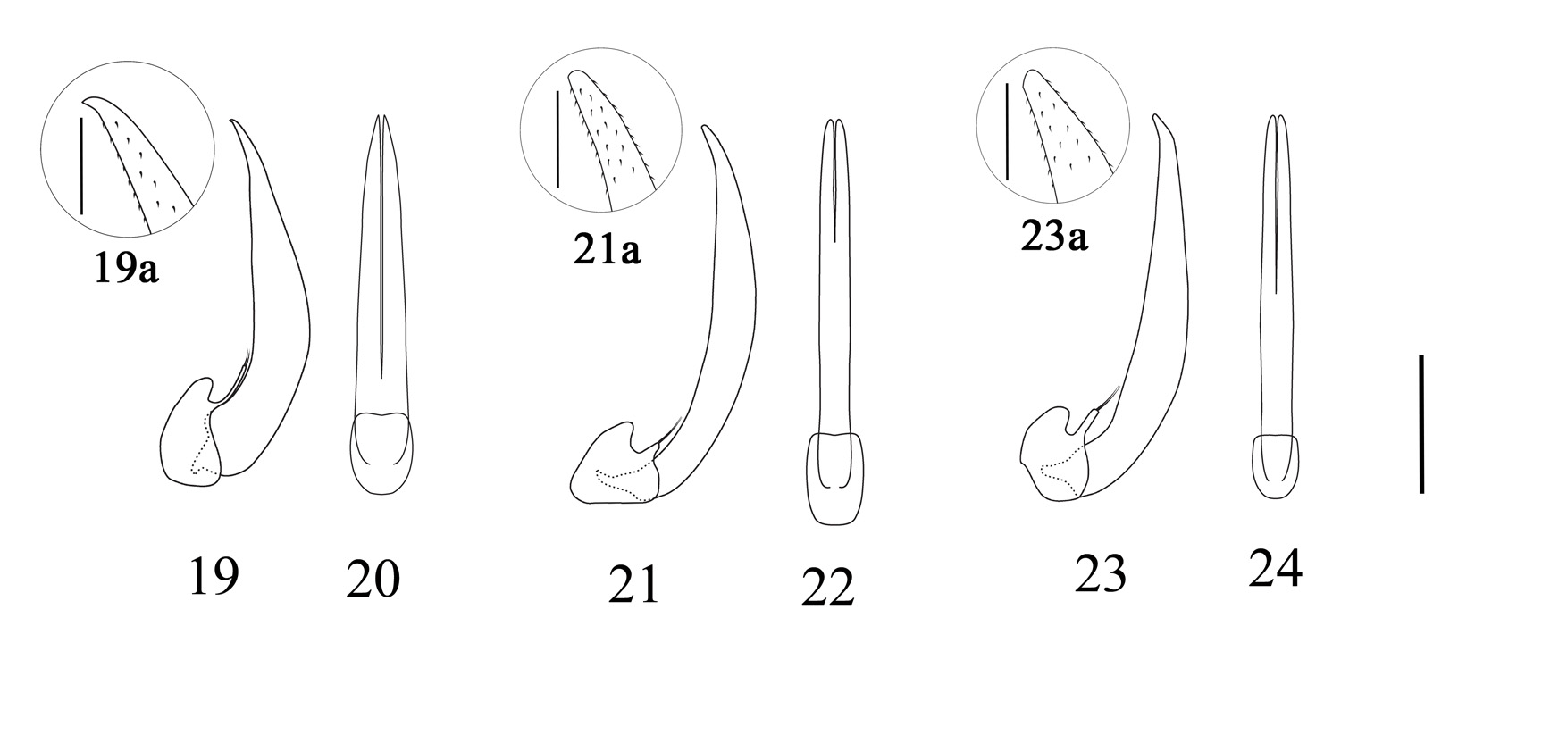
Diagnosis. Claudiella jeaneae sp. n. can be distinguished from other species of Claudiella by the following combination of characteristics: tooth-like projection on hind edge of metatrochanter conspicuous ( Fig. 8 View FIGURES 7 – 9 ); interstices strongly convex; left mandible with outer edge projected; RP vein reaching the oblongum cell in the middle of the rp-mp1 vein ( Fig. 26 View FIGURES 25 – 27 ); by the body size (length: 1.93–2.15 mm) and the shape of the male genitalia, with the median lobe, in dorsal view, split longitudinally only in the apical third; in lateral view, with the apex digitiform and continuous ( Figs. 21–22 View FIGURES 19 – 24 ).
Description. Habitus. ( Fig. 3). Shape oval, convex dorsally. Lateral sides of pronotum rounded. Elytra regularly tapering towards the apex. Lateral outline discontinuous between pronotum and elytra.
Color. Dorsal surface black with shiny golden colored punctures; ventrally reddish, legs dark reddish ( Figs. 3–4).
Measurements (n = 16). BL: 1.93–2.15 mm; BW: 1.12–1.28 mm; BR: 1.66–1.77; PL: 0.3 8– 0.47 mm; PW: 0.81–0.95 mm; PR: 1.89–2.44; EL: 1.4–1.58 mm; EW: 1.12–1.28 mm; ER: 1.19–1.29; ELPWR: 1.58–1.75.
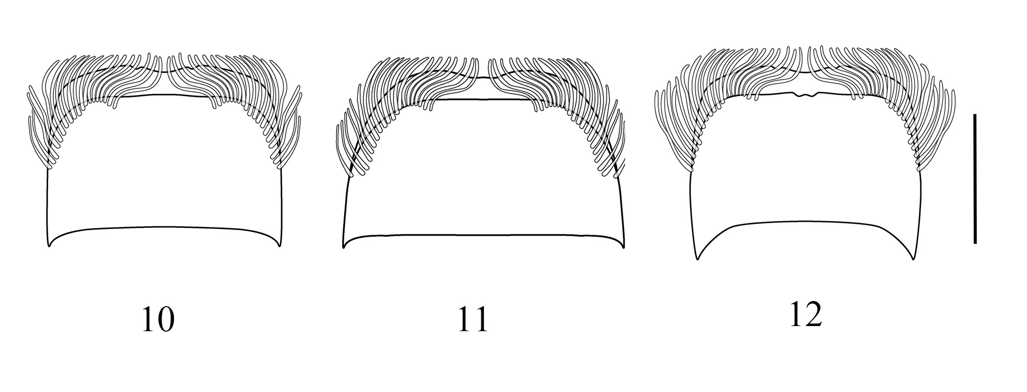
Head. Frons bearing a pair of longitudinal keels that run from the anterior edge of the eyes to the clypeus. Antennae with nine antennomeres; last antennomere a little shorter than antennomeres 6–8 together. Anterior edge of clypeus truncate and continuous; posterior edge convex; lateral edge parallel; anterolateral angles oblique, almost rounded. Labrum large, with very long recumbent setae anteriorly; anterior edge straight on dorsal surface, slightly sinuous on ventral surface, fringed with long fine filiform setae, absent in the median area; lateral edge curved ( Fig. 11 View FIGURES 10 – 12 ). Mandibles asymmetrical: left mandible with a bifid apical non-articulated tooth and a bifid articulated subapical tooth and right mandible with only a bifid apical non-articulated tooth; left mandible with outer edge projected, right mandible with outer edge convex ( Figs. 15–16 View FIGURES 13 – 18 ). Maxilla: mala without suture dividing it; with three apical laminar processes and a row of subapical setae; palpus with three palpomeres, palpomere I short, palpomeres II and III longer, similar in length; palpomere III bearing sensilla apically. Labium with fused glossae and paraglossae, bearing long setae along its entire length; prementum short and wide, densely pilose; labial palpi short, with elongated basal segment and two small apical segments.
Thorax. Pronotum coarsely punctured with surface microrugose; regularly widened towards the basal third; basal edge with median emargination with two small incisions in the middle; anterior edge strongly sinuous; lateral edges convex, slightly serrate. Scutellar shield very small, triangular. Elytra coarsely punctured with surface microrugose; wider than the base of the pronotum, widest before the middle; punctures arranged in 13 complete clearly marked striae, 12 dorsal and one sublateral, marginal stria; striae of punctures and interstices uneven; interstices strongly convex; humeral callus strongly protuberant; lateral edges slightly serrate only at the base, regularly narrowed towards the apex; apex slightly acute. Metathoracic wings ( Fig. 26 View FIGURES 25 – 27 ) fringed around the anterior and posterior edges, except on the costal margin; RP2 vein not reaching the apex of the wing; r1 vein well developed, short, distinctly oblique; ScP vein not distinctly curved, clearly separated from the RA vein; R3 vein extends towards the apex; RP vein slightly marked, reaching the r1 vein. The CuA3+4 vein joins the AA3 vein without change of direction; oblongum cell present; RP vein reaching the oblongum cell in the middle of the rpmp1 vein. Posterior edge of the prosternal process slightly sinuous, almost truncate. Metasternal carina present, extending to the posterior edge of the metaventrite; metaventrite without tubercles. Metacoxae with posterior edge covering the trochanter; metatrochanter with tooth-like projection on hind edge, conspicuous ( Fig. 8 View FIGURES 7 – 9 ), with inner lateral edge convex; femora and tibiae with a row of long setae on dorsal face; femora ventrally grooved for reception of tibiae; metafemur with middle third as wide as basal third; tarsal formula 4-4-4, tarsomeres 1–2 short, tarsomeres 3–4 four times longer than tarsomere 2.
Abdomen. Ventrites 1–4 with longitudinal carina. Semilunar depression of last abdominal ventrite simple, not prolonged towards the apex, in both sexes. Aedeagus ( Figs. 21–22 View FIGURES 19 – 24 ): phallobase with ventral process. Median lobe (dorsal view) split longitudinally but restricted to the apical third, with lateral edges parallel, converging towards the apex, starting from the middle; in lateral view, slightly curved, almost straight, gradually narrowed from base to apex; dorsal face convex, ventral face sinuous and greater width between the middle and the apex; apex digitiform, continuous, with very short spines ( Fig. 21 View FIGURES 19 – 24 a). Parameres broad, very short and small, shorter than the phallobase process, with two thin apical setae.
Etymology. This species is named in honor of Jeane Marcelle Cavalcante do Nascimento (INPA/CBIO), a good friend and colleague in the field and laboratory, in gratitude for her help in collecting material during the fieldwork.
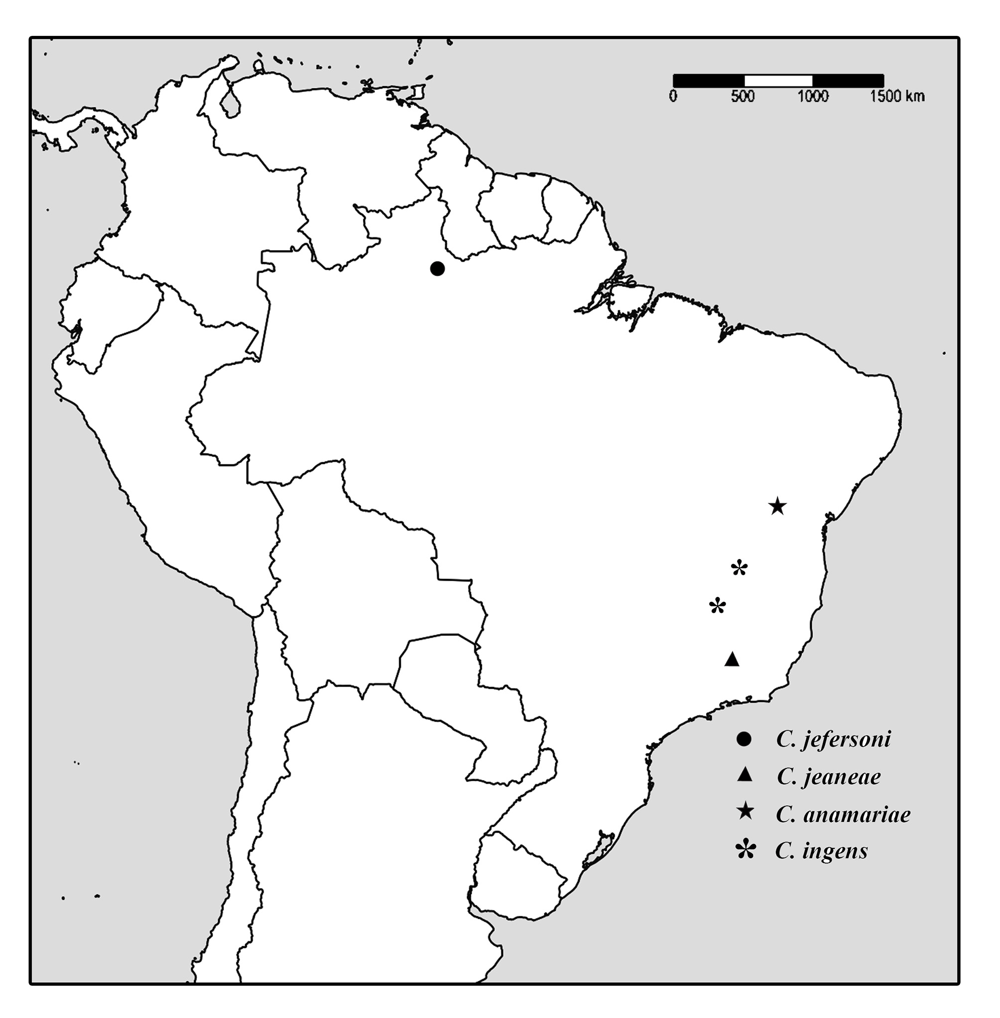
Distribution and habitat. Southeast Brazil, currently only known from the state of Minas Gerais ( Fig. 28 View FIGURE 28 ). Specimens were collected in a stream in the cerrado (central Brazilian savanna) biome, with width = 4 m, water pH = 6.5 and water temperature = 16.6°C.
Taxonomic comments. This species differs from C. jefersoni sp. n. and I. trombetensis by having the toothlike projection on the hind edge of the metatrochanter pronounced ( Fig. 8 View FIGURES 7 – 9 ), not pronounced in C. jefersoni sp. n. ( Fig. 7 View FIGURES 7 – 9 ) and I. trombetensis ; by its large size (average body length: 2.06 mm) and by the shape of the male genitalia ( Figs. 21–22 View FIGURES 19 – 24 ). Claudiella jeaneae sp. n. differs from C. ingens and I. quadridentatus in the shape of the tooth-like projection of the metatrochanter ( Fig. 8 View FIGURES 7 – 9 ), more conspicuous and well-marked in C. ingens and I. quadridentatus . The new species differs from I. quadridentatus in having two thin setae on the apex of each paramere ( Fig. 21 View FIGURES 19 – 24 ), while in I. quadridentatus the parameres have only one seta. C. jeaneae sp. n. differs from C. ingens by the shape of the male genitalia in dorsal view, with lateral edges of the median lobe parallel, converging towards the apex, starting from the middle ( Fig. 22 View FIGURES 19 – 24 ), while in C. ingens the lateral edges are parallel and converge only at the apex.


No known copyright restrictions apply. See Agosti, D., Egloff, W., 2009. Taxonomic information exchange and copyright: the Plazi approach. BMC Research Notes 2009, 2:53 for further explanation.
|
Kingdom |
|
|
Phylum |
|
|
Class |
|
|
Order |
|
|
Family |
|
|
Genus |