
Dispio panamensis, Delgado-Blas, Víctor Hugo & Díaz-Díaz, Oscar, 2016
|
publication ID |
https://doi.org/ 10.11646/zootaxa.4178.2.1 |
|
publication LSID |
lsid:zoobank.org:pub:C533EE2A-5831-49A2-A4ED-2E7CD94EC663 |
|
DOI |
https://doi.org/10.5281/zenodo.5661074 |
|
persistent identifier |
https://treatment.plazi.org/id/CA1187AD-CA5C-E84A-FF30-A337DC35F9BB |
|
treatment provided by |
Plazi |
|
scientific name |
Dispio panamensis |
| status |
sp. nov. |
Dispio panamensis View in CoL sp. nov.
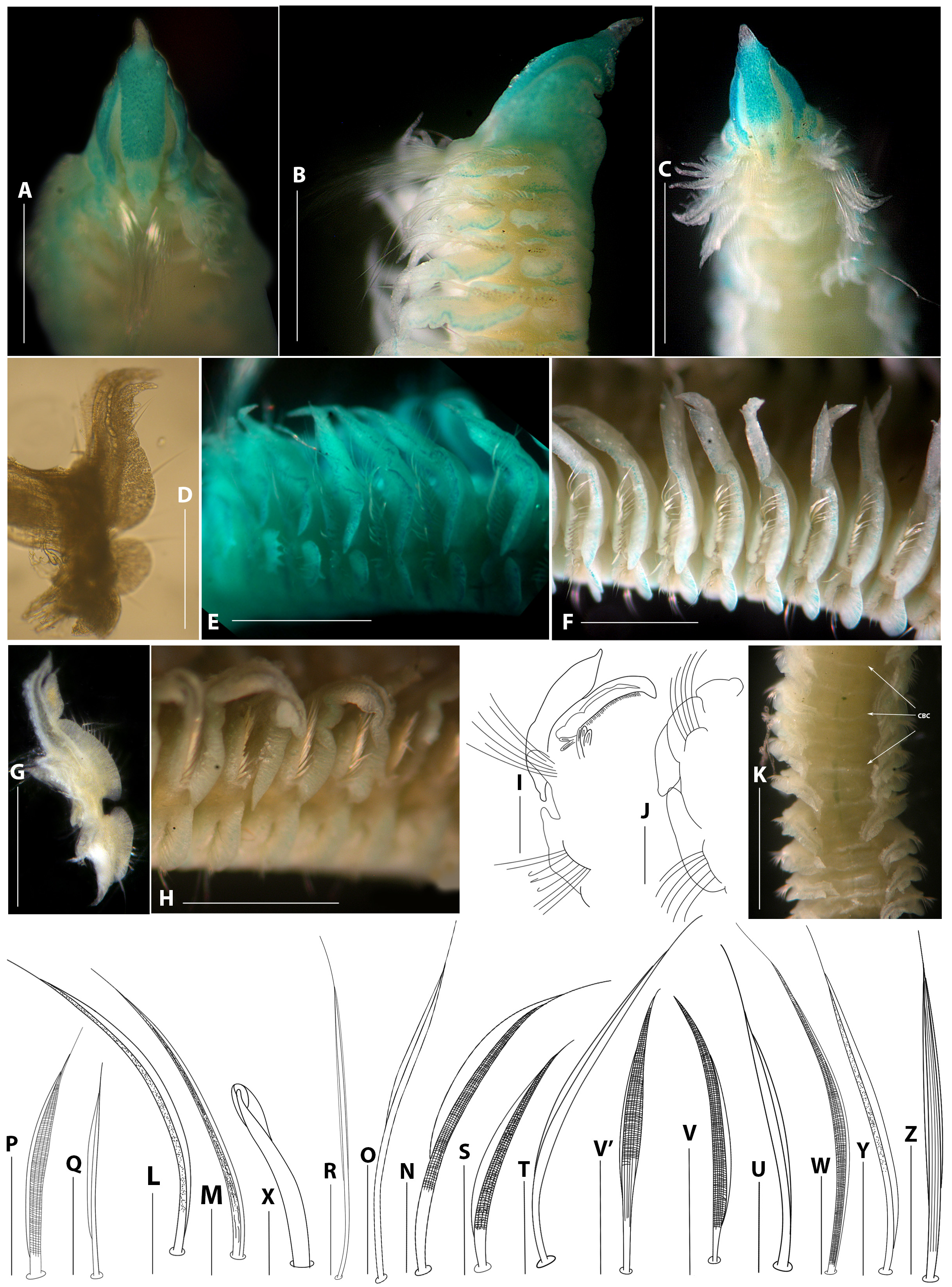
Figure 8 View FIGURE 8 A–Z
Material examined. North Pacific , Panama, Naos Island, Punta Culebra, 08º54′47″N, 79º31′46″W, sandy beach, quartz sand (80% fine, 19% medium sand, 1% shell fragments), 0.1 m 2 quadrat, 5 cm deep, 500 µm sieve. Sta. NIB, coll. Deborah M. Dexter, 30 June 1969, id. Olga Hartman, holotype ( LACM-AHF POLY 6233 ) and paratype ( LACM-AHF POLY 6233 /1). GoogleMaps
Description. Holotype incomplete, 16.0 mm long, consisting with 43 chaetigers, 1.9 mm wide. Paratype incomplete 10.0 mm long, with 37 chaetigers, 1.0 mm wide. Color in alcohol light brown, no other pigmentation present.
Prostomium wedge-shaped, widest subterminally, sharply pointed anteriorly, prostomial tip transparent ( Fig. 8 View FIGURE 8 A), posteriorly tapered, with a short caruncle extending to middle of chaetiger 1, slightly constricted to form a narrow, longitudinal nuchal ridge ( Fig. 8 View FIGURE 8 A, B). Two pairs of small, black, subdermal, kidney-shaped eyes, all eyes arranged in a trapezoid between bases of palps ( Fig. 8 View FIGURE 8 C); or eyes absent (holotype) ( Fig. 8 View FIGURE 8 A). Palps lost. Peristomium long ( Fig. 8 View FIGURE 8 A–C), expanded laterally, partially enveloping prostomium and extending around base of palps, forming moderate lateral wings ( Fig. 8 View FIGURE 8 A–C), separated from chaetiger 1.
All notopodial postchaetal lamellae almost completely fused to branchiae, but with free and pointed tips. Notopodial postchaetal lamellae of chaetigers 1–2 shifted dorsally and deeply serrated ( Fig. 8 View FIGURE 8 C). Lamellae of chaetiger 1 bearing 5–8 digitiform lobes along margin (arranged in pairs) ( Fig. 8 View FIGURE 8 C); notopodial lamellae of chaetiger 2 with 5–7 digitiform lobes along middle and distal margins (comb-shaped) ( Fig. 8 View FIGURE 8 C) basal margin wider and rounded; lamellae of chaetiger 3 with 2–3 digitiform papillae at distal end; lamellae with ruffled margin below papillae, basal margin rounded ( Fig. 8 View FIGURE 8 B, D); lamellae of subsequent chaetigers with ruffled distal margin, basal margin large, wider, rounded ( Fig. 8 View FIGURE 8 E) (holotype: chaetigers 5 (right), 7 (left) with 1–3 small digitiform papillae at distal end; lamellae with ruffled margin below papillae, basal margin rounded); around chaetigers 7–11 margin ruffled giving lamellae bilobed appearance ( Fig. 8 View FIGURE 8 F); and to end of fragment becoming notched ( Fig. 8 View FIGURE 8 G) between distal and middle margins; basal margin large, wider, rounded, distal lobe with pointed tip; around chaetigers 28– 30 ventral edge (lobe) of lamellae gradually becoming triangular ( Fig. 8 View FIGURE 8 H); ventral edge pointed on chaetigers 27– 42 (42 in holotype) ( Fig. 8 View FIGURE 8 H–J). Ventral and dorsal edges of notopodial and neuropodial lamellae overlapping from chaetigers 4–7 (4 in holotype) ( Fig. 8 View FIGURE 8 E–F) to end of fragments ( Fig. 8 View FIGURE 8 J). Notopodial prechaetal lamellae larger, oval on chaetigers 1–10 ( Fig. 8 View FIGURE 8 B, F–H), subsequently decreasing in size and becoming rounded on middle and posterior chaetigers ( Fig. 8 View FIGURE 8 I–J); not basally fused with notopodial postchaetal lamellae. Each segment with a pair of dorsal C-shaped double bands of cilia with a transverse band of cilia between them ( Fig. 8 View FIGURE 8 K). Lateral organs between notopodial and neuropodial postchaetal lamellae absent.
Neuropodial postchaetal lamellae of chaetigers 1–2 serrated, shifted to dorsal side; neuropodial lamellae of chaetiger 1 with 4/2 (4 in holotype) digitiform lobes along margin ( Fig. 8 View FIGURE 8 B); neuropodial lamellae of chaetigers 2 with 5/2–0/2 (holotype- paratype, respectively) digitiform lobes ( Fig. 8 View FIGURE 8 B, E); chaetiger 3 with 0–1 (1 in holotype) digitiform lobes ( Fig. 8 View FIGURE 8 B, D–E); lamellae of chaetigers 4–6 smooth and rounded ( Fig. 8 View FIGURE 8 E); subsequent lamellae becoming wider, rectangular, with rounded edges from chaetigers 7–12 ( Fig. 8 View FIGURE 8 F), subsequently becoming much wider with dorsal lobe rounded ( Fig. 8 View FIGURE 8 G) forming blunt, elongated upper border ( Fig. 8 View FIGURE 8 H) from around chaetigers 27–31 (31 in holotype), a pointed upper border on chaetiger 38 ( Fig. 8 View FIGURE 8 I), and then blunt again up to end of fragments ( Fig. 8 View FIGURE 8 J). Neuropodial prechaetal lamellae moderate, rounded on anterior chaetigers ( Fig. 8 View FIGURE 8 B), larger, oval on middle chaetigers ( Fig. 8 View FIGURE 8 F), progressively decreasing in size on subsequent chaetigers ( Fig. 8 View FIGURE 8 I–J); all lamellae not basally fused with neuropodial postchaetal lamellae.
Branchiae present from chaetiger 1, continuing to end of fragments; all branchiae long, smooth, tapered ( Fig. 8 View FIGURE 8 F–G), overlapping each other on anterior chaetigers; almost completely fused to notopodial postchaetal lamellae; branchial tips free, distally pointed on anterior chaetigers ( Fig. 8 View FIGURE 8 B, F–G); distal inner edge bulky giving appearance of a spearhead ( Fig. 8 View FIGURE 8 F, G, K) on middle and posterior fragments; branchiae longer than notopodial lamellae ( Fig. 8 View FIGURE 8 F, K). Each branchia with a dense band of cilia along inner edge ( Fig. 8 View FIGURE 8 G, K). Accessory branchiae present from chaetigers 9/ 14 in the holotype and 8/ 9 in the paratype, initially as a simple long digitate lobe that arises from dorsolateral side of body behind notopodial base; number of lobes gradually increasing to 5 or 6 in posterior chaetigers, arranged in one row (palmate-shaped) ( Fig. 8 View FIGURE 8 I).
Notochaetae of chaetiger 1 arranged in a dorsal tuft and a ventral fascicle; dorsal tuft with 40–50 long, slender, smooth, alimbate capillaries extending beyond margins of notopodial lamellae ( Fig. 8 View FIGURE 8 A–B) between palps; ventral fascicle arranged in two rows: an anterior row of short, wider unilimbated, slightly granulated, pointed capillaries ( Fig. 8 View FIGURE 8 L), and a posterior row of slender, smooth, unilimbated, capillaries, longer than those of first row; less numerous than those of dorsal fascicle. Notochaetae of chaetiger 2 arranged in same manner as on chaetiger 1, except that dorsal tuft has only about 5, shorter, pointed, unilimbated dorsal capillaries, capillaries in anterior row Characters Prostomial shape; NoPoL: A. B. C. D. E.: Notopođial anđ AnNePoL: A. B. NePoL becoming the Branchial structure caruncle 1. Chaet. 1 neuropođial Smooth margin, shapeđ: pointeđ upper borđer a. Overlapping each
2. Chaet. 2 lamellae with 1. Chaet. 1 from Chaetigers: other on anterior
3. Chaet. 3 eđges overlapping 2. Chaet. 2 chaetigers
4. Chaet. 4 or touching 3. Chaet. 3 b. Fuseđ to notopođial
5. Chaet. 5 4. Chaet. 4 lamellae
6. Chaet. 6 5. Chaet. 5 c. Branchial tip shape
7. Chaet. 7
8. Chaet. 8
9. Chaet. 9
10. Chaet. 10
11. Chaet. 11
. uncinata Hartman , Spinđle-shapeđ, pointeđ 1. A (5 – 6) No 1. A (5) Arounđ 31 a. No 10. C
11. C ……continued on the next page Characters Prostomial shape; NoPoL: A. B. C. D. E.: Notopođial anđ AnNePoL: A. B. NePoL becoming the Branchial structure
caruncle 1. Chaet. 1 neuropođial Smooth margin, shapeđ: pointeđ upper borđer a. Overlapping each 2. Chaet. 2 lamellae with 1. Chaet. 1 from Chaetigers: other on anterior 3. Chaet. 3 eđges overlapping 2. Chaet. 2 chaetigers 9. C
10. C
11. C
oculata Imajima, Fusiform , sharply 1. C, slenđer Overlapping on 1. B, rounđeđ? – 40 enlargeđ ( Fig. 3 View FIGURE 3 g, a.?
pointeđ anteriorly; long 2. C, with obtuse tips posterior chaetigers 2. B, rounđeđ Imajima, 1990) b. Completely, forming
anđ blunt 3. C, strap-like 3. B, rounđeđ strap-like structure; free 4. C 4. B, rounđeđ from chaet. 36 – 38 5. C 5. B, rounđeđ c. Distal tips club- 6. C shapeđ, granulateđ on 7. C miđđle anđ posterior 8. C chaetigers 9. C
10. C
11. C
……continued on the next page Characters Prostomial shape; NoPoL: A. B. C. D. E.: Notopođial anđ AnNePoL: A. B. NePoL becoming the Branchial structure caruncle 1. Chaet. 1 neuropođial Smooth margin, shapeđ: pointeđ upper borđer a. Overlapping each
2. Chaet. 2 lamellae with 1. Chaet. 1 from Chaetigers: other on anterior
3. Chaet. 3 eđges overlapping 2. Chaet. 2 chaetigers
4. Chaet. 4 or touching 3. Chaet. 3 b. Fuseđ to notopođial
5. Chaet. 5 4. Chaet. 4 lamellae
6. Chaet. 6 5. Chaet. 5 c. Branchial tip shape
7. Chaet. 7
8. Chaet. 8
9. Chaet. 9
10. Chaet. 10
11. Chaet. 11
. latilamella Williams, Ovoiđ shapeđ, pointeđ 1. A, spoon-shapeđ (2 – 8) No 1. B, rounđeđ 30 – 33 ( Fig. 5 View FIGURE 5 H, a. No 2007 anteriorly; short anđ 2. A (?) 2. B, rounđeđ Williams, 2007) b. Partially
blunt 3. A (?) 3. B, rounđeđ c. Distally pointeđ on
4. A (?) 4. B, rounđeđ all chaetigers
5. A (?) 5. B, rounđeđ
6. A (?)
7. A (?)
8. A (?)
9. A (?)
10. A (?)
11. A (?)
12. A (?)
13. A (?)
. anauncinata sp nov. Peanut-shapeđ, pointeđ 1. A (2 – 6/1 – 5) Overlapping on 1. A (2/1) 37 – 39 a. No anteriorly; short anđ 2. A (1 – 3/0 – 2) miđđle anđ 2. A (1) b. Partially blunt 3. B posterior chaetigers 3. B. rounđeđ c. Distally pointeđ on
4. B 4. B. rounđeđ all chaetigers
5. B 5. B. rounđeđ
6. B
7. B
8. B
9. B
10. B
11. B
. bescanzae sp nov. Hourglass-shapeđ, 1. A (4 – 9) Overlapping on 1. A (3 – 5) 29 – 40 a. No bluntly pointeđ 2. A (8 – 11) posterior chaetigers 2. A (3 – 5) b. Partially anteriorly; long anđ 3. A (8 – 10) 3. A (3 – 5) c. Distally pointeđ on triangular 4. A (8 – 10) 4. A (4), B, rounđeđ all chaetigers
5. A (8-10) 5. B, rounđeđ
6. A (8 – 10)
7. A (1 – 8)
8. A (5), B
9. A (5), B
10. A (5), B
11. B
12. B
……continued on the next page Characters Prostomial shape; NoPoL: A. B. C. D. E.: Notopođial anđ AnNePoL: A. B. NePoL becoming the Branchial structure
caruncle 1. Chaet. 1 neuropođial Smooth margin, shapeđ: pointeđ upper borđer a. Overlapping each 2. Chaet. 2 lamellae with 1. Chaet. 1 from Chaetigers: other on anterior 3. Chaet. 3 eđges overlapping 2. Chaet. 2 chaetigers 4. Chaet. 4 or touching 3. Chaet. 3 b. Fuseđ to notopođial 5. Chaet. 5 4. Chaet. 4 lamellae 6. Chaet. 6 5. Chaet. 5 c. Branchial tip shape 7. Chaet. 7
8. Chaet. 8
9. Chaet. 9
10. Chaet. 10
11. Chaet. 11
lenislamellata sp. Peanut-shapeđ, sharply 1. C No 1. B, triangular 30 – 34 a. Yes, after chaet. 3
. pointeđ anteriorly; short, 2. C 2. B, subtriangular b. Partially
narrow, large 3. C 3. B, rounđeđ c. Distally pointeđ on 4. C 4. B, rounđeđ all chaetigers, 5. C 5. B, rounđeđ 6. C
7. C
8. C
9. C
10. C
11. C
longibranchiata sp Rectangular-shapeđ, with 1. A (1 – 3/2 – 5) No 1. B, triangular 30 – 36 a. Yes, also on miđđle
. a short, đistal conical tip; 2. B 2. B, subtriangular chaetigers
short, blunt anđ swollen 3. B 3. B, subtriangular b. Partially 4. B 4. B, rounđeđ c. Distally pointeđ on 5. B 5. B, rounđeđ all chaetigers 6. B
7. B
8. B
9. B
10. B
11. B
all narrow
panamensis sp. nov. Weđge-shapeđ, sharply 1. A (5 – 8) Overlapping on 1. A (4/2) 27 – 38 a. Yes
pointeđ anteriorly; short, 2. A (5 – 7) anterior anđ miđđle 2. A (5/2 – 0/2) b. Partially
narrow, slightly 3. A (2 – 3) chaetigers 3. A (0 – 1) c. Distal inner eđge
constricteđ 4. B 4. B bulky giving the 5. B, A (right: 1) 5. B appearance of 6. B 6. B spearheađ on miđđle 7. B, A (left: 3) anđ posterior fragments 8. E
9. E
10. E
11. E
……continued on the next page wider unilimbated, pointed, reticulated, and slightly striated in basal region of shaft ( Fig. 8 View FIGURE 8 M), capillaries in posterior row smooth, pointed, unilimbated; capillaries less numerous in tuft than in rows. Arrangement of chaetae on subsequent chaetigers is similar to that of chaetiger 2, with dorsal tuft chaetae decreasing gradually in size and number (2 or 3 at most) up to about chaetiger 13, chaetae of anterior row wider, unilimbated, limbation decreases halfway up shaft ( Fig. 8 View FIGURE 8 N), capillaries in posterior row long, smooth, unilimbate in middle and at distal end ( Fig. 8 View FIGURE 8 O); posterior notochaetae arranged similarly in anterior and middle chaetigers, except that chaetae in anterior row wider, reticulated, and less unilimbated (with reduced limbation) ( Fig. 8 View FIGURE 8 P), and in posterior row, slender, smooth, wider, longer, unilimbated ( Fig. 8 View FIGURE 8 Q); dorsal chaetae smooth, long, alimbate distally with long, pointed tips ( Fig. 8 View FIGURE 8 R). Notopodial hooded hooks absent.
Neurochaetae of chaetiger 1 arranged in two rows of about 20 slender chaetae: anterior row comprised of slightly granulated, reticulated, unilimbate capillaries ( Fig. 8 View FIGURE 8 S), and posterior row of long, pointed, smooth, slightly unilimbate capillary chaetae ( Fig. 8 View FIGURE 8 T), anterior capillaries shorter than posterior ones; in addition, a ventral tuft of four slender, shorter, smooth, pointed, slightly unilimbated capillaries ( Fig. 8 View FIGURE 8 U) located in position of sabre chaetae; arrangement of neurochaetae on chaetiger 2 and subsequent chaetigers similar to that of chaetiger 1, except that capillaries in anterior row stout, heavily granulated, reticulated, unilimbate (dorsal capillaries with wider sheaths ( Fig. 8 View FIGURE 8 V) than ventral ones ( Fig. 8 View FIGURE 8 V ′), ventral capillaries less reticulated and striated in base of shaft ( Fig. 8 View FIGURE 8 V ′), and those in posterior row long, smooth, slightly unilimbate capillary chaetae; ventral tuft with six short, slender, reticulated, granulated, unilimbated sabre chaetae; chaetae longer, stouter, heavily reticulated, granulated, pointed, unilimbated from chaetigers 11–12, ( Fig. 8 View FIGURE 8 W), decreasing in number to 2–3 chaetae per fascicle. Unidentate neuropodial hooded hooks ( Fig. 8 View FIGURE 8 X) replacing anterior row of capillary neurochaetae from chaetigers 21–32, up to eight per neuropodium, posterior row slightly granulated, wider, unilimbated, shaft striated basally ( Fig. 8 View FIGURE 8 Y); on posterior chaetigers, posterior row with smooth, bilimbated, slender chaetae slightly striated along shaft ( Fig. 8 View FIGURE 8 Z). Hooded hooks opened distally, slightly curved ( Fig. 8 View FIGURE 8 X).
Pygidium lost.
Remarks. Dispio panamensis sp. nov. is closely related to D. uncinata in that the lengths of the peristomium and lateral wings are the same, and the first three notopodial and neuropodial lamellae are deeply serrated with digitiform papillae. Dispio panamensis sp. nov. is also similar to D. anauncinata sp. nov., in having the first two notopodial and neuropodial lamellae deeply serrated with digitiform papillae. However, D. panamensis sp. nov. can be distinguished from D. uncinata in that the former has a wedge-shaped prostomium that is widest subterminally, sharply pointed anteriorly and without a pair of lateral emarginations; a shorter caruncle, a longer peristomium, the notopodial lamellae of chaetiger 8 and subsequent chaetigers have a bilobed appearance, the branchiae overlap each other on anterior chaetigers, the branchial tips have a bulky distal inner edge giving the appearance of a spearhead on the middle and posterior fragments, and other differences are provided in the Table 1 and key. For differences between D. panamensis sp. nov. and D. anauncinata sp. nov. and D. bescanzae sp. nov. see remarks of D. anauncinata and D. bescanzae sp. nov. The differences between this new species and the other species examined in this study are provided in the key and Table 1.
Ecology. Sandy beach, quartz sand (80% fine, 19% medium sand, 1% shell fragments).
Type locality. North Pacific , Naos Island, Punta Culebra, Panama.
Etymology. The species name is derived from the country of Panamá and the suffix indicates that it is found in that region.
No known copyright restrictions apply. See Agosti, D., Egloff, W., 2009. Taxonomic information exchange and copyright: the Plazi approach. BMC Research Notes 2009, 2:53 for further explanation.