
Echinoderes levanderi Karling, 1954
|
publication ID |
https://doi.org/ 10.5852/ejt.2018.436 |
|
publication LSID |
lsid:zoobank.org:pub:37D0E672-FD6F-47B5-8768-E34677E02D94 |
|
persistent identifier |
https://treatment.plazi.org/id/B80F87C3-FFCE-AB49-FDEF-7FE1D129FC32 |
|
treatment provided by |
Plazi |
|
scientific name |
Echinoderes levanderi Karling, 1954 |
| status |
|
Echinoderes levanderi Karling, 1954
Figs 2–4 View Fig. 2 View Fig. 3 View Fig. 4 , Tables 1–3
Emended diagnosis
Echinoderes with middorsal spines on segments 4–8, and spines in lateroventral positions on segments 6–9. Lateroventral spines on segment 9 considerably longer in females than in males, projecting beyond segment 11. Tubes present occasionally in subdorsal and always in ventrolateral positions on segment 2, in sublateral positions on segments 4 and 8, in lateroventral positions on segment 5, and in laterodorsal positions on the posterior margin of segment 10. Glandular cell outlets type 2 not present. Segment 2 with weak indication of midventral fissure. Segment 9 with large oval sieve plates. Tergal extensions of segment 11 are short and pointed, with an additional point on inferior margins. Males with three penile spines; females with lateral terminal accessory spines.
Material examined
Neotype
BALTIC SEA: ♂, Tvärminne , [59°50′ N, 23°15′ E], “Tv1959”, 1959, Karling leg. ( SMNH 157575 )
GoogleMapsAdditional material
BALTIC SEA: 1 ♂, Henriksberg , Tvärminne [59°49′ N, 23°08′ E], depth 10 m, 24 Aug. 1936 ( SMNH 157569 ); GoogleMaps 1 ♀, Sandö , E of Stockholm, [59°17′ N, 18°54′ E], depth 11 m, 18 Aug. 1958, Karling leg. ( SMNH 157570 View Materials ) GoogleMaps ; 3 specimens [poor condition], same data as for neotyope ( SMNH 157571 View Materials ) GoogleMaps ; 1 ♂, same data as for neotype ( SMNH 157573 View Materials ) GoogleMaps ; 2 ♂♂, 2 ♀♀, 2 juvs, Askö, S of Södertälje , [58°46′ N, 17°42′ E], “st. 4”, depth 45 m, 31 Oct. 1973, Cederwall leg. ( SMNH 157577 View Materials ) GoogleMaps ; 4 ♂♂, 2 ♀♀, 1 juv., same data as for previous ( SMNH 157578 View Materials ) GoogleMaps .
GULF OF BOTHNIA – Bothnian Sea : 4 ♂♂, 7 ♀♀, 4 juvs, SE of Järnäshamn, 63°23′21″ N, 19°51′27″ E, “S095”, depth 64 m, May 26 2011, Albertsson leg. ( SMNH 128787 View Materials ); GoogleMaps 2 ♂♂, 2 ♀♀, NE of Ljusne , 61°26′21″ N, 17°37′41″ E, depth 60 m, 17 Apr. 1974, Martin leg. ( SMNH 157579 View Materials ); GoogleMaps 1 ♂, 2 ♀♀, E of Harnösand , 62°35′07″ N, 19°03′25″ E, depth 220 m, 17 Apr. 1974, Martin leg. ( SMNH 157580 View Materials ). – GoogleMaps Bothnian Bay : 1 ♂, 1 ♀, NE of Umeå, 64°11′15″ N, 21°23′20″ E, depth 44 m, 20 Aug. 1974, Elmgren GoogleMaps
leg. ( SMNH 157581 ); 1 ♂, SE of Skellefteå, 64°23′03″ N, 22°18′52″ E, depth 99 m, 1974, Elmgren leg. ( SMNH 157582 ) GoogleMaps .
Redescription
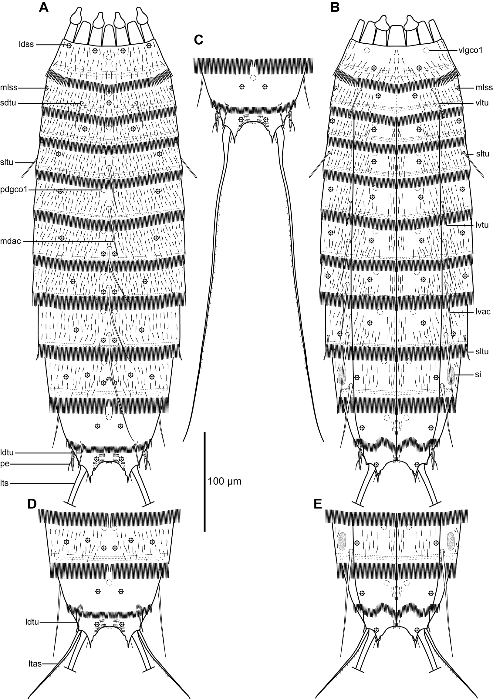
Adults with head, neck and eleven trunk segments ( Figs 2A–B View Fig. 2 , 3A View Fig. 3 , 4A View Fig. 4 ). For a complete overview of measures and dimensions, see Table 2. Distribution of cuticular structures, i.e., sensory spots, glandular cell outlets, spines and tubes, is summarized in Table 3.
The head is formed by a retractable mouth cone with nine outer oral styles and an introvert with scalids. The condition of the SEM specimens did not allow a detailed description of the scalid arrangement. The neck has 16 placids, measuring 23 µm in length. The midventral placid is broadest, measuring 20 µm in width at its base, whereas all others are narrower, measuring 13 µm in width at their bases. The trichoscalid plates are well developed, and located in introvert sectors 2, 4–5, 7–8, and 10. A trichoscalid is attached to each trichoscalid plate.
Segment 1 consists of a complete cuticular ring ( Figs 2A–B View Fig. 2 , 3A–C View Fig. 3 , 4A–D View Fig. 4 ). Sensory spots are located anteriorly on the segment in subdorsal and laterodorsal positions ( Figs 2A View Fig. 2 , 4B, D View Fig. 4 ), and more posteriorly in ventromedial positions ( Figs 2B View Fig. 2 , 4C View Fig. 4 ). On this and the following two segments, the sensory spots are medium sized and rounded or slightly oval, and made up by numerous micropapillae around a central pore. Glandular cell outlets type 1 are present in middorsal and ventrolateral positions. Cuticular hairs emerge through rounded perforation sites, covering the dorsal and lateral sides densely, whereas the ventral side has a large W-shaped area without hairs ( Fig. 4C View Fig. 4 ). The posterior segment margin terminates into a pectinate fringe with long and slender fringe tips.
Segment 2 consists of a complete cuticular ring, but with weak indications on a midventral fissure, visible both inside the cuticle ( Fig. 3C View Fig. 3 ) and on the surface ( Fig. 4E View Fig. 4 ). Pachycyclus of the anterior segment margin is of regular thickness and not interrupted. All specimens with tubes present in ventrolateral positions ( Figs 2B View Fig. 2 , 3C View Fig. 3 , 4E View Fig. 4 ); in addition, some ( Figs 2A View Fig. 2 , 3B View Fig. 3 , 4D View Fig. 4 ), but not all ( Fig. 4B View Fig. 4 ) with tubes in subdorsal positions; subdorsal tubes present in at least nine specimens, and certainly lacking in eight specimens, whereas eventual presence or absence could not be confirmed with certainty for the remaining specimens. Presence or absence of subdorsal tubes does not appear to be correlated with the specimens’ gender. Sensory spots are located in middorsal ( Fig. 4B View Fig. 4 ), midlateral ( Fig. 4D View Fig. 4 ) and ventromedial ( Fig. 4E View Fig. 4 ) positions. Glandular cell outlets type 1 are present in middorsal positions. Secondary pectinate fringe present as single fringe with rather short fringe tips on anterior part of segment. On this and all following segments, the cuticular hairs are bracteate. Hairs are densely distributed around the segment, except on the ventral-most halves of the sternal plates, that only have a few, short filiform hair-like extensions. Pectinate fringe of posterior margin as on preceding segment, but with slightly longer fringe tips.
Segment 3, and remaining segments, consisting of one tergal and two sternal plates ( Figs 2A–B View Fig. 2 , 3A, C, E View Fig. 3 , 4E View Fig. 4 ). Sensory spots are located in subdorsal, sublateral and ventromedial positions ( Figs 2A–B View Fig. 2 , 4D–E View Fig. 4 ). Glandular cell outlets type 1 present in middorsal and ventromedial positions ( Figs 2A–B View Fig. 2 , 3E View Fig. 3 ). Secondary pectinate fringe and cuticular hairs as on preceding segment. Pectinate fringe of posterior margin as on preceding segment, but with slightly longer fringe tips.
Segment 4 with acicular spine in middorsal position and tubes in sublateral positions ( Figs 2B View Fig. 2 , 3E View Fig. 3 , 4F View Fig. 4 ). Sensory spots present in laterodorsal and ventromedial positions ( Figs 2A–B View Fig. 2 , 4D–E View Fig. 4 ); from this segment and onwards, sensory spots are more elongate and droplet- to wedge-shaped. Glandular cell outlets type 1 located in paradorsal and ventromedial positions ( Figs 2A–B View Fig. 2 , 3E View Fig. 3 ). Pachycycli, secondary fringe, pectinate fringe of posterior margin and cuticular hairs as on preceding segment.
Segment 5 with acicular spine in middorsal position and tubes in lateroventral positions ( Figs 2A–B View Fig. 2 , 3E View Fig. 3 , 4F View Fig. 4 ). Sensory spots present in laterodorsal, sublateral and ventromedial positions ( Figs 2A–B View Fig. 2 , 4F View Fig. 4 ). Glandular cell outlets type 1, pachycycli, secondary fringe, pectinate fringe of posterior margin and cuticular hairs as on preceding segment.
Segments 6 and 7 are almost identical, with acicular spines in middorsal and lateroventral positions ( Figs 2A–B View Fig. 2 ), and sensory spots in paradorsal, laterodorsal (segment 6 only), subdorsal (segment 7 only), sublateral and ventromedial positions. Glandular cell outlets type 1 ( Fig. 3D View Fig. 3 ), pachycycli, secondary fringe, pectinate fringe of posterior margin and cuticular hairs as on preceding segment.
Segment 8 with acicular spines in middorsal and lateroventral positions. Tubes present in sublateral positions ( Figs 2B View Fig. 2 , 3F View Fig. 3 ). Sensory spots present in paradorsal, subdorsal, lateral accessory and ventromedial positions. Glandular cell outlets type 1 ( Fig. 3F View Fig. 3 ), pachycycli, secondary fringe, pectinate fringe of posterior margin and cuticular hairs as on preceding segment.
Segment 9 without middorsal spine, but with acicular spines in lateroventral positions. Length of lateroventral spines show sexual dimorphism, with the female spines being about twice as long as the male spines and hence extending beyond the posterior margin of segment 11 (see Figs 2A–E View Fig. 2 , 3H, 3J View Fig. 3 and Table 2). Sensory spots present in paradorsal, subdorsal, laterodorsal and ventrolateral positions. A cuticular depression or scar, lined with short hair, is present in middorsal position, in between the paradorsal sensory spots ( Figs 2A View Fig. 2 , 4G View Fig. 4 inset). Large oval sieve plates, with uniformly distributed pores present in sublateral positions ( Figs 2B View Fig. 2 , 3G View Fig. 3 , 4G View Fig. 4 ). Pectinate fringe of posterior segment margin as on preceding segment, except in the middorsal position, where the fringe tips are considerably shorter ( Figs 2A View Fig. 2 , 4I View Fig. 4 ). Glandular cell outlets type 1, pachycycli, secondary fringe and cuticular hairs as on preceding segment.
Segment 10 with laterodorsal tubes at the posterior segment margin: tubes are well-developed in both sexes ( Figs 2 View Fig. 2 , 3I View Fig. 3 , 4H–I View Fig. 4 ); a single specimen had one additional tube, but only in the right side ( Fig. 4I View Fig. 4 ). Sensory spots present in subdorsal ( Figs 2A View Fig. 2 , 4I View Fig. 4 ) and ventrolateral ( Figs 2B View Fig. 2 , 4J View Fig. 4 ) positions. Glandular cell outlets type 1 located in middorsal and ventromedial positions. The segment is completely devoid of cuticular hairs ( Figs 4H–J View Fig. 4 ). Pectinate fringe of posterior segment margin present, but with considerably shorter fringe tips compared to preceding segments. Pachycycli and secondary fringe as on preceding segment.
Segment 11 with long lateral terminal spines ( Figs 2C View Fig. 2 , 3A View Fig. 3 ) that almost equal the trunk length (see lts/TL ratio in Table 2). Females with thin but otherwise well-developed lateral terminal accessory spines ( Figs 2D–E View Fig. 2 , 3H–I View Fig. 3 ). Lateral terminal accessory spines lined with numerous, marginal hairs. Females with additional midlateral tufts of papillae, emerging from the transition between segment 10 and 11 ( Fig. 4H View Fig. 4 ). Males with three pairs of thin and tubular penile spines ( Figs 2A–C View Fig. 2 , 3J View Fig. 3 , 4J View Fig. 4 ). Sensory spots present in subdorsal ( Fig. 4I View Fig. 4 ) and ventrolateral ( Fig. 4J View Fig. 4 ) positions. Glandular cell outlets type 1 not present. The segment is completely devoid of cuticular hairs in both sexes, but is densely covered with short hair-like cuticular extensions in subdorsal positions and along the margin between the tergal extensions ( Fig. 4I View Fig. 4 ); ventral side with longer hair-like extensions in paraventral positions ( Fig. 4J View Fig. 4 ). Tergal extensions are short and pointed, with an additional point on each inferior margin ( Figs 2A–E View Fig. 2 , 3I –J View Fig. 3 ). Sternal extensions are triangular, and slightly shorter than tergal ones ( Fig. 4J View Fig. 4 ).


No known copyright restrictions apply. See Agosti, D., Egloff, W., 2009. Taxonomic information exchange and copyright: the Plazi approach. BMC Research Notes 2009, 2:53 for further explanation.
|
Kingdom |
|
|
Phylum |
|
|
Class |
|
|
Order |
|
|
Family |
|
|
Genus |