
Eclipidrilus microthecus, Fend, Steven V. & Lenat, David R., 2012
|
publication ID |
https://doi.org/ 10.5281/zenodo.210008 |
|
DOI |
https://doi.org/10.5281/zenodo.6165718 |
|
persistent identifier |
https://treatment.plazi.org/id/BC6B87A2-FFD1-7631-FF5B-F9F82BD8FE3E |
|
treatment provided by |
Plazi |
|
scientific name |
Eclipidrilus microthecus |
| status |
sp. nov. |
Eclipidrilus microthecus n. sp.
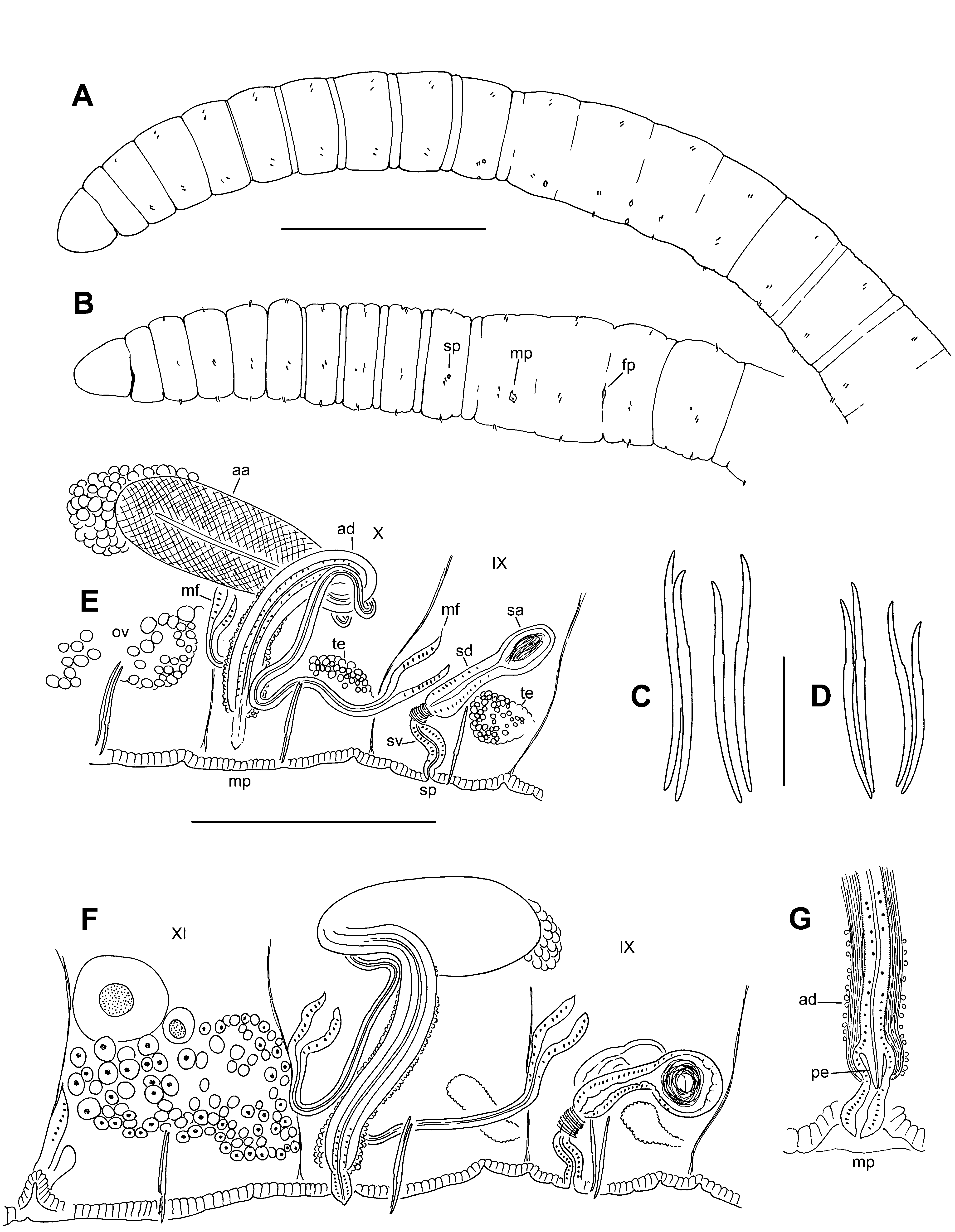
( Figures 3–4 View FIGURE 3 View FIGURE 4 )
Holotype: USNM 1156960. A whole, slide-mounted worm in Canada balsam.
Type locality: North Carolina, Richmond Co., unnamed tributary to Naked Creek at Forest Service road upstream of SR 1003, 28 January 2009. Collected by D.R. Lenat.
Paratypes: From the type locality. USNM 1156961-63: 10 March 2010, 1 sagittally sectioned, 2 whole mounts. Collected by D.R. Lenat.
Etymology: Named for the unusually small spermathecae, compared to other Eclipidrilus species.
Other material: North Carolina, Carteret Co., Pettiford Creek, 19 January 2010, 1 whole mount. Hoke Co., Flat Creek at Manchester Road, 12 February 2009, 1 whole mount. 8 October 2009, 1 whole mount. 11 February 2010, 1 whole mount. 18 February 2010, 2 whole mounts. 6 March 2010, 1 whole mount. 25 March 2010, 3 whole mounts. 22 September 10, 1 whole mount. 5 October 2010, 4 whole mounts. Richmond Co., unnamed tributary to Naked Creek at Forest Service road upstream of SR 1003, 28 January 2009, 1 whole mount. 10 March 2010, 1 sagittally sectioned, 2 whole mounts. 18 March 2010, 2 whole mounts. All collected by D.R. Lenat.
Description. Small, thin worms, length (preserved) 7.7–9.5 mm, 48–64 segments; width 0.20–0.30 mm in X, maximum width 0.24–0.3 mm. Segmentation usually obscured in clitellum, weakly expressed in some post-clitellar segments; secondary segmentation sometimes weak, a narrow anterior ring from about VI–X ( Fig. 3 View FIGURE 3 A–B). Clitellum X–XII. Chaetae sigmoid, simple-pointed, with nodulus 35–40 % of chaeta length from tip ( Fig. 3 View FIGURE 3 C–D). Chaeta length 58–84 μm in mid-body; 55–80 μm in posterior segments; proportions similar in anterior and posterior segments. Dorsal and ventral chaetae approximately equal in length; within each bundle, the outer (more lateral) chaeta may be slightly shorter than the inner. Prostomium rounded-conical, length slightly less than width; prolobous.
Brain in the peristomium, strongly lobed. Pharynx in II–IV; dorsal wall with columnar, ciliated cells; ventral wall very thin, with cuboidal, non-ciliated cells. Pharyngeal glands in (IV)V–VII(VIII), relatively small, irregular lobes, dorsal and/or lateral to gut. Longitudinal muscle layer 12–14 μm thick in preclitellar segments; circular muscle layer 3 μm. Septa 1/2 and 2/3 inconspicuous. Epidermis 4–12 μm thick anterior to clitellum; to about 12–15 μm in clitellum; 4–5 μm in post-clitellar segments; to 12–27 μm in prostomium. Lateral, commissural blood vessels in at least some preclitellar segments; these vessels thin and convoluted, typically joining ventral vessel 1 segment behind junction with dorsal vessel. No lateral blood vessels in middle or posterior segments. Dorsal blood vessel separate in anterior segments; on top of gut posterior to about XI. Chloragogen cells begin in about VII. First nephridia usually paired on 6/7, the next on 12/13; nephridia in few posterior segments, usually on one side only. Each nephridium has a small anteseptal funnel and a granular, narrow postseptal thickening 30–40 μm long; the posterior duct forms a loop that extends ventrally, entering one or more posterior segments, and terminating in a short ectal duct in the originating segment. Nephropores anterior to ventral chaetae, inconspicuous; ectal ducts without distinct vesicles.
Spermathecae paired in IX; pores small, on ventral chaetal lines, just behind the ventral chaetae; accessory glands weak or absent ( Fig. 3 View FIGURE 3 E–F). Ectal vestibule of duct is narrow and usually curved, about 30–50 μm long by up to 14 μm wide; ental 1/2–1/3 narrower, surrounded by a ring of circular (transverse) muscle at the junction with the main duct ( Figs. 3 View FIGURE 3 E–F, 4A–C). Main part of spermathecal duct 38–60 μm long, 18–22 μm wide in ectal part, narrowing to 12–17 μm entally, with densely- packed, columnar epithelium and a narrow lumen; spermathecal duct well differentiated from the ampulla ( Fig. 3 View FIGURE 3 E–F). Spermathecal ampullae entirely in IX; ovate and compact, to 37– 64 μm long by 25–40 μm wide. Ampullar epithelium thin (3–4 μm) ( Fig. 4 View FIGURE 4 D). In mated specimens the sperm is in an unordered bundle.
Testes paired in IX and X, small to medium size, extending at most to mid-segment. Ovaries paired in XI; usually extending through XI, sometimes into XII. Sperm sacs not developed (sperm in testicular segments); egg sacs usually paired; may extend to XIV. Female funnels up to 50 μm tall; female pore intersegmental, on 11/12.
Male funnels single on 9/10 and 10/11; anterior and posterior similar in size (height 35–48 μm), directed anteriad within IX and X. Both anterior and posterior male funnels simple, conical; functional, with associated sperm. Both anterior and posterior vasa deferentia narrow (7–10 μm diameter); they approach the atrial duct near the male pore, then loosely follow the duct, joining the atrium near the base (ectal end) of the ampulla. Posterior vas deferens enters X directly, without penetrating 10/11 ( Fig. 3 View FIGURE 3 E–F)
Male pore inconspicuous; single, slightly lateral to ventral midline in X, near 10/11 ( Fig. 3 View FIGURE 3 A–B). Atrial duct widens slightly at the ectal end ( Fig. 4 View FIGURE 4 E), subtending a small, narrowly conical penis within narrow sac (length 20– 33 μm, width 10–16 μm); the entire structure terminates in a small, conical papilla, which may be contained within a shallow depression ( Fig. 3 View FIGURE 3 G, 4I). A few small (to 20 μm) accessory glands may be present at male pore; absent or inconspicuous in most specimens. The main atrial duct extends dorsally around one side of gut, then usually loops posteriorly and widens abruptly to form the ampulla ( Fig. 3 View FIGURE 3 E–F, 4E). Duct length 180–295 μm, width to 25–29 μm near male pore, narrowing entally to 17–25 μm. Duct musculature more or less longitudinal, 5–7 μm thick in ectal part; lumen narrow ectally, widening to 6 μm entally; epithelium very thin. Atrial ampulla narrowly ovate ( Fig. 3 View FIGURE 3 E– F, 4F); length 152–215 μm, width at middle 52–98 μm. Ampulla with a very thick, cross-hatched muscle layer, to 29–31 μm, composed of about 7–10 layers of fibers in alternating spirals, oriented at about 50–60˚ from the longitudinal axis ( Fig. 4 View FIGURE 4 F–H). Epithelium thin and indistinct, as little as 2 μm thick; lumen very narrow, usually less than 5 μm ( Fig. 4 View FIGURE 4 F, H). Most of ampulla without prostate glands, although a poorly defined, decumbent clump of cells is visible at the ental end in most specimens ( Fig. 3 View FIGURE 3 E–F, 4F). Atrium usually extends into XI.
Remarks. All E. microthecus specimens had the same configuration of reproductive organs, which resembles that of Eclipidrilus pacificus Fend, 2005 . The unpaired anterior and posterior male ducts are unusual in the family, and contrast with the paired arrangement in other species having median atria (e.g., E. pacificus ; E. lacustris ; Tatriella slovenica Hrabë ), but are perhaps similar to Eclipidrilus asymmetricus ( Smith, 1896) . The very small spermathecal ampullae and the possible single prostate gland are unusual within the genus.
Over 20 specimens appeared sexually mature, with sperm on the male funnels and in the spermathecae. Length and number of segments were based on 9 complete worms; blood vessels and nephridia were difficult to see in most specimens, so these observations were limited. Although most of these specimens had mature eggs in the egg sacs, there did not appear to be well-developed sperm sacs at any stage, and only a small amount of free sperm was visible in the coelom of testicular segments.

| USNM |
Smithsonian Institution, National Museum of Natural History |
No known copyright restrictions apply. See Agosti, D., Egloff, W., 2009. Taxonomic information exchange and copyright: the Plazi approach. BMC Research Notes 2009, 2:53 for further explanation.