
Eidmanacris putuhra Campos, 2017
|
publication ID |
https://doi.org/ 10.5281/zenodo.897058 |
|
DOI |
https://doi.org/10.5281/zenodo.6001796 |
|
persistent identifier |
https://treatment.plazi.org/id/AB7EC101-852B-E870-82B4-FB1DFDC4FAE5 |
|
treatment provided by |
Plazi |
|
scientific name |
Eidmanacris putuhra Campos |
| status |
sp. nov. |
Eidmanacris putuhra Campos , sp. nov.
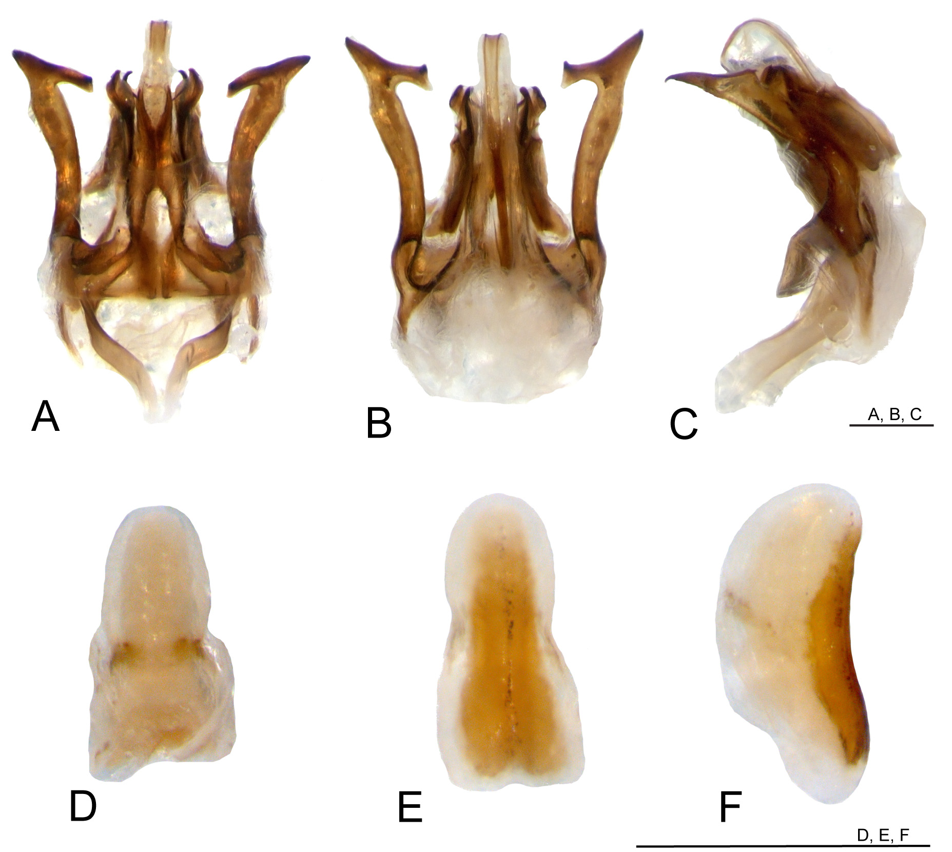
( Figs. 52–54 View FIGURE 52 View FIGURE 53 View FIGURE 54 )
http://lsid.speciesfile.org/urn:lsid: Orthoptera .speciesfile.org:TaxonName:499171
Type material. Holotype male, allotype, 4 males paratypes and 6 females paratypes (MZSP); 2 males paratypes and 2 females paratypes (UBTU). Holotype male, allotype, 2 males paratypes and 2 females paratypes labeled: “ BRASIL (M.G.)— Mun. Viçosa—Jardim Botânico da UFV— 4.II.97 — C. F. Sperber . BRASIL (MG), Viçosa, Fragmento: BIO, Data : 09/iii/2000, Sperber leg., Código: #5282” . 4 males paratypes and 6 females paratypes labeled: “ BRASIL (MG), Viçosa, Fragmento: BIO #6154 View Materials , Data : 16/iii/2002, Coleta Noturna: C/ Aveia, Mendes, M. H. & Rocha A . BRASIL (MG), Viçosa, Fragmento: MBIO, Data : 10/i/2002, Sperber leg., Código: Trilha #6031”. Specimens preserved in ethanol 80%.
Type locality. Brazil, state of Minas Gerais, municipality of Viçosa.
Etymology. This species was formerly described and named by Dr. Carina Mews in her Master’s dissertation, but it was not published ( Mews, 2006). In the language of the Brazilian indigenous tribe Puri, Putuhra means “yellow”, referring to the outer coloration of FIII.
Diagnosis. This species can be distinguished from other Eidmanacris by the following characters: antennae with bands composed of four whitish antenomeres, the bands that follow increasing in number, interspersed with one whitish antenomere; pseudepiphallic arm slightly curved outwards, apex curved inwards in dorsal and ventral views; anterior projection of pseudepiphallic sclerite curved inwards in dorsal view; PsP2 distal portion convex outside, concave inside, forming posterior spine curved inwards; ectophallic apodeme laterally flattened; endophallic apodeme short; copulatory papilla cylindrical, down curved, more sclerotized ventrally.
Description. Head. Dorsum pubescent, different shades of brown. Occiput reddish brown, with light brown band going from occiput to margin of each eye ( Fig. 52C View FIGURE 52 ); vertex reddish brown, with three lines light brown, vertical, well discernible. Fastigium reddish brown, longer than wide, with two rows of bristles, slightly narrowed at apex, narrower than scape; separated from vertex by transverse line forming “v” ( Fig. 52C View FIGURE 52 ). Frons light brown, two vertical bands dark brown below eyes, extending to mandibles, one central, lighter band ( Fig. 52A View FIGURE 52 ). Three ocelli present, well developed, rounded ( Figs. 52A, B, C View FIGURE 52 ); eyes with unpigmented small area on supero-internal angle ( Fig. 52C View FIGURE 52 ). Maxillary palpi joints 3 and 4 medium to dark brown, joint 5 light to medium brown, long, thin; joints 4 and 5 almost same-sized, apex of joint 5 whitish, slightly upcurved ( Fig. 52G View FIGURE 52 ). Gena light brown ( Figs. 52A, B View FIGURE 52 ); frontoclypeal suture medium to dark brown, centrally dark. Clypeus whitish, with two vertical median stripes light brown; labrum whitish, lower portion light brown. Mandible dark brown, posterior margins light brown. Antennal scape light brown, inner surface darker ( Figs. 52A, B, C View FIGURE 52 ); antennae with bands composed of four whitish antenomeres, the following bands increasing in number, interspersed by one whitish antenomere.
ThOrax. Pronotum DD reddish brown, wider than long, inflated, divided by vertical line light yellow almost no discernible, and four lateral lines, in dorsal view; cephalic margin slightly concave, caudal margin almost straight ( Fig. 52C View FIGURE 52 ); ventro cephalic angle rounded, margin light brown, ventro caudal margin darker, gradually ascendant ( Fig. 52B View FIGURE 52 ).
Legs. FI and II yellowish, annulated with medium brown. TI and II light yellow, annulated with dark brown; TI with two same-sized apical spurs, TII with two inner apical spurs, one outer, smaller. FIII yellowish brown, dorsally reddish brown, several thin stripes reddish brown on outer surface, apical third reddish brown ( Fig. 52F View FIGURE 52 ). TIII medium to dark brown, lighter at apical third; subapical spurs 4/4, serrulation above and between subapical spurs; apical spurs 3/3, more developed on inner surface; inner apical spurs: median one longer (iam), dorsal shorter (iad), ventral smallest (iav) (iam>iad>iav); outer apical spurs: median one longer (oam), dorsal (oad) little longer than ventral (oav) (oam>oad>oav). Basitarsi I, II and III yellowish.
AbdOmen. Medium brown, marbled, sub-cylindrical, with several dark spots and dark vertical lines, divided by light brown thin, sagittal line almost no discernible ( Fig. 52D View FIGURE 52 ).
Male. Small to medium-sized body, general coloration reddish brown, marbled, with dark spots and maculae. FWs medium to dark brown, rounded, reticulated, apex connected to single vein that divides the external part of FW as lateral field, inner and posterior margins light brown, posterior margin without glandular thickening ( Figs. 52B, C View FIGURE 52 ); inner margins not touching each other, covering metanotal gland area, posterior margin not surpassing posterior metanotum border ( Fig. 52C View FIGURE 52 ). Metanotal gland present, with anteromedian crest triangular, apex elliptical, line of bristles anteriorly; lateral projections short, cylindrical, top rounded, close each other; ( Figs. 52H, I View FIGURE 52 ). Supra-anal plate light to medium brown, posterior margin dark brown; anterior margin slightly concave, posterior margin somewhat concave; lateral-distal projection elongate, lighter ( Fig. 52J View FIGURE 52 ). Subgenital plate light brown, lateral and posterior borders darker, longer than wide, anterior margin sub-straight, posterior margin somewhat convex forming “v” centrally ( Fig. 52K View FIGURE 52 ).
Phallic cOmPlex ( Figs. 53A–C View FIGURE 53 ; 54A–C). Pseudepiphallus: base of pseudepiphallic sclerite weakly sclerotized, dorso-ventrally flattened, centrally narrow, with central concavity posteriorly; pseudepiphallic arm sclerotized, narrow, upcurved, slightly curved outwards, apex curved inwards in dorsal and ventral views, with dorsal line of bristles on dorsal surface; apex of pseudepiphallic arm bifid, forming superior and internal projections, pointing posteriorly; inferior projection present, apex pointed in ventral view; anterior projection of pseudepiphallic sclerite elongate, thin, curved inwards in dorsal and ventral views, surpassing median part of pseudepiphallus; PsP2 well sclerotized, elongate, not surpassing posterior extremity of pseudepiphallic arms, distal portion convex outside, concave inside, forming posterior spine curved inwards, with membranous sphere on concavity; PsP1 longer than wide, thin, well sclerotized, posteriorly pointed in ventral view. Ectophallic invagination: Ectophallic apodeme elongate, upcurved, laterally flattened; ectophallic arc short, located posteriorly to base of pseudepiphallic sclerite; dorsal projections well sclerotized, elongate, apex pointed, curved inwards, posterior margin concave almost forming “v”, not surpassing PsP2 posteriorly in dorsal view; ventro-posterior projection less sclerotized, not so elongate. Endophallus: median-posterior projection of pseudepiphallic sclerite elongated, surpassing pseudepiphallic arms apex; lateral-posterior lobes of endophallic sclerite very short; endophallic apodeme relatively short, its limits not surpassing ectophallic apodeme anterior margin.
Female. Larger than male, general coloration medium brown, marmored ( Fig. 52E View FIGURE 52 ). Supra-anal plate yellowish brown, posterior margin dark brown, posterior margin rounded with long bristles ( Fig. 52L View FIGURE 52 ). Subgenital plate light brown, marbled, posterior margin slightly convex, ( Fig. 52M View FIGURE 52 ). Ovipositor as in figs. 52N and 52O.
COPulatOry PaPilla ( Figs 54D–F View FIGURE 54 ). Cylindrical, longer than wide, down-curved, more sclerotized ventrally, anterior third little wider in dorsal and ventral views, posterior margin rounded.
Measurements (mm). Male (n=4): Hw, 2.34 ± 0.14 (2.23–2.54); iod, 1.25 ± 0.06 (1.17–1.3); Lpron, 2.4 ± 0.09 (2.35–2.54); awpron, 2.37 ± 0.14 (2.23–2.54); pwpron, 2.76 ± 0.13 (2.6–2.91); wpron, 3.25 ± 0.06 (3.22– 3.34); LFW, 1.55 ± 0.11 (1.42–1.67); wFW, 1.58 ± 0.16 (1.36–1.73); LFIII, 12.3 ± 0.5 (11.7–12.9); wFIII, 2.58 ± 0.07 (2.55–2.7); LTIII, 13.42 ± 0.92 (12.3–14.55); Ltars 1-III, 3.25 ± 0.24 (3.25–4.05).
Female (n=6): Hw, 2.73 ± 0.12 (2.6–2.91); iod, 1.37 ± 0.07 (1. 3–1.48); Lpron, 2.75 ± 0.07 (2.66–2.85); awpron, 2.55 ± 0.05 (2.48–2.6); pwpron, 3.31 ± 0.12 (3.16–3.47); wpron, 3.72 ± 0.18 (3.53–3.96); LFIII, 14.37 ± 0.9 (13.35–15.75); wFIII, 2.97 ± 0.16 (2.7–3.15); LTIII, 15.09 ± 0.93 (14.4–16.65); Ltars 1-III, 4.08 ± 0.16 (3.9–4.2); OL, 15.36 ± 0.5 (14.55–15.9).


| BIO |
University of the Basque Country |
No known copyright restrictions apply. See Agosti, D., Egloff, W., 2009. Taxonomic information exchange and copyright: the Plazi approach. BMC Research Notes 2009, 2:53 for further explanation.