
Eigenmannia muirapinima, Peixoto & Dutra & Wosiacki, 2015
|
publication ID |
https://doi.org/ 10.1111/zoj.12274 |
|
persistent identifier |
https://treatment.plazi.org/id/03B08780-FFFC-3A19-FC0B-A2FDFB05FEF8 |
|
treatment provided by |
Felipe |
|
scientific name |
Eigenmannia muirapinima |
| status |
sp. nov. |
EIGENMANNIA MUIRAPINIMA SP. NOV.
( FIGS 15 View Figure 15 , 16 View Figure 16 ; TABLE 3)
Diagnosis: Eigenmannia muirapinima can be distinguished from other species in the E. trilineata species group, except E. desantanai , E. guairaca , and E. microstoma , by the ii,11–12 pectoral-fin rays (versus ii, 13–14 in E. antonioi and E. pavulagem ; ii, 16–17 in E. matintapereira and E. trilineata ; ii, 15–17 in E. vicentespelaea ; and ii, 13–15 in E. waiwai ). Eigenmannia muirapinima differs from E. desantanai and E. microstoma by the pattern of premaxillary dentition with eight to ten teeth distributed in two rows (outer row with three to five teeth; inner with four to six teeth) [versus 24–25 teeth distributed in four rows (outermost row with five teeth; second row with six to eight teeth; third row with six to seven teeth; innermost row with seven to eight teeth); and 16 teeth distributed in three rows (outermost row with five teeth; middle row with six; innermost with five teeth), respectively]. Eigenmannia muirapinima also differs from E. desantanai by depth of the inferior medial stripe of two or three scales deep (versus one scale deep). Eigenmannia muirapinima can be further distinguished from E. microstoma by: suborbital depth 18.7– 28.4% HL (versus 29.9–40.8%); length of anterodorsal process of maxillar equal to 50% of the width of the posterior nostril (versus equal to the width of posteri- or nostril); and coronomeckelian bone length equal to 20% of the length of Meckel’s cartilage (versus 45% of the length of Meckel’s cartilage). Eigenmannia muirapinima also differs from E. guairaca by the number of total anal-fin rays 170–198 (versus 151– 170).
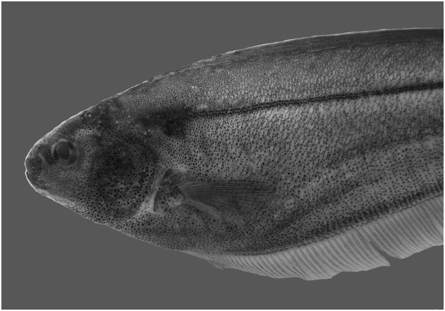
Description: Morphometric data are presented in Table 3. Body elongate and laterally compressed. Dorsal profile of body nearly straight from rear of head to vertical through middle of anal fin, and then posteroventrally aligned with distal portion of caudal filament. Ventral profile of body posteroventrally aligned from anterior margin of dentary to anal-fin rays 15–30, and then posterodorsally aligned with last anal-fin ray. Ventral margin of caudal filament straight. Greatest body depth at vertical through distal margin of pectoral fin.
Head laterally compressed with greatest width at opercular region and greatest depth at posterior margin of supraoccipital. Dorsal profile of head slightly convex from upper lip to vertical through branchial opening. Ventral profile of head slightly concave from anterior margin of lower lip to branchial opening. Snout rounded in profile. Mouth terminal. Upper lip slightly overlapping lower lip or jaws of same length. Premaxilla teeth, 8(1), 9(2), or 10(1), distributed in two rows [outer row with 3(1), 4(1), or 5(1) teeth; inner row with 4(2), 5(2), or 6(1) teeth]. Maxilla with sickle-shaped anterodorsal process equal to 50% of width of posterior nostril. Dentary teeth 11(2), 13(1), 15(1), or 16(1), distribut- ed in one or two rows [outer row with 7(1), 8(1), 9(1), 10(1), or 11(1) teeth; inner row with 4(1) or 5(3) teeth]. Dentary teeth increasing abruptly in size from fifth, sixth, or seventh teeth of outer row towards rictus. Coronomeckelian bone equal to 20% of length of Meckel’s cartilage. Endopterygoid with 8(2) or 9(3) teeth in two series. Mouth rictus at vertical through anterior nostril or in region between nares. Anterior naris tube-like, with posterior margin located at vertical through posterior margin of or in median portion of rictus. Posterior naris elliptical, without tube, located closer to anterior margin of eye than snout tip. Eye approximately circular, covered by skin, laterally located on anterior half of head. Antorbital and infraorbitals 1–4 in form of enlarged, partial cylinders with slender osseous arches. Fifth and sixth infraorbitals slender and tubular. Depth of posterodorsal expansion on infraorbitals 1 + 2 equals total length of infraorbitals 1 + 2. Branchial opening moderately elongate. Branchial membrane joined to isthmus. Anus and urogenital papilla shifting anteriorly ontogenetically. Anus and urogenital papilla at vertical through posterior margin of orbit in mature specimens.
Cycloid scales present from immediately posterior to head to distal portion of caudal filament. Lateral line complete, with 100(2), 101(1), 113(1), 117*(4), 119(2), 120(1), 121(1), 126(1), 128(3), 129(2), or 140(1) perforated scales to vertical through end of anal fin. Longitudinal series of scales above lateral line, 8(2), 9(4), 10*(5), 11(3), 12(2), or 13(3). Scales over anal-fin pterygiophores approximately one-half the size of others.
Pectoral-fin rays ii,11*(11) or ii,12(8). Distal margin of fin slightly rounded. Tip of pectoral fin margin reaching vertical through base of anal-fin rays 18–21. Analfin origin located immediately posterior to vertical through pectoral-fin base; total anal-fin rays, 170– 198 (179*, N = 18; Table 2). Distal margin of anal fin approximately concave. Caudal filament cylindrical, tapering gradually distally, relatively short and approximately 30% of LEA in mature specimens.
Precaudal vertebrae 13(4) or 14(1). Anterior vertebrae 11(5). Transitional vertebrae 2(4) or 3(1). Displaced haemal spines 2(1) or 3(4).
Coloration in alcohol: Background colour darkened. Head dark dorsally and gradually becoming lighter ventrally. Lips and suborbital region dark yellow. Body dark brown dorsally, gradually becoming lighter to region overlying anal-fin pterygiophores. Four longitudinal dark stripes along body. Lateral-line stripe thin, one scale deep, extending from first perforated lateral-line scale to distal portion of caudal filament. Superior medial stripe thick, two or three scales deep, tapering from vertical between base of anal-fin rays 21–30 to posterior one-third of body. Inferior medial stripe moderately thick, two or three scales deep, extending from vertical between base of anal-fin rays 12–22 to posterior one-third of body. Anal-fin base stripe thick, two scales deep, extending from vertical between base of anal-fin rays 1–10 to last anal-fin ray. Pectoral and anal fins hyaline, with scattered tiny chromatophores on interradial membranes.
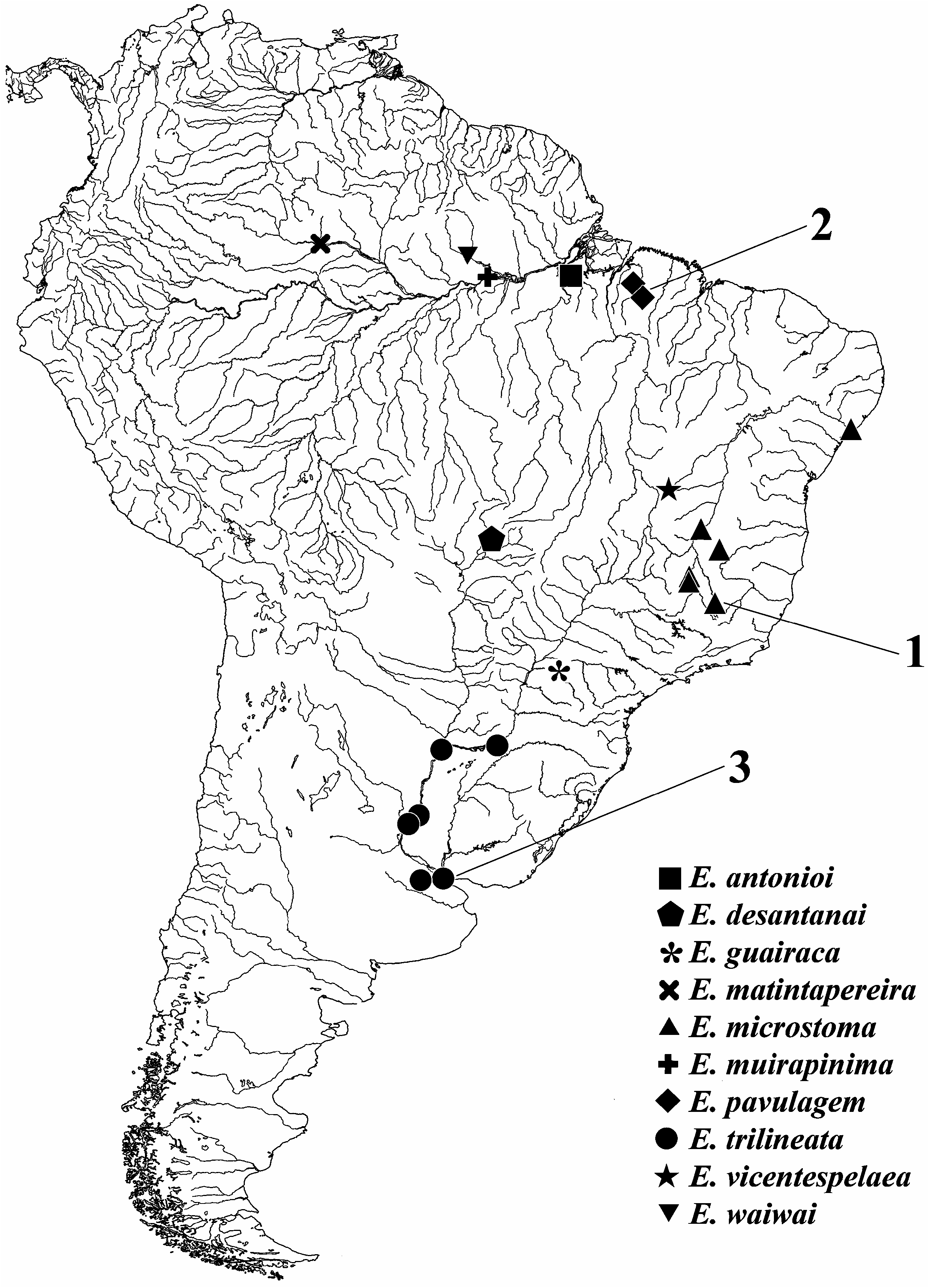
Distribution: Eigenmannia muirapinima sp. nov. is known from Igarapé Santo Antônio and Lago Jará, both tributaries of Rio Amazonas, Rio Amazonas basin, Pará, Brazil ( Fig. 6 View Figure 6 ).
Etymology: The epithet ‘ muirapinima ’ is a tribute to the indigenous people of the tribe Muirapinima , who inhabit the region near the type locality of the species.
Material examined
Holotype: Brazil. Pará: MPEG 21778 View Materials , 98.7 mm LEA, Igarapé Santo Antônio, tributary of Rio Amazonas, Rio Amazonas basin, 2°09′15.9″ S, 56°05′17.9″ W, collect- ed by W. Wosiacki, R. Raiol, and M. Mendonça, 6 October 2011. GoogleMaps
Paratypes: Brazil. Pará: MPEG 21777 View Materials , 1 View Materials + 3 CS, 84.6– 98.5 mm LEA, Lago Jará, tributary of Rio Amazonas, Rio Amazonas basin, 2°12′45″ S, 56°00′45.4″ W, col- lected by W. Wosiacki, 6 October 2011 GoogleMaps . MPEG 22163 View Materials , 1 View Materials , 86.7 mm LEA, Lago Jará, tributary of Rio Amazonas, Rio Amazonas basin, 2°09′15.9″ S, 56°05′17.9″ W, collected by L. Peixoto, 5 October 2011 GoogleMaps ; MPEG 29489 View Materials , 9 View Materials , 76.2–97.7 mm LEA; MZUSP 116796 View Materials , 2 View Materials + 2CS, 80.0– 96.9 mm LEA, collected with holotype GoogleMaps .

| LEA |
University of Lethbridge |
No known copyright restrictions apply. See Agosti, D., Egloff, W., 2009. Taxonomic information exchange and copyright: the Plazi approach. BMC Research Notes 2009, 2:53 for further explanation.