
Enchodelus (Paraenchodelus) thornei Baqri & Jairajpuri (1974)
|
publication ID |
https://doi.org/ 10.5281/zenodo.8145041 |
|
DOI |
https://doi.org/10.5281/zenodo.8144952 |
|
persistent identifier |
https://treatment.plazi.org/id/03D687A0-E568-FFD3-DD6C-F507F99EFB09 |
|
treatment provided by |
Carolina |
|
scientific name |
Enchodelus (Paraenchodelus) thornei Baqri & Jairajpuri (1974) |
| status |
|
Enchodelus (Paraenchodelus) thornei Baqri & Jairajpuri (1974) View in CoL
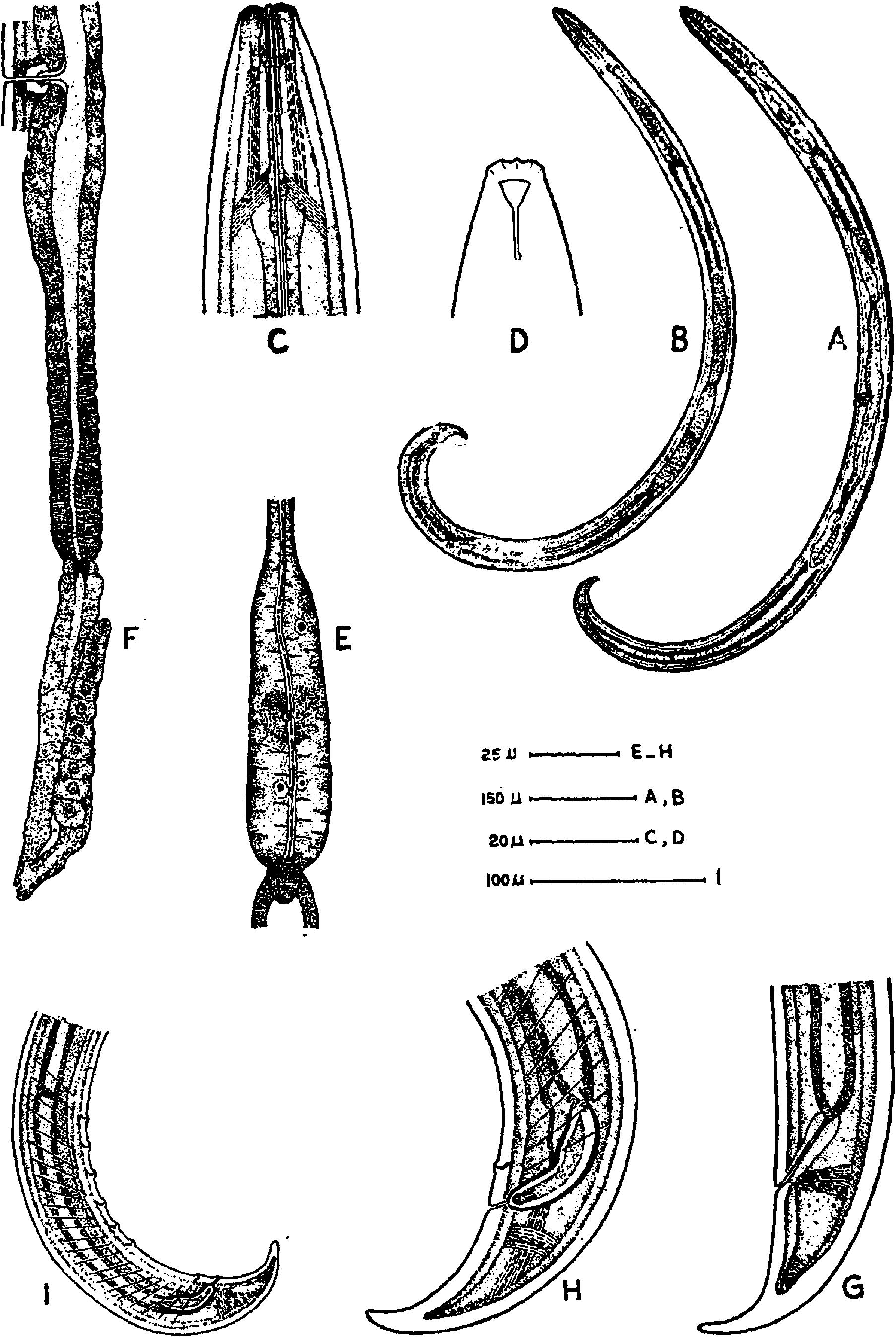
WI‘Y" ‘I I I I Y 1110 ( Fig. 8 View Fig , A-I)
Dimensions
Female L Ls==: 1 1.' 23 mm a =31; b= 4.7; 0:24; cl =2.2 V= 52; I. \tllICI-IV fu AA _ IN 1 G IL __ Ill' Iv íIX u1 ——/.u, ug— 1 u.
Male L =1.38 mm; a =35; b =5.2; c =28; cl = l.5; T=52.
Description
Female Body ventrally curved upon fixation, more strongly in posterior half, tapering gradually in neck region. Cuticle finely striated, its thickness on body 3 μm and on tail tip 13 μm. Dorsal, ventral and llull-VI lateral QI. U body 09] P » l @ LI indistinct.
Lip region set off from body by a slight depression, about 1 /3rd body-width at base of oesophagus. Amphids cup-shaped with curved slit-like apertures occupying about 2/3rd of corresponding body-width. Odontostyle 17 μm or 1.4 head-widths long, its aperture about 1 /8th of its length. Guiding ring one head-width from anterior end. Odontophore 25 μm long or 1.4 times the odontostyle length.
Basal expanded part of oesophagus occupying about 40% of neck length. Location of oesophageal gland nuclei and their orifices as given in Table III. Nerve ring surrounding the anterior slender part of oesophagus at 50% of neck length from anterior end of body. Cardia tongue-shaped, surrounded by intestinal tissues. Oesophago-intestinal disc present. Prerectum about four and rectum about one anal body-width long.
Vulva a transverse slit. Vagina extending inwards about 1 /3rd of corresponding body-width, encircled by cuticularization. Gonads amphidelphic. Uterus divided into a proximal glandular and distal muscular part. Oviduct and uterus separated by a distinct sphincter. Ovaries reflexed, oocytes arranged in a single row except in growth regıon.
Tail 2.2 anal body-widths long with two caudal pores on each side, conoid, ventrally arcuate, with rounded terminus.
Male Supplements an adanal pair and 6 ventromedians, the latter spaced nearly at regular intervals. Spicules about 1.3 anal body-widths along median axis. Lateral guiding pieces well developed. Copulatory muscle bands occupy the area up to last ventromedian Supplement. Prerectum about 4 anal body-widths long, extending up to the last ventromedian supplement.
Habitat and locality Collected from soil around roots of deodar, Cedrus deodara from Naggar (altitude approx. 1,825 m), district l\ Kain UI l), Himachal ııııııayııaı Pradesh.
Remarks The above study is based on a female and a male specimens from the Nematode Collection of the Department of Zoology. Aligarh Muslim University.
No known copyright restrictions apply. See Agosti, D., Egloff, W., 2009. Taxonomic information exchange and copyright: the Plazi approach. BMC Research Notes 2009, 2:53 for further explanation.