
Epipsylla hainanana Yan
|
publication ID |
https://doi.org/ 10.11646/zootaxa.3986.1.8 |
|
publication LSID |
lsid:zoobank.org:pub:F98EF2B7-6314-421D-A16F-65C4C5C4B7F2 |
|
DOI |
https://doi.org/10.5281/zenodo.6095811 |
|
persistent identifier |
https://treatment.plazi.org/id/DB2F057D-FFE9-FFD2-FF69-FDB4F3125CA9 |
|
treatment provided by |
Plazi |
|
scientific name |
Epipsylla hainanana Yan |
| status |
|
Epipsylla hainanana Yan View in CoL g & Li
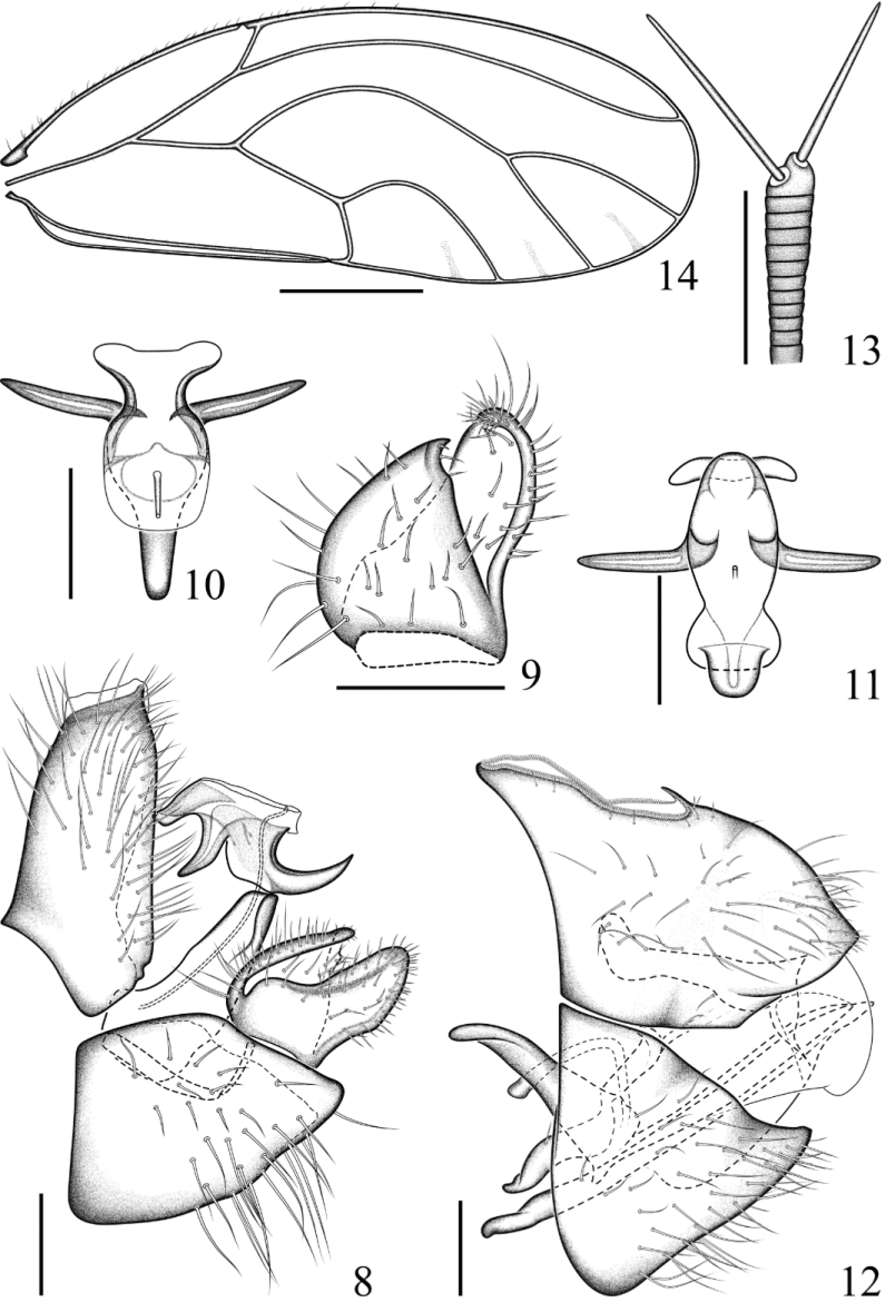
( Figs. 8–14 View FIGURES 8 – 14 , 17–18 View FIGURES 15 – 18 )
Epipsylla hainanana Yang & Li, 1983: 308 View in CoL ; Li, 2011: 563.
Material examined. China: 8 ♂, 10 ♀, Hainan Province, Ledong , Jianfengling , 5–6.ix.2013, coll. Bin Li and Meng Jiao ( GUGC, NHMB, dry mounted).
Description. Adult. Coloration. Body yellow to ochreous and cyan. Vertex ( Fig. 17 View FIGURES 15 – 18 ) cyan in the middle, yellow to ochreous laterally and anteriorly. Genal processes greenish yellow to yellow at base, ochreous in apical half. Compound eyes dark purplish red; median ocellus yellow to orange, lateral ocelli red. Antenna yellowish brown, with black apices on segments 3–7, segments 8–10 entirely black. Pronotum yellow to ochreous; mesoprescutum with anterior half ochreous, posterior half cyan; mesoscutum mostly ochreous; mesoscutellum and parapteron cyan; metascutum yellow; Metascutellum cyan medially, yellow to ochreous laterally; metapostnotum ochreous medially and yellow laterally. Head and thorax ventrally yellow to light brown. Pro and mesofemora yellow green to dark brown, metafemora yellow green; tibiae and tarsi brown to dark brown. Forewing membrane light brown, transparent, veins brown. Abdomen green to bluish, tergite 4 ochreous medially and yellow laterally. Male terminalia yellowish, apex of inner lobe of paramere and sclerotised appendages on distal portion of aedeagus dark brown or black. Female proctiger light cyan, yellowish green dorsally and brown apically; subgenital plate light cyan to yellow, brown apically.
Structure. Body medium-sized ( Table 1 View TABLE 1 ). Head ( Figs. 17, 18 View FIGURES 15 – 18 ) with fine, moderately long setae on vertex and long and conspicuous setae on genal processes. Genal processes inclined from plane of vertex; 1.2–1.4 times as long as vertex along mid-line; axes of genal processes subparallel or slightly converging to apex; subacute apically. Antenna 4.8–5.3 times head width; terminal antennal setae ( Fig. 13 View FIGURES 8 – 14 ) subequal in length, about as long as antennal segment 10. Metatibia with 7 apical spurs. Forewing ( Fig. 14 View FIGURES 8 – 14 ) narrowly oval, widest in the middle; pterostigma almost absent; fore margin with fine long setae, gradually getting shorter toward wing apex where they become inconspicuous; surface spinules present in all cells, leaving broad spinule-free stripes along the veins, radular spinules forming narrow stripes in the middle of cells m1, m2 and cu1 along wing margin. Abdominal sternites covered with dense, long setae.
Male terminalia. Proctiger ( Fig. 8 View FIGURES 8 – 14 ) tubular, almost straight posteriorly; covered with long setae except for base. Paramere ( Figs. 8, 9 View FIGURES 8 – 14 ) bifid, shorter than proctiger; in profile, with broad base, in basal third strongly, in apical two thirds weakly narrowing to apex which is subacute and slightly curved; outer surface beset with moderately long setae in apical half; inner lobe about four fifth as long as outer lobe; inner lobe triangula, wide at base, relatively evenly narrowing to subacute apex which is sclerotised, anterior margin of inner lobe relatively evenly curved, with long setae, posterior margin straight, inner surface of inner lobe with some long setae; inner surface of outer lobe with some long setae, lacking dark sclerotisation apically. Proximal portion of aedeagus ( Fig. 8 View FIGURES 8 – 14 ) almost straight, massif at base, narrowing to apex. Distal portion of aedeagus ( Figs. 8, 10, 11 View FIGURES 8 – 14 ) short and robust, with base forming hook-like process extending caudad; apex curved, blunt apically, with one pair of moderately long sclerotised processes ventrally ( Figs. 10, 11 View FIGURES 8 – 14 ); sclerotised end tube of ductus ejaculatorius weakly curved, short. Subgenital plate ( Fig. 8 View FIGURES 8 – 14 ) with long setae in apical two thirds.
Female terminalia ( Fig. 12 View FIGURES 8 – 14 ). Proctiger short, irregularly hemispherical, bearing long setae subapically and laterally; circumanal ring oval, slightly less than half as long as proctiger, consisting of two unequal rows of pores.
Subgenital plate short, cuneate, densly covered in long setae. Lateral valvula irregularly curved posteriorly; dorsal valvula cuneate; ventral valvula straight, pointed apically.
Measurements in Table 1 View TABLE 1 .
Immature. Unknown.
Host plant. Unknown.
Remarks. The relatively short original description of E. hainanana is supplemented here with more detail, particularly concerning the male and female terminalia. The species is similar to E. rubrofasciata in the colour pattern on head and thorax, the relatively narrow forewings as well as the male and female terminalia. It differs in the shorter distal portion of the aedeagus ( E. hainanana : 0.4 times as long as basal portion, E. rubrofasciata : 0.8 times as long) and the apically narrowly rounded or subacute outer lobe of paramere which is truncate in E. rubrofasciata .

| NHMB |
Naturhistorisches Museum, Basel |
No known copyright restrictions apply. See Agosti, D., Egloff, W., 2009. Taxonomic information exchange and copyright: the Plazi approach. BMC Research Notes 2009, 2:53 for further explanation.