
Epora biprolata Men & Qin
|
publication ID |
https://doi.org/ 10.5281/zenodo.206608 |
|
DOI |
https://doi.org/10.5281/zenodo.6188150 |
|
persistent identifier |
https://treatment.plazi.org/id/039087B2-FFDA-FFC0-1FB9-FDB7FD2DDD6E |
|
treatment provided by |
Plazi |
|
scientific name |
Epora biprolata Men & Qin |
| status |
sp. nov. |
Epora biprolata Men & Qin View in CoL , sp. nov.
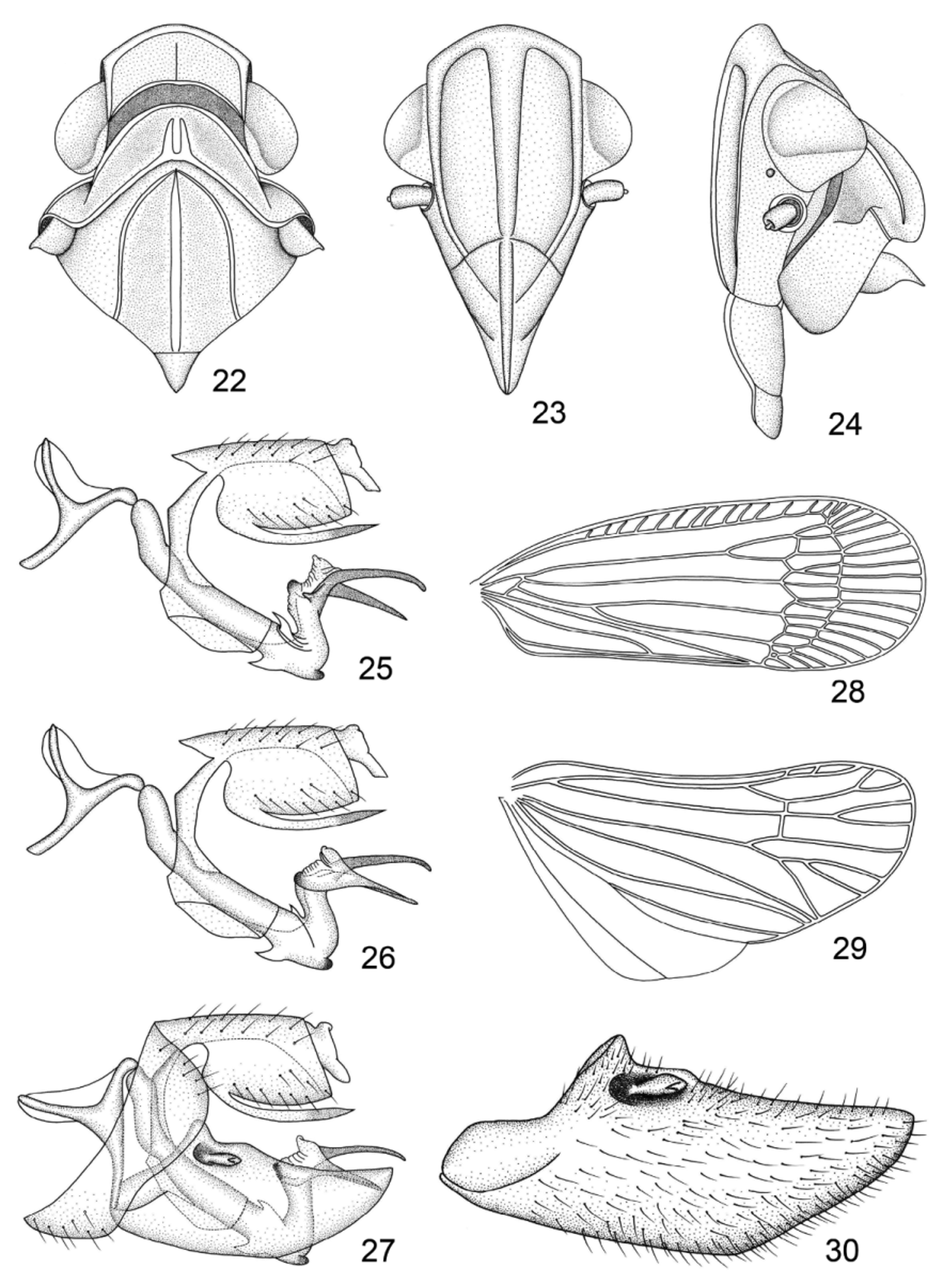
( Figs 3, 4 View FIGURES 1 – 6 , 22–30 View FIGURES 22 – 30 )
Description. Male. Body length 9.5-9.7 mm (n=2).
General color green to yellowish green ( Figs 3, 4 View FIGURES 1 – 6 ). Vertex with apical and lateral margins yellowish brown. Eyes brown, ocelli yellow. Pronotum, mesonotum and abdomen green. Male genital segment pale green. Forewings with veins dark green, posterior cells of clavus orange yellow. Legs green with apices of spines on tibiae and tarsi black.
Head including eyes narrower than pronotum, slightly produced in front of eyes ( Figs 3, 4 View FIGURES 1 – 6 , 22, 24 View FIGURES 22 – 30 ). Vertex broader at base than long in midline (about 2.3: 1.0), anterior margin arched and thickened, lateral margins parallel and distinctly ridged, posterior margin concave, sharp-edged, disc of vertex slightly depressed, median sulcus single, distinct, not reaching anterior margin, anterior margin of vertex and base of frons meeting in a broad callus ( Figs 4 View FIGURES 1 – 6 , 24 View FIGURES 22 – 30 ). Frons longer in midline than maximum width (about 1.4: 1.0), anterior margin convex and ridged, lateral margins ridged, subparallel except where convergent terminally from antennal base, median carina thickened ( Fig. 23 View FIGURES 22 – 30 ). Compound eyes oval ( Fig. 24 View FIGURES 22 – 30 ), lateral ocelli small ( Fig. 24 View FIGURES 22 – 30 ). Clypeus distinctly ridged centrally ( Fig. 23 View FIGURES 22 – 30 ).
Pronotum with anterior margin arched, posterior margin obtusely excavated; dorsal disc distinctly broader than vertex at base, lateral carinae of the disc sinuately converging cephalad, two median carinae not attaining anterior margin; with one distinct, short, lateral carina on either side between eye and tegula ( Figs 3 View FIGURES 1 – 6 , 22, 24 View FIGURES 22 – 30 ). Mesonotum tricarinate; median carina straight, reaching the distinct transverse suture separating the dorsal angle; lateral carinae sinuate, extending to hind margin; pronotum and mesonotum together about 4.6 times as long as vertex in midline ( Figs 3 View FIGURES 1 – 6 , 22 View FIGURES 22 – 30 ). Forewings semitransparent, rounded apically, widest near apex; costal cell with 14–15 short transverse veinlets; Sc+R forking at basal 3/5, Cu1 forking at basal 1/4, claval apex beyond mid-point of forewing, claval veins meeting at basal 2/5 of forewing; two ranks of transverse veinlets in membrane forming numerous apical cells and subapical cells ( Figs 3 View FIGURES 1 – 6 , 28 View FIGURES 22 – 30 ). Hindwings hyaline ( Fig. 29 View FIGURES 22 – 30 ). Hind tibia with 3 lateral spines and 6 apical spines, metatarsomere I with 5 distal spines.
Male pygofer symmetrical and quadrangular in profile ( Fig. 27 View FIGURES 22 – 30 ). Anal tube stubby, not reaching the end of aedeagus; ventro-basally with two ribbon-like processes surpassing the apex of anal segment; anal style very short ( Figs 25–27 View FIGURES 22 – 30 ). Genital styles lobe-shaped, with apical 1/3 strongly narrowed caudad, its dorsal margin triangularly produced dorsad at basal 1/4, with a hook-shaped process with acute and curved apex on dorsal edge at basal 1/4 ( Figs 27, 30 View FIGURES 22 – 30 ). Aedeagal shaft tubular and elongate, with two asymmetrical triangular processes at apical 1/3: one on dorsal side and another one on ventral side and distad of which aedeagus bent dorsad; apex membranous, with two strongly sclerotised processes distally, the right one relatively long and directed caudoventrad, the left one having a triangular process at base ( Figs 25–27 View FIGURES 22 – 30 ). Periandrium well developed, symmetrical, fused with ventral base of anal segment and surrounding aedeagal shaft from base to middle, with a flaky process on ventral side ( Figs 25–27 View FIGURES 22 – 30 ).
Material examined. Holotype male, China: Jianfengling, Hainan Province, 7 Jul. 2007, coll. Lijun Cai ( NWAFU). Paratype. 1 male, China: Diaoluoshan, Hainan Province, 16 Aug. 2010, coll. Chaozhong Jiang ( NWAFU).
Etymology. The name of the new species is derived from the Latin prefix ‘ bi -’ and Latin root ‘ prolatus ’ with the feminine termination - a, referring to its aedeagal shaft with two apical processes.
Distribution. China (Hainan Province).
Remarks. Epora biprolata sp. nov. is similar to E. bilemisca Qin & Men, 2010 , but differs from the latter in the presence of the single lateral carina between eye and tegula (with two lateral carinae in E. bilemisca ), the male anal segment with two ventro-basal processes (with two ventro-distal processes in E. bilemisca ), the periandrium long and smooth on dorsal side (short, with processes on dorsal side in E. bilemisca ), and the aedeagal shaft with two triangular processes in apical third and two long processes at apex (with two lateral flaky processes in apical third and one apical process at apex in E. bilemisca ).

No known copyright restrictions apply. See Agosti, D., Egloff, W., 2009. Taxonomic information exchange and copyright: the Plazi approach. BMC Research Notes 2009, 2:53 for further explanation.
|
Kingdom |
|
|
Phylum |
|
|
Class |
|
|
Order |
|
|
Family |
|
|
Genus |