
Exsuperantia archipelagus, Carvalho & Pisera, 2019
|
publication ID |
https://doi.org/ 10.11646/zootaxa.4613.1.7 |
|
publication LSID |
lsid:zoobank.org:pub:6E25D4D4-00EF-4D37-B701-8509FBD645DD |
|
persistent identifier |
https://treatment.plazi.org/id/6C499E3A-FFD6-9B00-17AB-B6E3EF9DF94E |
|
treatment provided by |
Plazi |
|
scientific name |
Exsuperantia archipelagus |
| status |
sp. nov. |
Exsuperantia archipelagus View in CoL sp. nov.
Figures 1 View FIGURE 1 –4; Table 3 View TABLE 3
urn:lsid:zoobank.org:act:7ADC8D28-86CF-446F-A3AD-E33B6822125D
Synonymy. Racodiscula clava sensu Topsent (1892, 1904 , 1928); Exsuperantia sp. Carvalho et al. (2015).
Type locality. Terceira island, Azores archipelago, Portugal.
Type material. Holotype. MNHN DT-782/1 (dry specimen), locality: Terceira island, Azores archipelago, Por- tugal (38º52’50’’N, 27º23’05’’W), depth: 599 m GoogleMaps . Paratypes. MNHN DT-782/2 (dry specimen), locality: Terceira island, Azores archipelago, Portugal (38º52’50’’N, 27º23’05’’W) depth: 599 m GoogleMaps ; DOP1976 (specimen preserved in 90% ethanol), locality: Azores Bank, Azores archipelago, Portugal (38º05’59’’N, 29º08’59’’W), 168–594 m depth. GoogleMaps
Additional material. Exsuperantia archipelagus sp. nov.: DOP 5883/6212/6248/6255, Azores archipelago; HBOM 003:02023 (BMR 29-V-91 -3-003), Madeira, HBOM 003:00660 (BMR 09-VI-91 -4-008), Canaries (see table 1 for more details).
Comparative material examined. Exsuperantia clava Schmidt 1879 — Syntype MCZ 6436 (orig. 275), Gulf of Mexico.
External morphology. Columnar to ficiform sponges, that can possess lateral protuberances or branches ( Fig. 1 View FIGURE 1 ). Small, 20–30 mm tall and 10–20 mm thick, attached to the substrate by the entire base. Surface is smooth, with marked water canals on the surface of the choanosomal skeleton. Oscules or pores are not visible to the naked eye. Color is beige to whitish in ethanol and when dry.
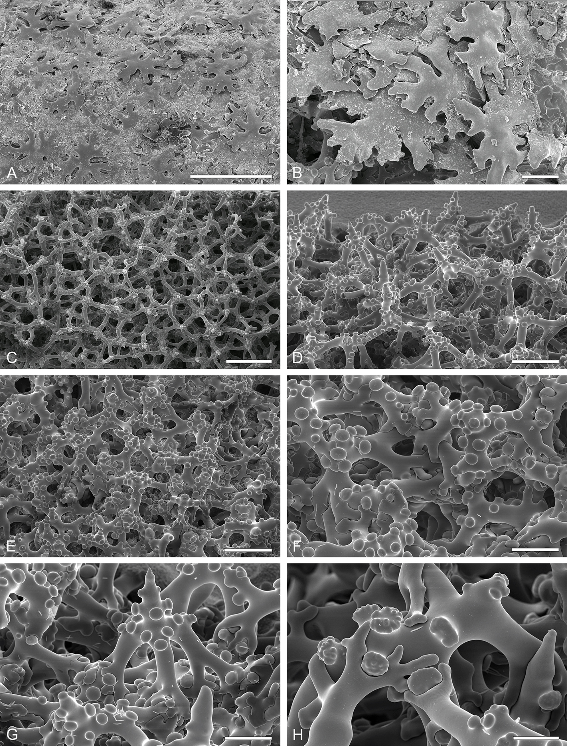
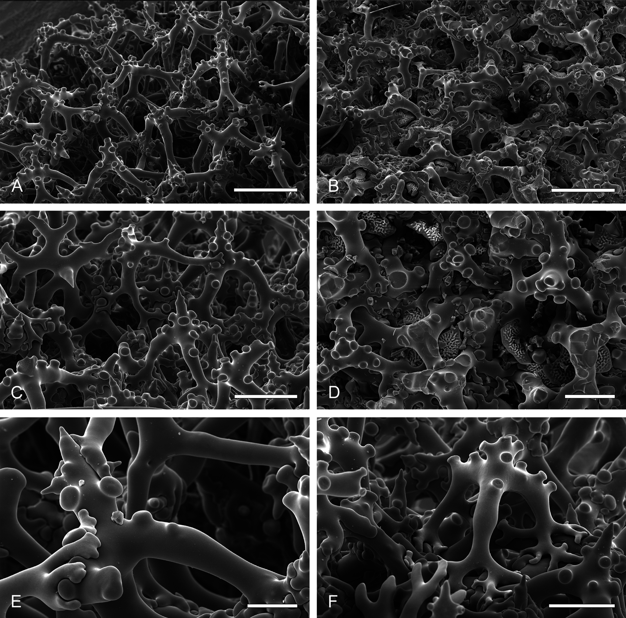
Skeleton. The ectosome is composed by a layer of overlapped phyllotriaenes ( Figs. 2 View FIGURE 2 A–B), numerous acanthomicroxeas and acanthorhabds, and some amphiasters. Pores are surrounded by these microscleres. The choanosome has regular trider-type desmas with smooth and/or tuberculated tubercles; the sculpture of the tubercles is related to the stage of formation of the desma: smooth tubercles in earlier stages and tuberculated when older; the extremities of the desmas also varies, spine-like and smooth when young, or with a tubercle on the top when older ( Figs. 2 View FIGURE 2 C–H, Fig. 3 View FIGURE 3 ). Subtylostyles to tylotes are transverse to the surface, and cross both parts of the skeleton. Acanthorhabds and acanthomicroxeas are very abundant and spread through the entire skeleton; amphiasters are few and dispersed.
In addition, we have found some spicules with a strange appearance in the lower part of the sponge in the paratype DOP1976 ( Fig. 3D View FIGURE 3 ). They resemble irregular disco- to phyllotriaenes with a strong sculpture that have been developed in the lower part and merged with the desma skeleton. This skeleton formation, previously observed in other lithistid demosponges, may have the purpose of consolidating the basal skeleton of the sponge.

No known copyright restrictions apply. See Agosti, D., Egloff, W., 2009. Taxonomic information exchange and copyright: the Plazi approach. BMC Research Notes 2009, 2:53 for further explanation.
|
Kingdom |
|
|
Phylum |
|
|
Class |
|
|
Order |
|
|
Family |
|
|
Genus |