
Fitchiella zahniseri, de Freitas & Dietrich & Takiya, 2020
|
publication ID |
https://doi.org/ 10.5852/ejt.2020.717.1097 |
|
publication LSID |
lsid:zoobank.org:pub:A03063E4-23C7-4084-BDB6-7495687FFDC5 |
|
DOI |
https://doi.org/10.5281/zenodo.4330353 |
|
persistent identifier |
https://treatment.plazi.org/id/24D79AE7-EA07-4A0D-AF6D-FFBA6D5A9107 |
|
taxon LSID |
lsid:zoobank.org:act:24D79AE7-EA07-4A0D-AF6D-FFBA6D5A9107 |
|
treatment provided by |
Valdenar |
|
scientific name |
Fitchiella zahniseri |
| status |
sp. nov. |
Fitchiella zahniseri View in CoL sp. nov.
urn:lsid:zoobank.org:act:24D79AE7-EA07-4A0D-AF6D-FFBA6D5A9107
Figs 13–15 View Fig View Fig View Fig , 26F View Fig
Diagnosis
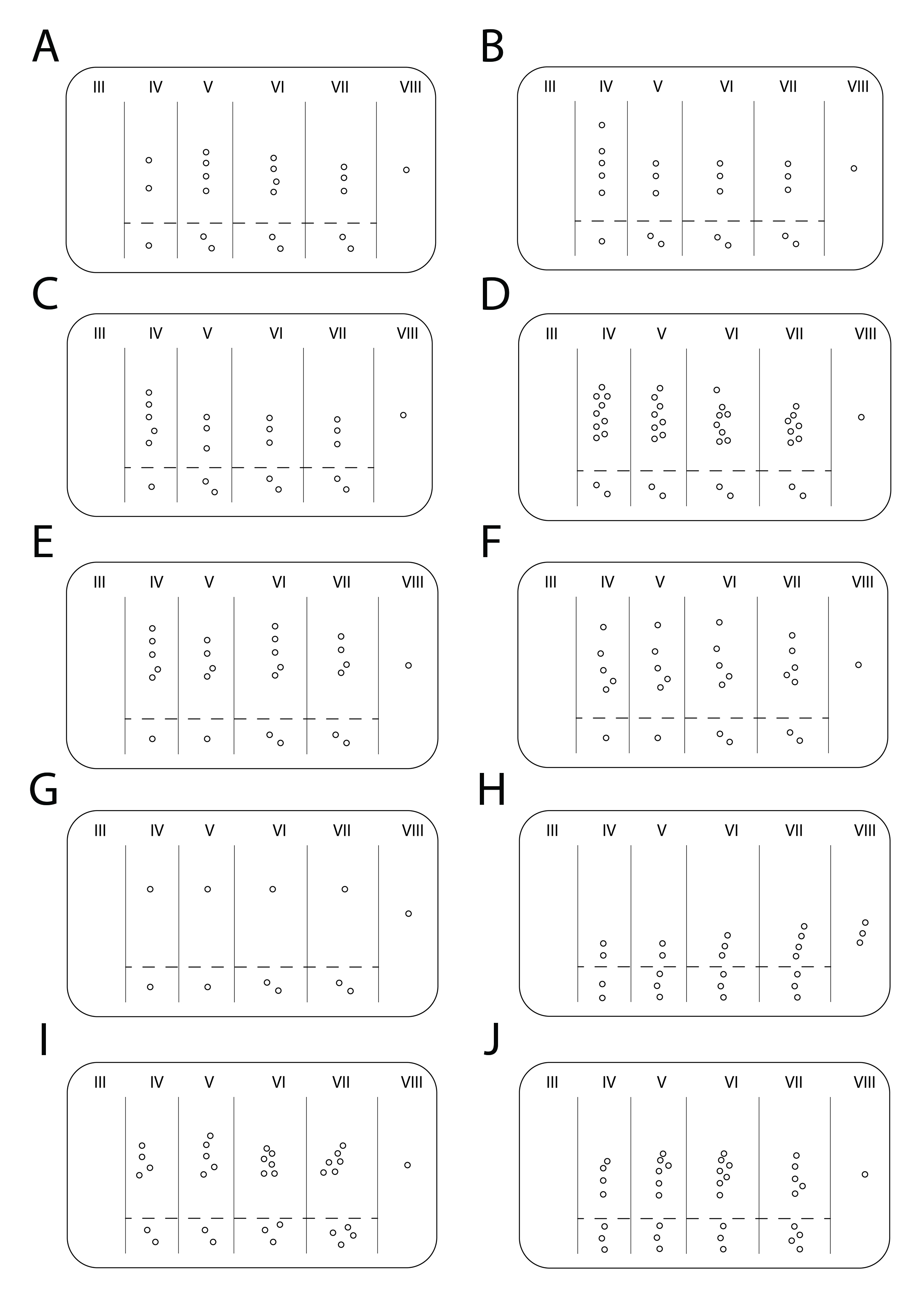
Body mainly stramineous with several dark brown maculae and sensory pits bordered by dark brown ( Fig. 13 View Fig ); snout longer than interocular distance (measured from base to tip) with apex rounded in frontal and lateral view ( Fig. 13A View Fig , C–D, F); lateral lobe of pronotum with four sensory pits arranged in a row ( Fig. 13C View Fig ); forewings with reticulated venation ( Fig. 13 View Fig B–C, E–F); abdominal tergites ( Figs 13C, F View Fig , 26F View Fig ) with row of sensory pits followed by single isolated ventral sensory pit (tergite IV and V) or isolated pair of diagonally aligned ventral sensory pits (tergites VI to VII).
Etymology
The specific name ʻ zahniseri ʼ is in honor of Dr James Zahniser (USDA = US Department of Agriculture) who collected the type series of this species in Panama and kindly sent us photographs of holotypes of Caliscelidae deposited at the NMNH.
Material examined
Holotype
PANAMA • ♂; Verugas, Pan-Am Highway , 40 km W of Santiago; 08.20298° N, 081.18768° W; 166 m a.s.l.; Aug. 2005; J.N. Zahniser leg.; sweep; DNA voucher ENT4204; NMNH. GoogleMaps
Paratypes
PANAMA • 1 ♀; same collection data as for holotype; INHS GoogleMaps • 1 ♀; same collection data as for holotype; DZRJ GoogleMaps .
Description
BODY LENGTH. Male = 2.7 mm; females = 3.5 mm.
COLORATION. Body mainly stramineous with several dark brown maculae and sensory pits bordered by dark brown; dark brown maculae darker and more extensive in males than females ( Fig. 13 View Fig A–C vs Fig. 13 View Fig D–F). Vertex ( Fig. 13B, E View Fig ) with pair of large dark brown maculae. Frons ( Fig. 13A, D View Fig ) with pair of dark brown stripes bordering sublateral carinae and dark brown stripe covering median carina; side of frons ( Fig. 13C, F View Fig ) dark brown where sensory pits are lacking. Gena ( Fig. 13C, F View Fig ) with large dark brown macula. Clypeus in lateral view ( Fig. 13C, F View Fig ) with dorsal portion extended anteriorly dark brown, median portion stramineous, ventral portion dark brown. Lateral lobe of pronotum ( Fig. 13C, F View Fig ) with anterior portion dark brown. Mesonotum ( Fig. 13B, E View Fig ) region between lateral carinae stramineous with pair of elongated dark brown maculae near lateral carinae. Forewings ( Fig. 13 View Fig B–C, E–F) light brown with veins white; white stripes within cells. Legs stramineous with some elongated dark brown maculae. Abdomen ( Fig. 13 View Fig B–C, E–F) with several elongated dark brown maculae starting from sensory pits and extending posteriorly; dark brown maculae forming continuous longitudinal line between row of sensory pits and isolated one.
HEAD AND THORAX. Vertex ( Fig. 13B, E View Fig ) hexagonal, as long as half its width, as long as half of pronotum length, with slight median carina; posterior margin slightly elevated. Frons ( Fig. 13A, D View Fig ) with median carina and pair of sublateral carinae; sublateral carinae convergent and fused to each other ventrally ( Fig. 13A, D View Fig ); central plate ( Fig. 13A, D View Fig ) longer than wide at widest portion, visible in dorsal view ( Fig. 13B, E View Fig ), not extending anteriorly beyond sublateral carinae in lateral view ( Fig. 13C, F View Fig ); sides of frons partially visible in frontal view ( Fig. 13A, D View Fig ), fused above clypeus, with two rows of sensory pits in lateral view: anterior row with eight sensory pits, five dorsal ones grouped together, followed by three isolated ones ventrally to concavity on anterior margin of frons; posterior row with four sensory pits. Clypeus ( Fig. 13C, F View Fig ) not swollen, with dorsal portion extending anteriorly, consequently pushing frons and producing moderately produced snout; snout in dorsal view ( Fig. 13B View Fig ) longer than interocular distance (measured from base to tip), rounded in frontal and lateral view ( Fig. 13C, F View Fig ), with median carina. Ocelli absent. Eye oblong. Antenna short, with several small circular structures visible on pedicel. Pronotum ( Fig. 13B, E View Fig ) semicircular, shorter than half its width; posterior margin straight; with median carina; surface of disc almost completely covered by 15 sensory pits on each side; lateral lobe of pronotum ( Fig. 13C, F View Fig ) with four sensory pits arranged in a row. Mesonotum ( Fig. 13B, E View Fig ) with slight median carina and pair of lateral carinae; region between lateral carinae depressed, without sensory pits; region outerad of lateral carinae with 11 to 12 sensory pits on each side. Brachypterous, with reticulated venation. Legs simple, with carinae and setae; tibia III with single median spine.
ABDOMEN. Terga with longitudinal carina. Tergite III ( Figs 13C, F View Fig , 26F View Fig ) without sensory pits. Tergite IV ( Figs 13C, F View Fig , 26F View Fig ) with one row of four to seven (four in holotype) sensory pits, with penultimate one slightly displaced, followed by single isolated ventral one. Tergite V ( Figs 13C, F View Fig , 26F View Fig ) with one row of four to five (five in holotype) sensory pits, with penultimate one slightly displaced, followed by single isolated ventral one. Tergites VI and VII ( Figs 13C, F View Fig , 26F View Fig ) with one row of four to five (five in holotype) sensory pits, with penultimate one slightly displaced, followed by isolated ventral pair aligned diagonally. Tergite VIII ( Fig. 26F View Fig ) with one sensory pit.
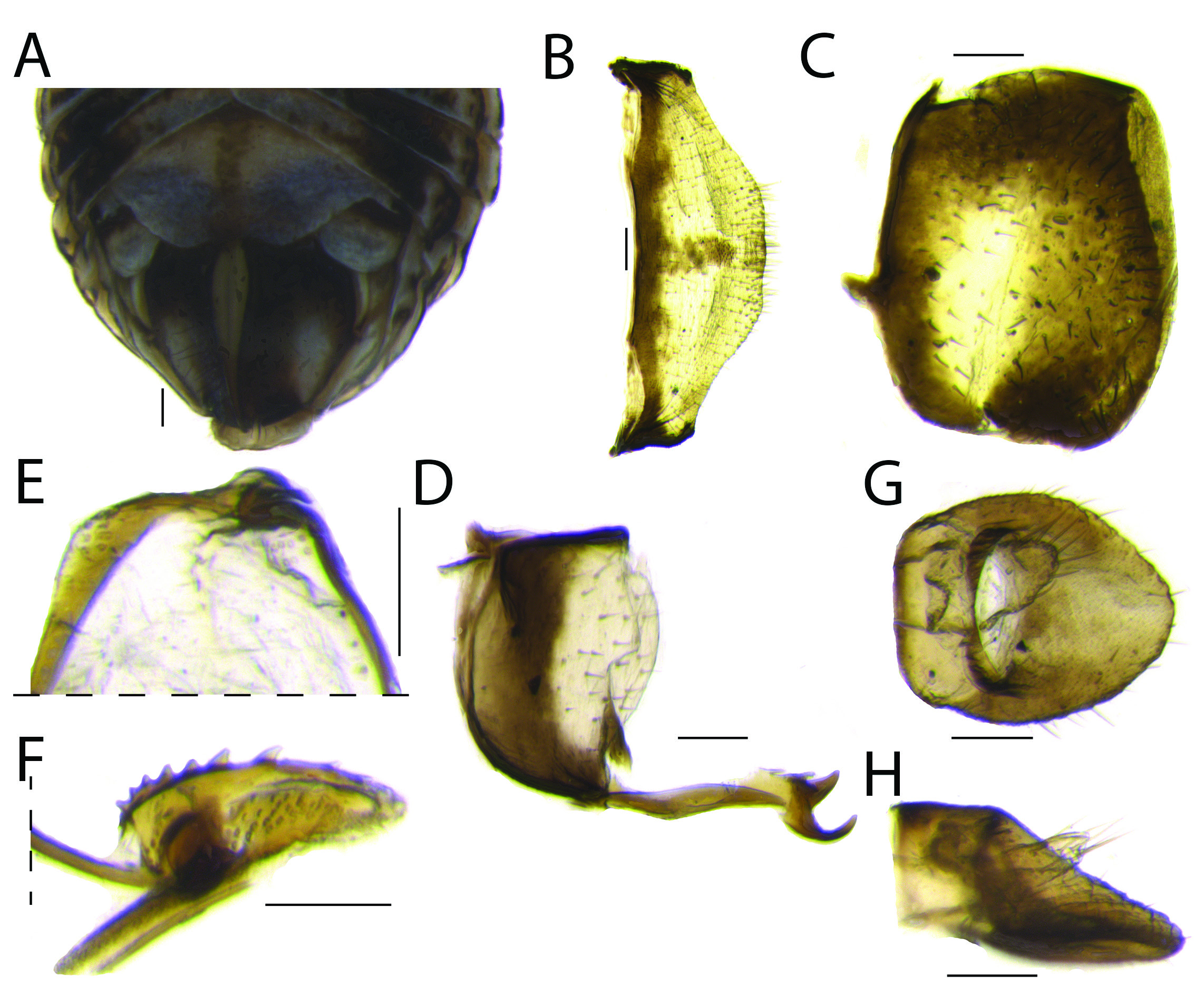
MALE TERMINALIA. Pygofer ( Fig. 14A View Fig ) narrow, with anterior margin deeply concave; posterior margin almost straight, except for slight concavity near ventral portion. Connective ( Fig. 14B View Fig ) inverted Y-shaped, with support bridge with dorsal lap. Style ( Fig. 14 View Fig C–D) hook-like; anterior portion pointed; caudal portion strongly curved anterodorsally and converging towards the other in dorsal view ( Fig. 14C View Fig ); dorsal margin ( Fig. 14D View Fig ) with slight protuberance on median third; ventral margin ( Fig. 14D View Fig ) with straight angle between anterior and middle third, after that mostly rounded; median portion ( Fig. 14D View Fig ) longer than high, setose; apex serrated. Phallobase ( Fig. 14 View Fig E–H) sclerotized, symmetrical, with two defined lobes; apex with pair of lobes truncate in dorsal view ( Fig. 14 View Fig E–F), rounded in lateral view ( Fig. 14 View Fig G–H), with dorsal process near apex in lateral view ( Fig. 14 View Fig G–H) surrounding apical half of aedeagus; with flap covering aedeagal hook in lateral view ( Fig. 14 View Fig G–H). Aedeagus ( Fig. 14 View Fig E–F) apex narrow and open dorsally; with pair of hooks, one curved anterodorsally, other curved posteroventrally in lateral view ( Fig. 14 View Fig G–H). Suspensorium V-shaped. Segment X of anal tube ( Fig. 14 View Fig I–J) as long as wide; posterior margin ( Fig. 14I View Fig ) rounded; segment XI of anal tube almost reaching posterior margin of segment X; setose.
FEMALE TERMINALIA. Posterior margin of sternite VII ( Fig. 15B View Fig ) with median portion produced, setose. Gonoplac ( Fig. 15C View Fig ) sclerotized, sub-rectangular with apex truncate, setose. Anterior connective lamina of gonapophysis VIII ( Fig. 15D View Fig ) with two conspicuous apical teeth: innermost narrower and longer than outer one; with two small spines laterad of outer tooth. Posterior connective lamina of gonapophysis IX ( Fig. 15 View Fig E–F) distal part in lateral view ( Fig. 15F View Fig ) with longitudinal row of nine to 10 wide, triangular, inclined spines; middle portion with several pits ( Fig. 15F View Fig ); apex setose ( Fig. 15F View Fig ). Segment X of anal tube ( Fig. 15 View Fig G–H) longer than wide at widest portion; posterior margin ( Fig. 15G View Fig ) with apex rounded; setose.
Remarks
The new species resembles Fitchiella robertsoni (type species) and F. brachyrhina sp. nov., sharing the following characteristics: (1) pattern of coloration ( Figs 11 View Fig , 13 View Fig ); (2) brachypterous with reticulated venation and with stripes within cells ( Figs 11B, E View Fig , 13B, E View Fig ); (3) overall shape of snout ( Figs 11C, F View Fig , 13C, F View Fig ), with anteroventrally produced clypeus, concavity on anterior margin of frons, and knobbed apex ( Figs 11C View Fig , 13C, E View Fig ); and (4) pattern of distribution of abdominal sensory pits ( Fig. 26 View Fig E–F). However, the new species can be distinguished from these two similar species by the following combination of characteristics: (1) snout ( Fig. 13C, F View Fig ) intermediate in length between the two previously described species, with apex less rounded and swollen than in F. robertsoni ; and (2) lateral lobe of pronotum with four sensory pits ( Fig. 13C, F View Fig ). Other species in Fitchiella , such as F. rufipes Lawson, 1933 and F. grandis Lawson, 1933 , among other, are easily distinguished from these species, F. robertsoni and F. brachyrhina sp. nov., by: (1) overall shape of snout, which is straight in some species, without concavity on anterior margin of frons and flattened laterally; (2) different pattern of coloration; (3) forewings without reticulated venation; (4) dorsoventrally expanded legs, more or less foliaceous in some species; and (5) different pattern of distribution of abdominal sensory pits (based on photographs of F. rufipes ). More studies are necessary to better define Fitchiella .


No known copyright restrictions apply. See Agosti, D., Egloff, W., 2009. Taxonomic information exchange and copyright: the Plazi approach. BMC Research Notes 2009, 2:53 for further explanation.
|
Kingdom |
|
|
Phylum |
|
|
Class |
|
|
Order |
|
|
Family |
|
|
Genus |