
Geophilus serbicus Stojanović, Mitić & Antić, 2019
|
publication ID |
https://doi.org/ 10.11646/zootaxa.4658.3.7 |
|
publication LSID |
lsid:zoobank.org:pub:7649EFF2-8908-4193-9D69-84645A36CFCC |
|
persistent identifier |
https://treatment.plazi.org/id/2276C812-C06B-CD03-FF03-FB94125EFA35 |
|
treatment provided by |
Plazi |
|
scientific name |
Geophilus serbicus Stojanović, Mitić & Antić |
| status |
sp. nov. |
Geophilus serbicus Stojanović, Mitić & Antić , new species
Material studied. Holotype: male (IZB ChG001), Babin Zub (43° 22.262′ N, 22° 36.532′ E; elevation about 1580 m a.s.l.; Fig. 1 View FIGURE 1 ), Stara Planina Mts. , Serbia, 20 July 2013, Fagetum moesiace montanum, under stones, D. Stojanović, I. Stojanović & K. Stojanović leg. GoogleMaps
Paratypes: two males (IZB ChG002 and IZB ChG003), four females (IZB ChG004–IZB ChG007), Babin Zub , Stara Planina Mts., Serbia: 1 ♂, 1 ♀, 21 June 2013, Fagetum moesiace montanum, under stones, D. Stojanović & K. Stojanović leg. ; 1 ♂, 3 ♀, 20 July 2013, Fagetum moesiace montanum, under stones, D. Stojanović, I. Stojanović & K. Stojanović leg.
Additional non-type material: 23 males (9 specimens on last adolescens stage) and 93 females (82 females with broods, nine without broods and only four adolescens specimens) (IZB ChG008–IZB ChG123), Babin Zub , Stara Planina Mts., Serbia, Fagetum moesiace montanum, leaf-litter, under stones and rotten stumps : 2 ♂, 8 ♀, 21 June 2013, D. Stojanović & K. Stojanović leg.; 5 ♂, 15 ♀, 6 July 2013, D. Stojanović, I. Stojanović & I. Stojanović leg.; 5 ♂, 16 ♀, 20 July 2013, D. Stojanović, I. Stojanović & K. Stojanović leg.; 4 ♂, 15 ♀, 3 August 2013, D. Stojanović, K. Stojanović, A. Petrović & J. Petrović leg.; 4 ♂, 17 ♀, 16 August 2013, D. Stojanović, I. Stojanović & K. Stojanović leg.; 3 ♂, 11 ♀, 10 July 2014, D. Stojanović, N. Zukanović & D. Antić leg.; and 11 ♀, 15 June 2016., D. Stojanović, D. Antić & S. Makarov leg.; 2 females (IZB ChG124 and IZB ChG125), Jabučko Ravnište (latitude 43° 21.724′ N; longitude 22° 35.008′ E; elevation about 1490 m a.s.l.; Fig. 1 View FIGURE 1 ), Stara Planina Mts., Serbia, 19 June 2013, Fagetum moesiace montanum, under stones, D. Stojanović, K. Stojanović, M. Kučinić & M. Živić leg. The GoogleMaps non-type material is used only to obtain data on variability of the number of leg-bearing segments, the number of coxal pores on coxopleura of the ultimate leg-bearing segments and total length of the specimens.
Etymology. The name is an adjective referring to the type locality.
Diagnosis: The new species clearly differs from its congeners by virtue of a unique combination of characters: small body size (less than 20 mm in adults); 41–47 leg pairs; cephalic plate ca. 1.1–1.2 times longer than wide; antennae ca. 3.1–4.5 times as long as the head; lappets present on first maxillary coxosternite and basal telopodite; pretarsus of the second maxillae apparently tapering gradually into a pointed tip, without spines or filaments; for- cipular coxosternite ca. 1.1–1.2 times wider than long, with anterior margin lacking denticles and with chitin-lines pointing lateral to condyles; basal denticles present only on the tarsungulum, all other forcipular telopodite articles without denticles; clypeus without a distinct clypeal area; ventral pore-fields present in all metasternites (from the first to the penultimate one), undivided only in anterior region of the body (up to ca. metasternites 12–13), all other metasternites with divided pore-fields; carpophagus pit inconspicuous; 3–7 (usually 4–5) coxal organs, all ventral, distinct and opening separately, most of them close to the margin of the ultimate metasternite, also a single one iso- lated posteriorly; apical claws of the walking legs long, with two accessory spines, ultimate pretarsi short, curved and without spines; anal pores present.
Geophilus serbicus sp. nov. can be clearly distinguished from the other western Palaearctic Geophilus species with a similarly number of leg-bearing segments, by a number of features presented in Table 1 View TABLE 1 and in the Discussion below.
Description: Holotype male, 41 pairs of legs, body length about 17 mm, maximum body width 0.68 mm. Paratype males with 43 and 45 pairs of legs, body length 14 mm, maximum body width 0.68 mm. Each of the four paratype females with 45 pairs of legs, body length 16.6–18 mm, and width 0.59–0.68 mm.
Colouration: From whitish-yellow to pale-yellow, with somewhat darker colouration of the head, mouthparts, forcipular segment and walking and ultimate legs ( Fig. 2A View FIGURE 2 ). Distal parts of the forcipular tarsungulum reddish brown.
Antennae: Elongated, ca. 3.1–4.5 (usually 3.5) times longer than the cephalic plate (in specimens from etha- nol, untreated with KOH). The basal article as long as wide; all other antennal articles longer than wide ( Fig. 3A View FIGURE 3 ). Terminal article ca. 1.5 times longer than penultimate article. There is no difference between dorsal and ventral chaetotaxy. All antennal articles with sensilla trichodea, their density higher in distal half of the antennae. Addition- ally, terminal article with sensilla basiconica (recorded in adult specimens of both sex) present opposite each other on the internal and external margins ( Fig. 3B View FIGURE 3 ). In holotype male, left and right antenna with 14 and 12 internal and nine and 11 external sensilla basiconica, respectively. Apex of terminal antennomeres completely rounded, without any recession on surface.
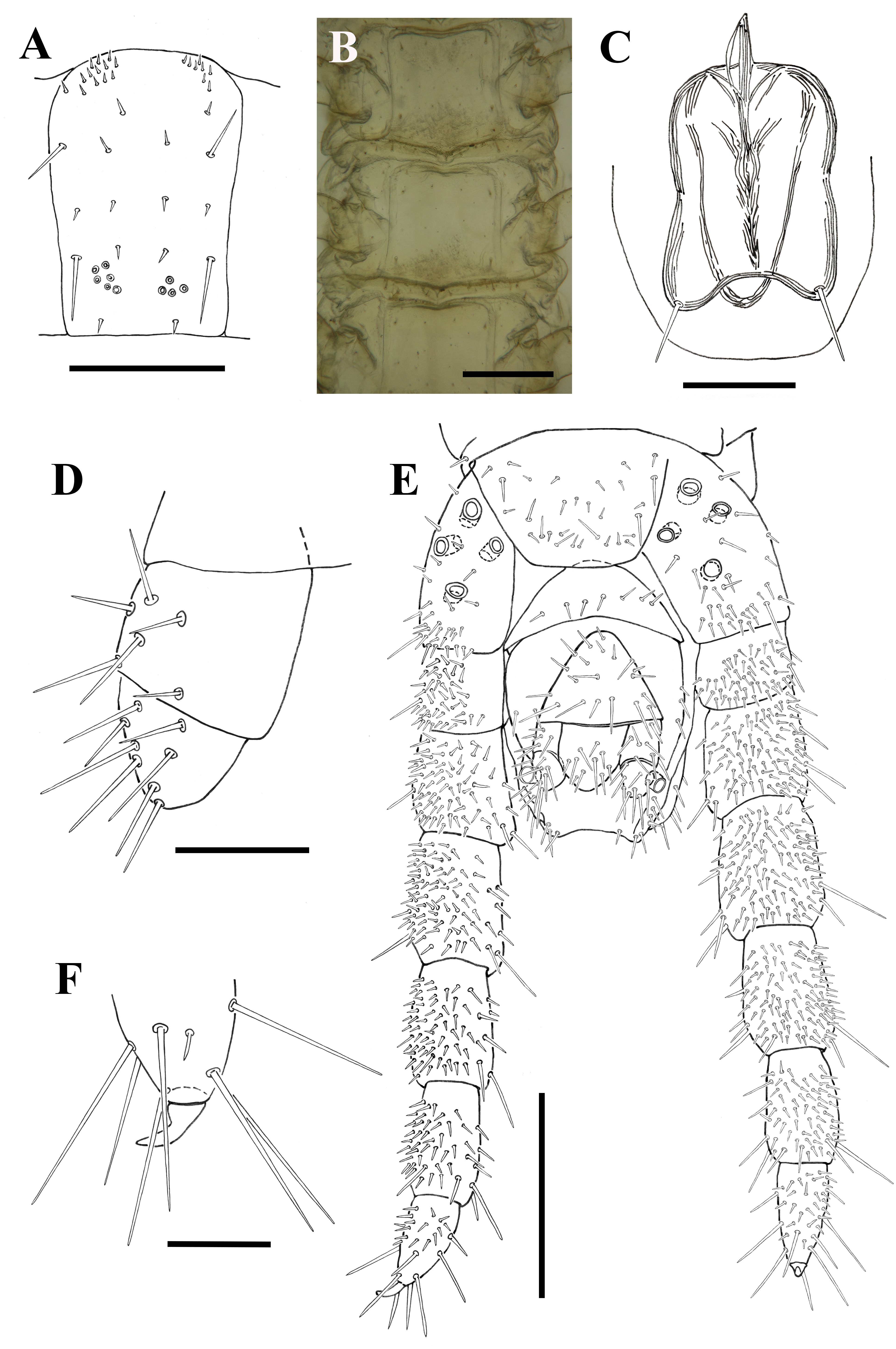
Cephalic plate: Longer than wide (length/width holotype 1.16:1; paratype males 1.11:1; paratype females 1.17:1, 1.14:1, 1.15:1 and 1.18:1). Maximum cephalic plate length and width in a holotype 0.57 mm and 0.49 mm; in paratype males length 0.51–0.52 mm, width 0.46–0.47 mm; in paratype females length 0.52–0.64 mm, width 0.44–0.56 mm. Posterior margin almost straight, laterally slightly rounded. Without sutures or paramedian sulci. Chaetotaxy as in Fig. 2C View FIGURE 2 .
Clypeus: Without clypeal area ( Fig. 2D View FIGURE 2 ). Surface clearly reticulated, with three pairs of setae, of which the post-antennary and intermediate pairs are somewhat larger, the posterior pair smaller (2/2/2; uniformly in all type specimens).
Labrum: Lateral and intermediate parts distinct. Intermediate part well developed, with eight symmetrically arranged tubercles, the two central ones significantly larger. Lateral parts with 7–8 bristles ( Fig. 2F View FIGURE 2 ).
Mandible: Shape of mandibles as in Figs 3D and E View FIGURE 3 . The slightly sclerotized pectinate lamella with ca. 15 hyaline teeth. Ventral ridge not expanded, continuous with pectinate lamella ( Fig. 3D View FIGURE 3 ). Condyle moderately chi- tinized.
First maxillae ( Fig. 2G View FIGURE 2 ): Coxosternite undivided, without setae, with a small lappet on the lateral margin. Telopodital lappets well developed and almost two times longer than the coxosternal ones. The ratio of their length is similar to that of the distal and basal articles of the first maxillary telopodite. Distal telopodital article apically rounded, with 2–3 long setae and 3–4 small sensilla. Basal telopodital articles without setae. Coxal projections sub- triangular, well developed, each with one long mesal seta and 3–4 short sensilla distally.
Second maxillae: Coxosternite undivided, without medial sutura or inner and foraminal processes; isthmus well sclerotized, indistinct statuminia; anterior margin concave, with three groups of short setae (3–5 per group) distributed along the border, and two separated paramedial setae ( Fig. 2G View FIGURE 2 ). Telopodite poorly setose. Terminal ar- ticle distally with four long, sclerotized setae ( Figs 2G View FIGURE 2 and 3C View FIGURE 3 ). Pretarsus well developed, moderately elongated, about half as long as terminal article, apparently tapering gradually into a pointed tip, without spines or filaments ( Fig. 3C View FIGURE 3 ).
Forcipular segment: Forcipular tergite distinctly narrowing anteriad. Forcipular coxosternite wider than long (width/length 1.1–1.3:1; usually 1.2:1), somewhat wider than the cephalic plate. Maximum forcipular coxosternite width in a holotype 0.53 mm, in paratype males 0.51–0.52 mm, and in paratype females 0.46–0.57 mm. Anterior margin of coxosternite slightly concave and without denticles ( Fig. 2B View FIGURE 2 ). Coxopleural sutures distinctly diverg- ing forwardly; chitin-lines present, pointing lateral to condyles, but not reaching them. Tarsungulum with almost straight basal denticle; without denticle-like projections along slender and moderately curved apical peak ( Fig. 2E View FIGURE 2 ). Trochanteroprefemur, femur and tibia without denticles. Calyx of poison gland cluster-associated, with almost straight duct opening externally near apical end of tarsungulum ( Fig. 2E View FIGURE 2 ). Chaetotaxy of the forcipular coxosternite and telopodites as in Fig. 2B View FIGURE 2 .
Trunk: Pore-fields present on all leg-bearing segments except metasternite of the ultimate leg-bearing segment. In the anterior half of the trunk, pore-fields are located in the posterior half, but near the middle of the metaster- nites (metasternites 1–7 in the holotype male, 1–12 in the paratype female). On all other metasternites, pore-fields closer to the posterior margin. Fields undivided on metasternites 1–12 in holotype male (metasternites 1–13 in one paratype female; IZB ChG007). The rest of the leg-bearing segments with divided fields. Number of sternal pores in holotype male as follows: metasternite 1 (4 pores), 2 (12), 3 (18), 4 (19), 5 (23), 6 (23), 7 (21), 8 (22), 9 (26), 10 (33), 11 (28), 12 (30), 13 (29: left 16/right 13), 14 (22: 12/10), 15 (28: 14/14), 16 (22: 12/10), 17 (20: 9/11), 18 (22: 9/13), 19 (18: 10/8), 20 (14: 8/6), 21 (15: 7/8), 22 (17: 8/9), 23 (17: 8/9), 24 (13: 5/8), 25 (15: 9/6), 26 (18: 11/7), 27 (18: 9/9), 28 (14: 5/9), 29 (14: 8/6), 30 (13: 5/8), 31 (17: 9/8), 32 (19: 10/9), 33 (18: 9/9), 34 (19: 10/9), 35 (15: 7/8), 36 (26: 14/12), 37 (21: 10/11), 38 (20: 9/11), 39 (15: 8/7) and metasternite 40 (10: 4/6). Number of sternal pores in paratype female (IZB ChG007) as follows: 1 (4), 2 (8), 3 (16), 4 (16), 5 (18), 6 (17), 7 (16), 8 (18), 9 (22), 10 (21), 11 (21), 12 (21), 13 (19), 14 (19: left 10/right 9), 15 (17: 9/8), 16 (14: 8/6), 17 (11: 3/8), 18 (12: 6/6), 19 (12: 6/6), 20 (12: 6/6), 21 (11: 7/4), 22 (10: 7/3), 23 (10: 5/5), 24 (7: 4/3), 25 (11: 5/6), 26 (7: 4/3), 27 (12: 6/6), 28 (9: 4/5), 29 (6: 3/3), 30 (14: 6/8), 31 (11: 4/7), 32 (10: 6/4), 33 (8: 4/4), 34 (10: 4/6), 35 (8: 4/4), 36 (11: 5/6), 37 (13: 6/7), 38 (14: 6/8), 39 (10: 4/6), 40 (11: 5/6), 41 (13: 8/5), 42 (12: 4/8), 43 (11: 7/4) and metasternite 44 (5: 2/3). Without distinct carpophagus pits, only some small median plateau in anterior part of trunk: metasternites 7–14 in holotype male ( Figs 4 View FIGURE 4 D–4H) and metasternites 4–12 in paratype female ( Fig. 5B View FIGURE 5 ). Chaetotaxy, shape of the pore-fields and their relative size on selected metasternites are presented in Figs 4 View FIGURE 4 and 5A View FIGURE 5 .
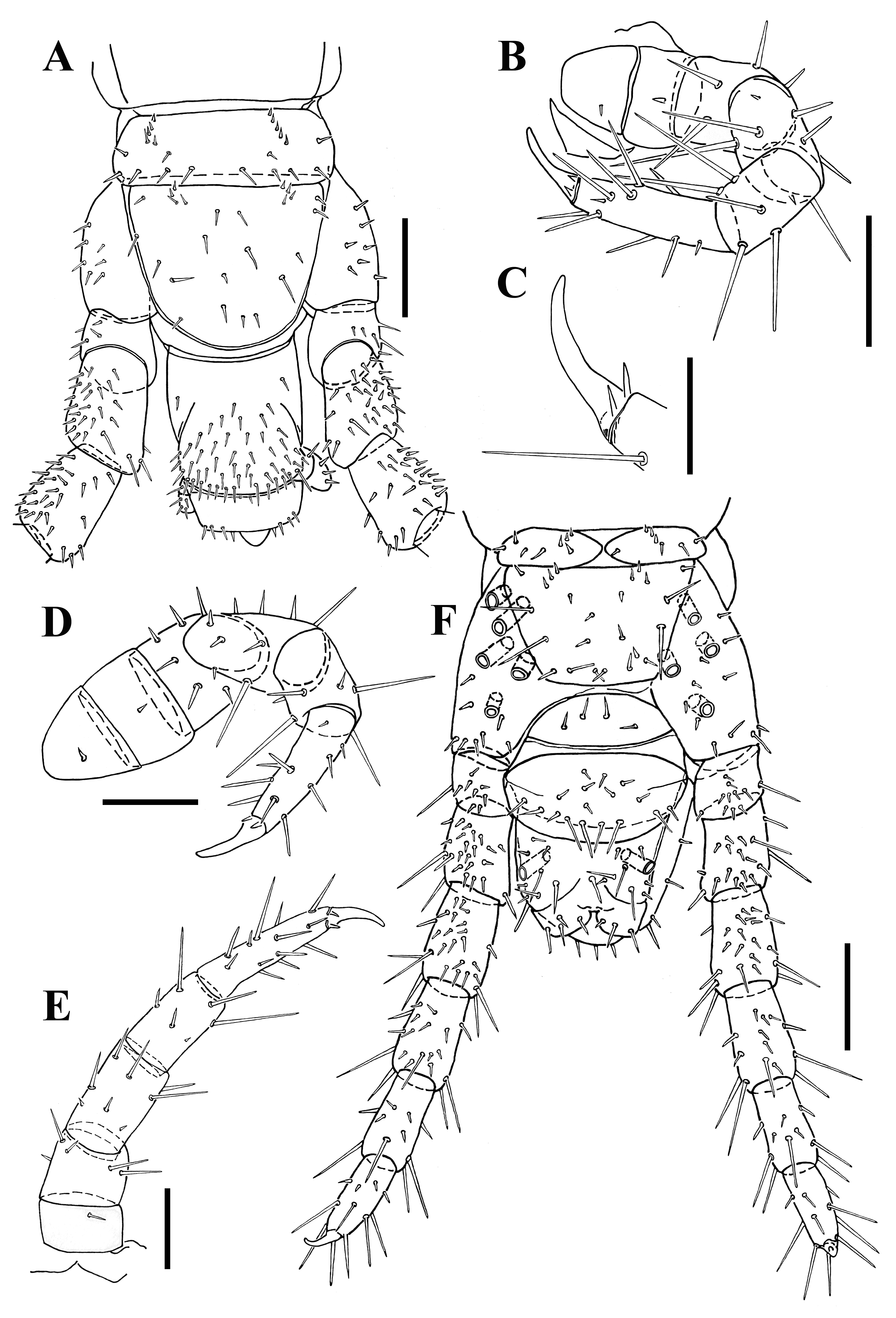
Walking legs: Chaetotaxy similar throughout the entire body length. Distribution and relative size of setae as in Figs 6B, 6D and 6E View FIGURE 6 . Apical claws curved, with two short accessory spines ventrobasally ( Fig. 6C View FIGURE 6 ).
Ultimate leg-bearing segment: Metasternite ca. 1.1–1.4 times wider than long (usually 1.2), trapezoid, ulti- mate presternite divided medially ( Fig. 6F View FIGURE 6 ). Coxopleuron slightly swollen, laterally rounded, ventrally with numer- ous small setae on the distal ventral margin, a few larger setae present on the remaining surface. Each coxopleuron with 3–7 (usually 4–5) coxal organs opening on the ventral surface separately; no ventral pits with multiple coxal organs; no dorsal pores or pits. In all collected specimens, one coxal pore displaced with a more posterior position and one more laterally; other coxal organs opening close to the metasternite margin ( Figs 5E View FIGURE 5 and 6F View FIGURE 6 ). In both sexes ultimate legs are much longer than the penultimate ones and are composed of six articles. Ultimate legs in female slender; somewhat thicker in males ( Figs 5E View FIGURE 5 and 6F View FIGURE 6 ). Ventral surface significantly more setose in males compared to females ( Figs 5A View FIGURE 5 and 6E View FIGURE 6 ). Also, the dorsal surface of the same pair of legs in males is with significantly lower density of setae than on their ventral surface in the same specimen ( Fig. 6A View FIGURE 6 ). Apical claws curved and shorter com- pared to walking legs, without accessory spines ( Fig. 5F View FIGURE 5 ).
Postpedal segments: Intermediate sternite in males with several setae arranged along the concave posterior margin ( Fig. 5E View FIGURE 5 ). Posterior margin in females more convex ( Fig. 6F View FIGURE 6 ). The first genital sternite in males triangular, with many setae almost uniformly distributed over the surface ( Fig. 5E View FIGURE 5 ). Male gonopods biarticulate, separated at their bases, with setae distributed mostly on external sides of articles; basal article with ca. six setae, apical article with ca. eight setae ( Fig. 5D View FIGURE 5 ). Penis as in Fig. 5C View FIGURE 5 . The second genital sternite with rounded posterior margin. The first genital sternite in females ellipsoid, with several short central setae, and longer setae on the posterior margin ( Fig. 6F View FIGURE 6 ). Female gonopods in a single lamina. Anal pores present in both sexes ( Figs 5E View FIGURE 5 and 6F View FIGURE 6 ).
Intraspecific variation: Taken altogether, the type and the additional non-type specimens show a variability of between 41 and 45 pairs of legs in males and between 45 and 47 pairs of legs in females. All specimens from the whole sample have a body length of less than 20 mm (the biggest collected female is around 18.6 mm long). The morphological differences noticed between adults males and females were in different morphology of the genital segments and ultimate legs, as well as in minor differences of size between them (somewhat higher length values in females) and antennae/cephalic plate length ratio [ca. 3.5 (3.1–4.5) in females; and ca. 3.7 (3.2–4.5) in males]. Measuring of specimens show average values of 12.98 mm (min 9.64 mm; max 16.7 mm) for males and 13.26 mm (min 11.54 mm; max 16.58 mm) for non-brooding females. The average body length of brooding females is 13.55 mm (the recorded extremes are: min 8.92 mm and max 18.56 mm). Some differences in nuances of colour are noticed between the more yellowish brooding females and the somewhat whitish females captured as free-living specimens.
Except the small differences in body size [average values of 11.07 (min 10.65 mm; max 11.94 mm) for young females and 11.06 mm (min 8.53 mm; max 12.71 mm) for young males] and antennae/cephalic plate length ratio [ca. 3.2 (3.1–3.7) in females and ca. 3.5 (3.2–4) in males], the collected juvenile specimens (all from the last adole- scens stage) show similar values as adults in all other proportions: cephalic plate length/width (ca. 1.1–1.2), forcipu- lar coxosternite width/length (ca. 1.1–1.2), and ultimate leg-bearing segment metasternite maximum width/length (ca. 1.1–1.4). Also, the similar number and arrangement of the coxal pores are recorded (each coxopleuron with 4 or 5 pores).


No known copyright restrictions apply. See Agosti, D., Egloff, W., 2009. Taxonomic information exchange and copyright: the Plazi approach. BMC Research Notes 2009, 2:53 for further explanation.
|
Kingdom |
|
|
Phylum |
|
|
Class |
|
|
Order |
|
|
Family |
|
|
Genus |