
Glomeridesmus spelaeus Iniesta & Wesener
|
publication ID |
https://doi.org/ 10.5281/zenodo.211959 |
|
DOI |
https://doi.org/10.5281/zenodo.6179710 |
|
persistent identifier |
https://treatment.plazi.org/id/5571F65F-9C14-DA2D-05D8-8B9DFB3F6E4A |
|
treatment provided by |
Plazi |
|
scientific name |
Glomeridesmus spelaeus Iniesta & Wesener |
| status |
sp. nov. |
Glomeridesmus spelaeus Iniesta & Wesener View in CoL , new species
Material examined: Holotype: 1 Male ( ISLA 1906) SLQ31 EU, Brazil, Pará, Curionópolis, iron cave SL 31, E 0650189m, N 9339714m, in bat guano pile far from entrance, Zampaulo, R.A. coll.
Paratypes: 1 M (fragmented, ZFMK MYR 0936), 1 F ( ZFMK MYR 0937), '1758', same data as holotype; 3 M (fragmented, ISLA 1758, ISLA 1759, fragmented ISLA 3837); 5 F ( ISLA 1928, ISLA 1929, ISLA 1930, ISLA 1931, fragmented ISLA 3838); 4 juv. ( ISLA 1932, ISLA 1933, ISLA 1934, ISLA 1935).
Other material examined: 3 F ( ISLA 3814, ISLA 3815, ISLA 3816), Brazil, Pará, Curionópolis, iron cave SL 31, E 0652243m/N 9339216m, Zampaulo, R.A. coll. same data as holotype; 2 F ( ISLA 3817, ISLA 3818), 1 M ( ISLA 3819) Brazil, Pará, Curionópolis, iron cave SL 31, E 0652243m/N 9339216m, Zampaulo, R.A. coll. same data as holotype; 4 F ( ISLA 3824, ISLA 3825, ISLA 3826, ISLA 3827), Brazil, Pará, Curionópolis, iron cave SL 58, E 0652243m/N 9339216m, Zampaulo, R.A. coll. 29.i.2011; 1 F ( ISLA 3820) Brazil, Pará, Curionópolis, iron cave SL 61, E 0 652512m /N 9338874m, Zampaulo, R.A. coll. 10.i.2011; 2 F ( ISLA 3828, ISLA 3829), Brazil, Pará, Curionópolis, iron cave SL 93, E 0 651386m /N 9341930m, Zampaulo, R.A. coll. 17.i.2011; 1 F, ( ISLA 3823), Brazil, Pará, Curionópolis, iron cave SL 95, E 651417m /N 9341906m, Zampaulo, R.A. coll. 03.ii.2011; 2 F, ( ISLA 3821, ISLA 3822), Brazil, Pará, Curionópolis, iron cave SL 97, E 0651411m/N 9339212m, Zampaulo, R.A. coll. 04.xii.2011;
Diagnosis. White, translucent body ( Fig. 2 View FIGURE 2 ) shared with G. sbordonii Shear, 1974 from Mexico, the only other known troglobitic species of the order. G. s p e l a e u s n. sp. differs from G. sbordonii in the presence of sclerotized knobs with mechanoreceptorical(?) setae on tergites (smooth and glabrous in G. sbordonii ), larger size (~ 5 mm body length in G. sbordonii ), antennae more densely pilose than in G. sbordonii (which could be a perspective error), joint 3 of telopod in oral (anterior) view not overlapping joint 4, as well as other telopod characteristic (provided drawing of S. sbordonii [ Shear 1974, fig. 2] difficult to compare with our SEM images). Location and shape of projection (immovable finger) of joint 3, posteriorly instead of laterally of joint 4, appears to be unique for the order ( Fig. 9 View FIGURE 9. G. s p e l a e u s n A, B, D), albeit telopods are only known from <1/3 of described Glomeridesmus species.
Etymology. Species epithet is a noun in apposition, as a reference to the Latin word spelaeus , meaning “cave”.
Description. Measurements: (largest) adult females (20+ AS tergites, 35+1 leg pairs): length: 8.6 mm; width (midbody): 2.3 mm.
Males (19+ AS tergites, 33+1+T leg pairs, holotype male): length: 7.2; width (midbody): 2.1; (32+1+T, n =2): length: 5.4, width: 1.6; (31+1+T, n=1): length: 6.3, width: 1.9.
Colour: translucent white.
Head: General shape: typical for the family ( Figs 3 View FIGURE 3. G. s p e l a e u s n C, E). Epicranium glabrous; frons, clypeus and labrum with several isolated setae. Both antennae widely separated by a distance (300 µm) longer than twice the width of the first antennal joint. Genae (area below the antennae) almost non-existant, but basal joint of mandible large and visible in dorsal view. Antennal base and organ of Tömösváry surrounded by sclerotized rim ( Fig. 3 View FIGURE 3. G. s p e l a e u s n D, E). Antennae consisting of seven joints, each covered with numerous setae ( Fig. 3 View FIGURE 3. G. s p e l a e u s n E). Joints 4–6 slightly flattened, apically much wider than basally, wider than long ( Fig. 3 View FIGURE 3. G. s p e l a e u s n C, E). Apical disc carrying four long (40 µm) apical cones; sensilla basiconicae seem to be absent ( Fig. 3 View FIGURE 3. G. s p e l a e u s n F). Tömösváry organs slightly larger than antennal base. Of well-rounded oval shape, interior covered by sclerotized plate ( Fig. 3 View FIGURE 3. G. s p e l a e u s n D). Gnathochilarium with very broad gula (hypostoma). Cardines very small, separated from basal part of gnathochilarium. Mentum basally towards gula with a transverse elevated area, completely fused with laminae linguales ( Fig. 3G View FIGURE 3. G. s p e l a e u s n ). Stipites laterally with single sclerotized ledge. Whole surface of gnathochilarium covered with few, isolated setae. Stipites apically with 2, lamello-mentum with 4 extra-long setae. Lateral palpi thin, carrying 4 or 5 sensory cones; inner palpi very wide, carrying several dozen long, tube-shaped sensory cones; central pads large, mesally touching one another, in ventral view also covered with numerous long, tube-shaped sensory cones.
Mandible: with single, densely pilose basal joint ( Fig. 3 View FIGURE 3. G. s p e l a e u s n C). Apical part not investigated.
Trunk: Collum (tergite 1): wider than head, shape similar to following tergites.
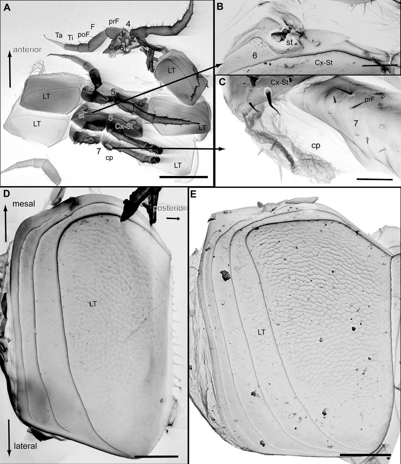
Tergites 2–19 (20): very thin, decalcified and difficult to distinguish from one another. Size of tergites first increasing until tergite 8, than slowly decreasing more posteriorly. Each tergite divided by 8–10 longitudinal striae, each stria projecting with a toothed margin above more posterior stria ( Fig. 6 View FIGURE 6. G B). Distances between striae expanding towards posterior margin ( Fig. 6 View FIGURE 6. G E). Posterior margin with single row of large sclerotized knobs ( Fig. 6 View FIGURE 6. G A, B, D). Knobs accompanied by longer sensorial setae ( Fig. 6 View FIGURE 6. G C). Surface of tergites covered by a sparse field of very short setae ( Fig. 6 View FIGURE 6. G B). Tergites at apico-lateral corner with 2 or 3 striae and denser rows of short setae ( Fig. 6 View FIGURE 6. G B). Tergite 12 onwards posterior-lateral edge pronounced into a sharp-edged tip, becoming a longer, spine like process at second last tergites. Last tergite at posterior margin towards anal shield without (male) or with (female) single row of short spines. Limbus (posterior margin of tergite) smooth, without any structures ( Fig. 5 View FIGURE 5. G D). Endotergum (underside of posterior margin of tergite) smooth area without any specific structures ( Figs 5 View FIGURE 5. G F). Anterior margin of tergite smooth, without any structures. ( Fig. 5 View FIGURE 5. G D, E). Latero-tergites of rectangular shape, posterior margin overlapping first third of subsequent latero-tergite ( Fig. 4 View FIGURE 4. G. s p e l a e u s n A). Anterior and posterior sides longer than mesal and lateral ones ( Fig. 4 View FIGURE 4. G. s p e l a e u s n D, E). Mesal side with a rounded angle towards coxosternite, marginal brim here extraordinarily thick; midbody latero-tergite with an elevated marginal brim engulfing all but posterior margin ( Fig.4 View FIGURE 4. G. s p e l a e u s n D, E). Posterior margin with a single row of short and slender spines. Marginal rim covering more than anterior third of latero-tergite, descending in 3 steps. Surface area not covered by marginal rim (posterior 2/3) reticulated. Whole surface of latero-tergite very sparsely covered by isolated setae ( Fig. 4 View FIGURE 4. G. s p e l a e u s n D, E). Latero-tergite similar to one another, but last latero-tergite on posterior margin with a well-rounded indentation, providing space for the movements of the last leg.
Trunk appendages: Coxosternite(?) serrated at apico-lateral margin ( Fig. 5 View FIGURE 5. G A, 4A). Sternal part more elevated, located basal and lateral of coxal part. Spiracle opening of anterior side in baso-lateral corner of coxosternite ( Fig. 4 View FIGURE 4. G. s p e l a e u s n A, B). Starting at coxosternite 7, each odd-numbered coxa up to pair 29 (male) or 31 (female) apically with an eversible coxal pouch posteriorly of prefemur ( Figs 4 View FIGURE 4. G. s p e l a e u s n A, C). Legs elongated and slender ( Fig. 4 View FIGURE 4. G. s p e l a e u s n A, 5A). Femur 2.5 times longer than wide, tarsus 5.5 times longer than wide. Coxa apically and mesally with few longer setae; prefemur apically, mesally and laterally with 3 or 4 longer setae; prefemur on mesal margin at midpoint with one very long seta, around 20 smaller setae oriented towards mesal margin; station of postfemur and tibia similar to prefemur ( Fig. 5 View FIGURE 5. G A). Tarsus with larger setae only in basal half, a pair of long spines present, tip with one apical spine, an elongated, weakly curved apical claw with basal flexible paranychium, twice as long as claw ( Fig. 5 View FIGURE 5. G C). Penultimate leg with coxosternite narrow, with stigma opening and sternal part located below instead of lateral of coxal part. Ultimate leg pair modified, consisting of coxosternite and 3 podomeres ( Fig. 7 View FIGURE 7. G. s p e l a e u s n A). Last two podomeres extending postero-laterally (confused with telopod by previous authors). Both legs widely separated from each other, connected by large plate (sternite?). Coxosternite large, almost as long as 3 podomeres combined. Podomere three apically with a large, long spine ( Fig. 7 View FIGURE 7. G. s p e l a e u s n A).
Anal shield: glabrous, with well-rounded edge ( Fig. 7 View FIGURE 7. G. s p e l a e u s n B). On lateral side with slightly serrated margin and line of small (sensorial?) setae ( Fig. 7 View FIGURE 7. G. s p e l a e u s n B). Subanal plate located behind last pair of legs, large and hyaline.
Sexual characters: Female: second coxae on posterior side with prominent ovipositors of medium length ( Fig. 3 View FIGURE 3. G. s p e l a e u s n B). Coxa protruding mesally as a short lobe. Ovipositors basally supported by an undivided plate (sternite?). Ovipositors of medium length, in largest females protruding posteriorly up to leg 7. Ovipositor divided into a basal part consisting of several layers of (eversible?) half-rings, each carrying isolated setae in a regular distance to one another, and an apical part with the opening ( Fig. 3 View FIGURE 3. G. s p e l a e u s n B). In the basal part, half rings are anteriorly and posteriorly interrupted by two different, more slender tubes, also consisting of rings, running along the whole length of the ovipositor. Apical part complex, consisting of apparently four larger plates, each arising out of a tube of rings. All four apical plates covered by longer setae ( Fig. 3 View FIGURE 3. G. s p e l a e u s n B).
Male: second coxa with gonopore located mesally ( Fig. 5 View FIGURE 5. G A, B). Gonopore consisting of two sclerotized, apically intertwined plates forming a slit-like opening in basal half ( Fig. 5 View FIGURE 5. G B). Apical part of both plates with 2 or 3 longer setae. Male telopod consisting of syncoxite with inner horns and 4 podomeres ( Fig. 8 View FIGURE 8. G. s p e l a e u s n A). Syncoxite engulfing the basal podomeres laterally and in anterior view, medially rising into a smooth glabrous process ( Fig. 8 View FIGURE 8. G. s p e l a e u s n A). Process apically with two long inner horns; horns basally only divided by suture, more apically completely separated, diverging and later overlapping one another ( Fig. 8 View FIGURE 8. G. s p e l a e u s n A). Each horn apically consisting of sclerotized lateral margins that work like a frame holding more membranous folds covering the median part of the tip ( Fig. 8 View FIGURE 8. G. s p e l a e u s n A).Whole surface of horns covered by isolated, minute setae.
Podomere 1 largest and most massive. Podomere 2 basally wide, apically tapering, mesally with a large, swollen, membranous area ( Fig. 8 View FIGURE 8. G. s p e l a e u s n A, B, 9A, B). Membranous area well rounded, in anterior view consisting of several inverse membranous folds ( Fig. 8 View FIGURE 8. G. s p e l a e u s n C), in posterior aspect with scale like and well-rounded sclerotized teeth ( Fig. 9 View FIGURE 9. G. s p e l a e u s n C). Podomere 3 long and slender, 2 times longer than wide, in anterior view smooth and glabrous ( Fig. 8 View FIGURE 8. G. s p e l a e u s n A, B). Posterior aspect of podomere 3 in apical part with slender, finger-shaped and well-rounded process, the immovable finger ( Fig. 9 View FIGURE 9. G. s p e l a e u s n A, B, D). Immovable finger protruding up to 3/4 of length of podomere 4, the movable finger ( Fig. 9 View FIGURE 9. G. s p e l a e u s n D). Movable finger slender and glabrous, at apex with single long spine ( Fig. 9 View FIGURE 9. G. s p e l a e u s n D).


| ZFMK |
Zoologisches Forschungsmuseum Alexander Koenig |
No known copyright restrictions apply. See Agosti, D., Egloff, W., 2009. Taxonomic information exchange and copyright: the Plazi approach. BMC Research Notes 2009, 2:53 for further explanation.