
Guelaguetzia cuicateca, Cruz-López & Francke, 2020
|
publication ID |
https://doi.org/ 10.11646/zootaxa.4748.3.2 |
|
publication LSID |
lsid:zoobank.org:pub:58F8C4B2-C336-461B-A7C0-FD9CA547F1AA |
|
DOI |
https://doi.org/10.5281/zenodo.3705614 |
|
persistent identifier |
https://treatment.plazi.org/id/837AAD42-FFAD-6A50-59A4-FA31FB05F80B |
|
treatment provided by |
Plazi |
|
scientific name |
Guelaguetzia cuicateca |
| status |
sp. nov. |
Guelaguetzia cuicateca View in CoL sp. nov.
urn:lsid:zoobank.org:act:E56EF24B-276E-439A-AAB7-F7B924E8A78E
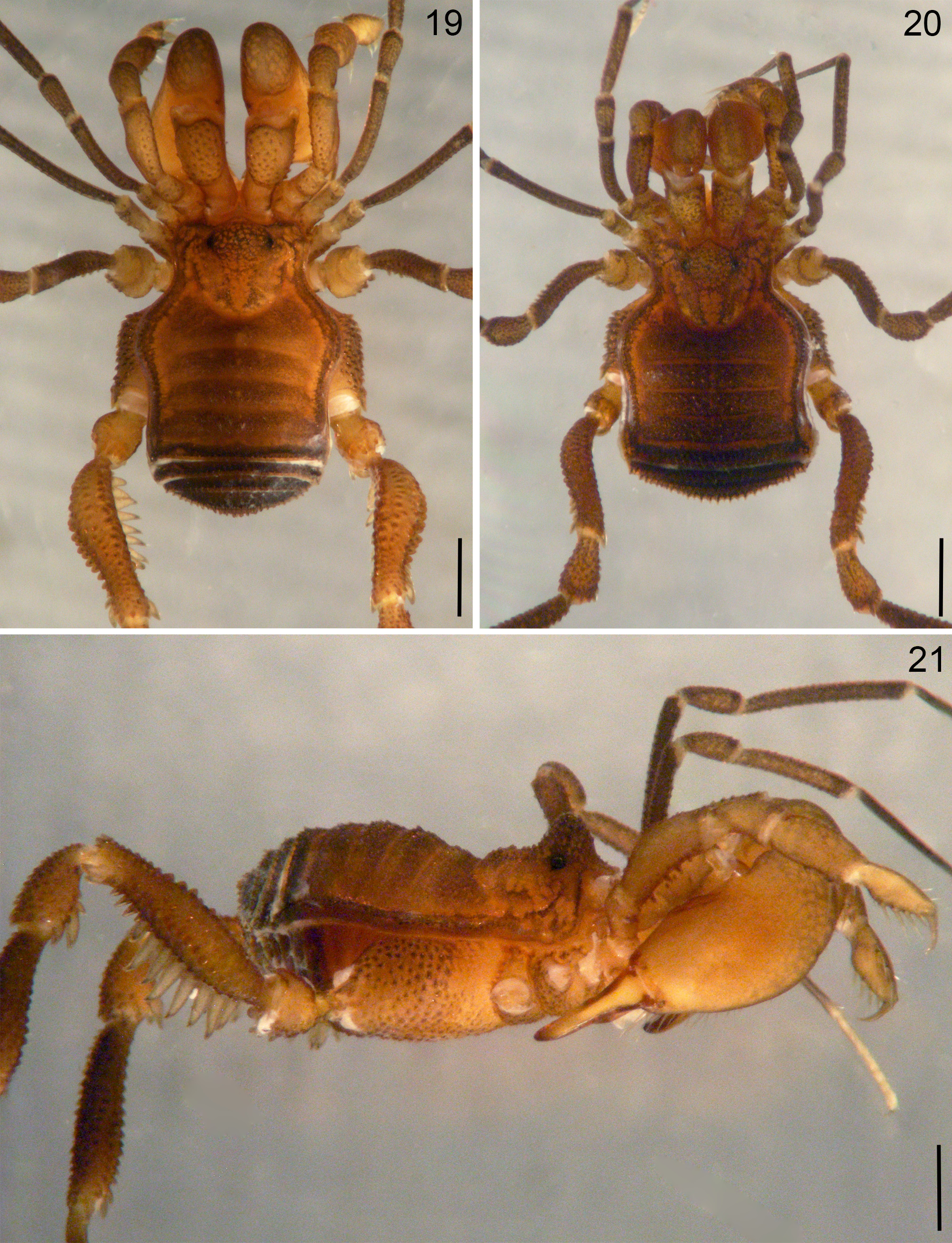
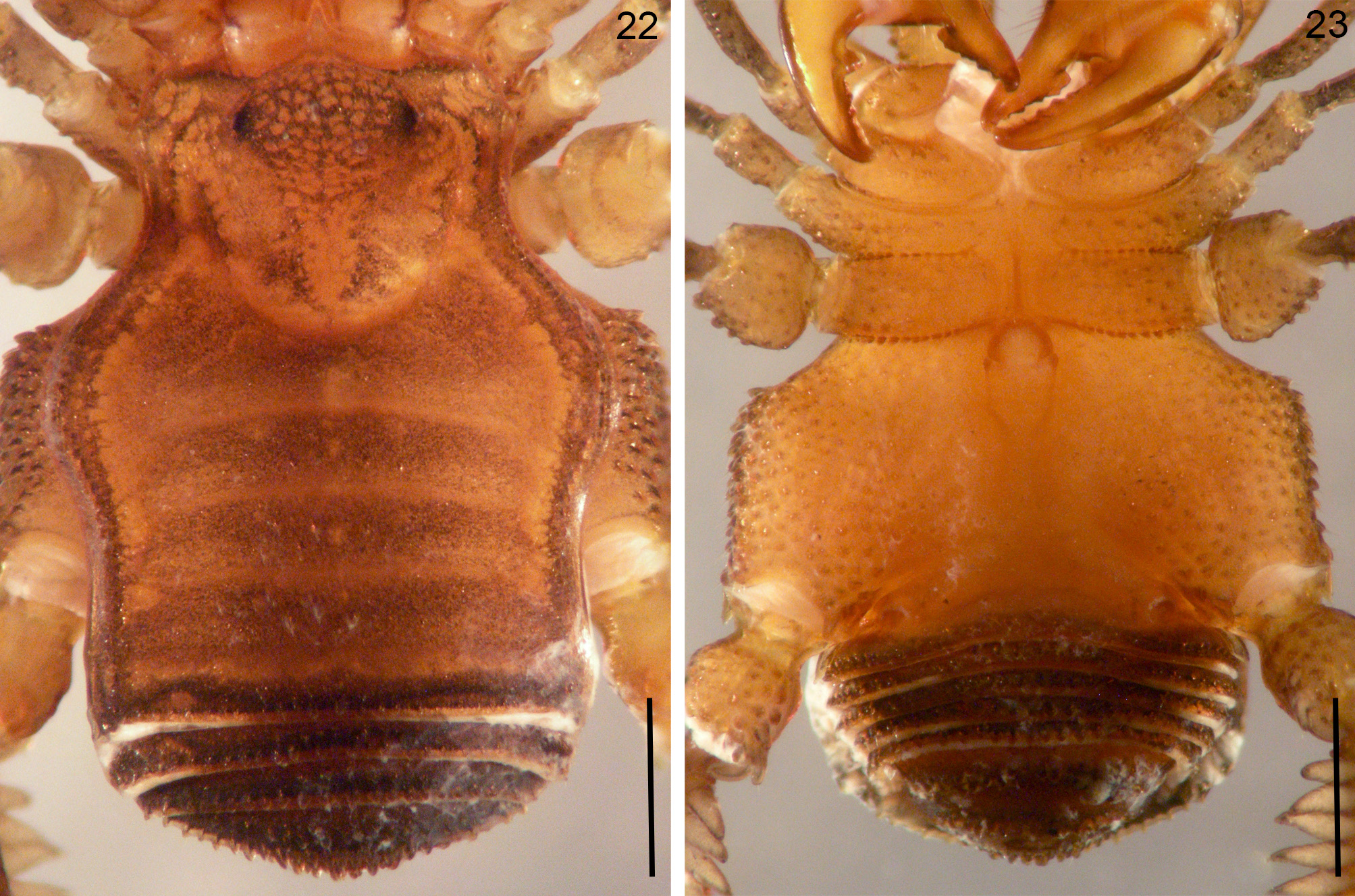
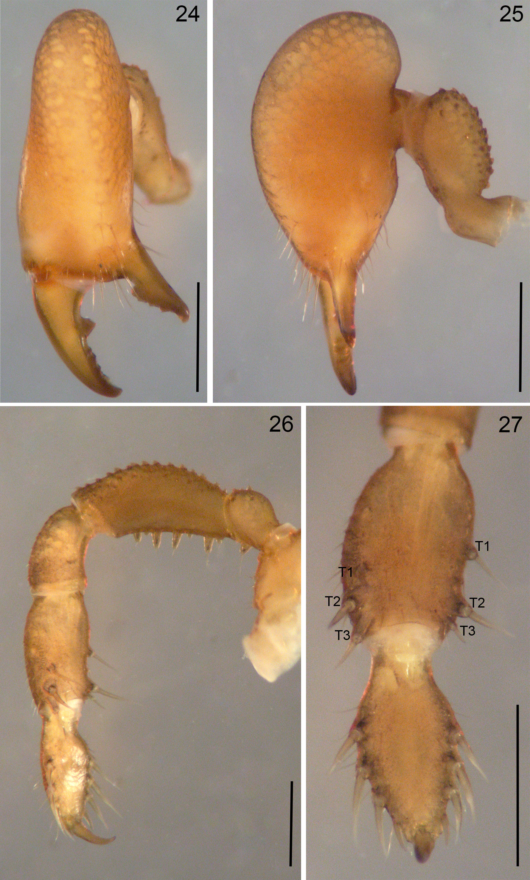
( Figs. 19–36 View FIGURES 19–21 View FIGURES 22, 23 View FIGURES 24–27 View FIGURES 28–33 View FIGURES 34–36 )
Type material. Holotype male ( CNAN-T01330 ), La Laguna , San Pedro Jocotipac, 17º43’37.7’’N, 97º6’6.1’’W, Oaxaca State, Mexico, 23.viii.2015, O. Francke, J. Cruz, D. Barrales, R. Monjaraz, J. Sánchez leg. GoogleMaps Five male and six female paratypes ( CNAN-T01331 ), same data as holotype GoogleMaps . Two male, two female and an immature paratypes ( CNAN-T01332 ), El Tanque , San Pedro Jocotipac, 17º44’34.5’’N, 97º6’20.2’’W, Oaxaca State, Mexico, 23.viii.2015, O. Francke, J. Cruz, D. Barrales, R. Monjaraz, J. Sánchez leg GoogleMaps . Five male and a female paratypes ( CNAN-T01333 ), La Joya , San Pedro Jocotipac, 17º45’58.1’’N, 97º6’4.2’’W, Oaxaca State, Mexico, 23.ix.2015, J. Sánchez leg. GoogleMaps
Etymology. The word ‘cuicateca’ means people from the region of Cuicatlán in northern Oaxaca, and where the type locality is found; the specific name is treated as an adjective in feminine form.
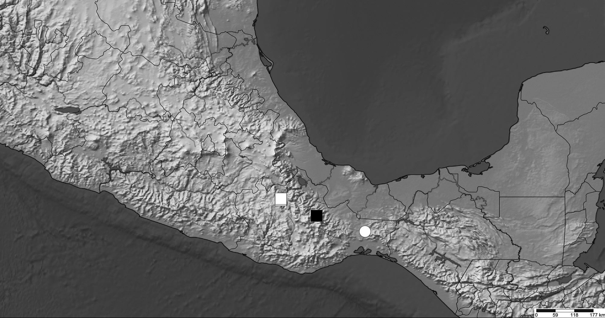
Distribution. Only known from the type locality and nearby records of the paratypes ( Fig. 57 View FIGURE 57 ).
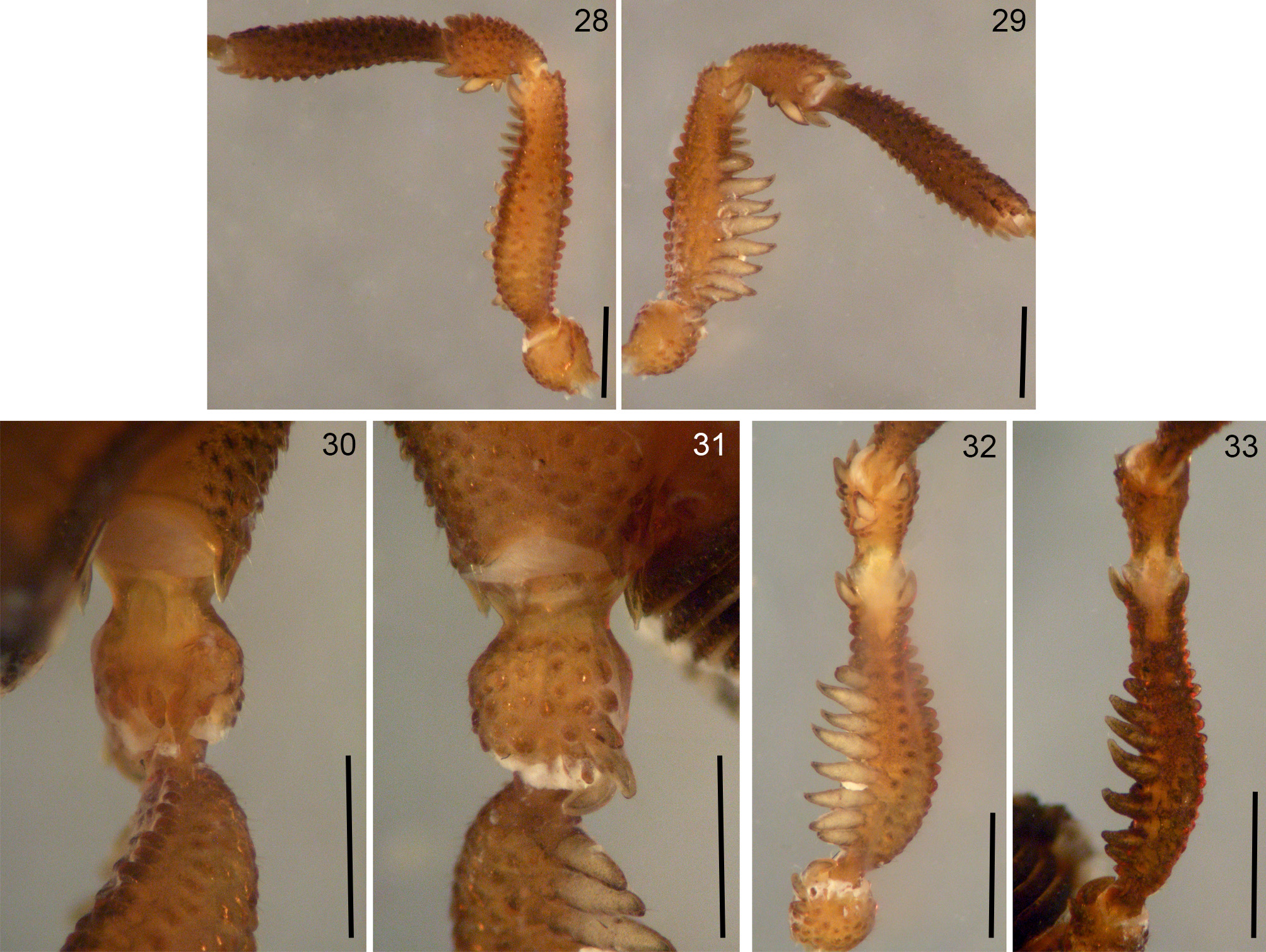
Diagnosis. Guelaguetzia cuicateca sp. nov. can be recognized from G. serrana sp. nov. by the following combination of characters: fixed finger of chelicera with a basal notch ( Fig. 24 View FIGURES 24–27 ), pedipalpal femur with four large major spiniform setiferous tubercles ( Fig. 26 View FIGURES 24–27 ), retrolateral apophysis of coxa IX spiniform ( Fig. 23 View FIGURES 22, 23 ), dorso-ectal apophysis on trochanter IV slightly larger than the central one ( Fig. 30 View FIGURES 28–33 ), ventro-distal apophysis on trochanter IV slender and very curved ( Fig. 31 View FIGURES 28–33 ), patella IV with two fused ventral apophyses ( Fig. 32 View FIGURES 28–33 ), ventro-prolateral row of tubercles of femur IV formed by small tubercles only ( Figs. 28 and 32 View FIGURES 28–33 ), and apical margin of flimsy lamina on male penis bilobed ( Fig. 36 View FIGURES 34–36 ).
Description. Measurements of the holotype, scutum length: 4.1, scutum width at level of the mid-bulge: 3.3, RSC: 1.1, RHW: 2.4.
Dorsum ( Figs. 19, 21 View FIGURES 19–21 , 22 View FIGURES 22, 23 ): Scutum type zeta (ζ) with the mid-bulge rounded, coda I deep and marked, coda II soft. Mesotergal areas with small central tubercles only, lateral margins of scutum with a continuous row of lateral pegs; two rows forming a “V” posterior to ocularium, the latter densely covered with small tubercles. Ocularium rounded and well-developed, prosoma with a soft hump behind ocularium. Free tergites with a transverse row of spiniform tubercles.
Venter ( Fig. 23 View FIGURES 22, 23 ): Venter densely covered with spiniform tubercles, slightly larger on coxa I. Coxa III slightly larger than coxa II. Coxa IV is the largest, occupying the greatest area in ventral view, sub-quadrangular shaped, with the ectal margins projected beyond the scutum margin; lateral sides with many spiniform tubercles, retrolateral apophyses spiniform. Stigmatic area short and compressed in the middle. Free sternites with a transverse row of spiniform tubercles, and anal plate covered with small tubercles.
Chelicerae ( Figs. 24, 25 View FIGURES 24–27 ): Basichelicerite with a long bulla, well-marked, dorsally covered by many spiniform tubercles. Cheliceral hand swollen, inserted with the basichelicerite near the middle portion. Cheliceral dentition heterogeneous, basal tooth of the movable finger blunt and displaced from the base of the finger, posteriorly with four small, contiguous teeth apically; fixed finger with a basal notch, followed by four contiguous teeth, the basalmost largest.
Pedipalps ( Figs. 26, 27 View FIGURES 24–27 ): Trochanter rounded, with a large ventral spiniform apophysis, dorsally with a few spiniform tubercles. Femur slightly compressed laterally and softly curved, dorsally ornate with many spiniform tubercles pointing forward, ventrally with a row of four long spiniform setiferous tubercles; between them are small setiferous tubercles. Patella unarmed, only with few spiniform tubercles dorsally. Tibia rectangular in cross section, with three (III, 1=2=3) and three (iIi, 2> 1> 3) major setiferous tubercles on both margins, respectively, these tubercles on ectal margin are confined to the apical portion, on the mesal margin the basalmost is in the middle, the last two apically and very close to each other. Both margins of tarsus with four major setiferous tubercles, decreasing in size distally. Tarsal claw shorter than tarsus.
Legs ( Figs. 28–33 View FIGURES 28–33 ): Measurements in Table 2 View TABLE 2 . Legs I and II slender, without armature and covered only by small tubercles. Trochanter III not globose, barely larger than trochanter II. Trochanter IV slightly globose, with two spiniform apophyses dorsally, the ectal slightly larger; ventrally with three mesodistal apophyses, increasing in size distally and gradually curved from the basal-most to the apical-most. Femur IV thickened, curved and ornate with longitudinal rows of tubercles, with the two ventral rows more prominent, the retrolateral row has eight very large spiniform tubercles, followed distally by four small tubercles and ending in apical spine; prolateral row formed by small tubercles similar in size and ending in a apical spine also. Patella with apical apophyses on retro- and prolateral faces, and with two fused spiniform apophyses ventrally. Tibia slightly curved in the middle, slightly swollen distally, covered by longitudinal rows of spiniform tubercles, the two ventral barely larger, both ending in an apical spine. Tarsal count: 5(2):9(3):6:6.
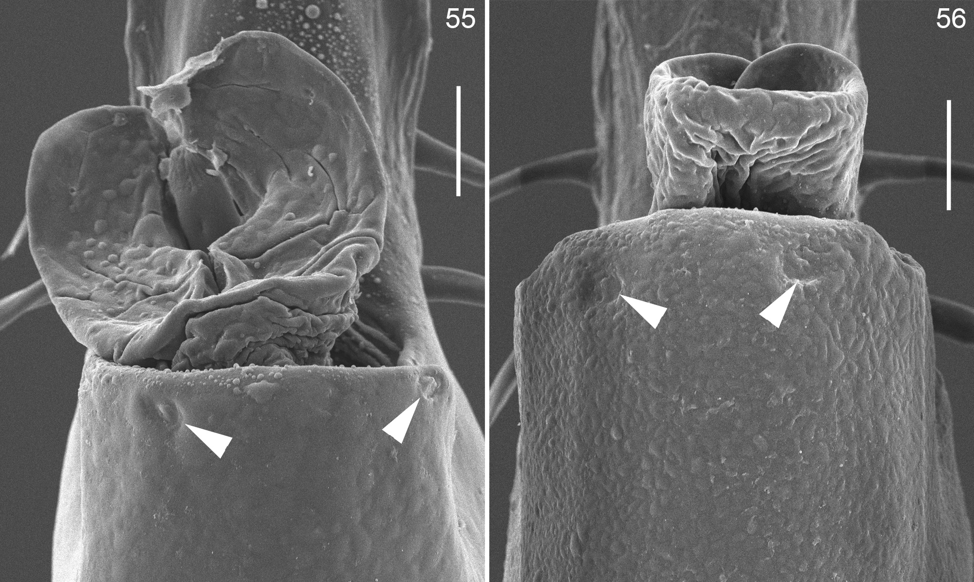
Male genitalia ( Figs. 34–36 View FIGURES 34–36 , 55 View FIGURES 55–56 ): Pars distalis compressed laterally, somewhat rectangular in lateral view, with the flimsy lamina contiguous, undifferentiated and very large, with the apical margin forming two small lobes. Follis emerging from the cavity formed at the base of flimsy lamina, hidden and with two rounded lobes covering the stylus. With a remnant pair of macrosetae D on the dorsal edge of pars distalis, in front of follis; with two pairs of filiform macrosetae C at the base of flimsy lamina; a pair of filiform macrosetae B in the middle of par distalis, slightly displaced to ventral portion; two pairs of filiform macrosetae A on the dorso-basal portion of par distalis; a pair of remnant sockets of macrosetae E on flimsly lamina, just distal to macrosetae C.
Female. Sexual dimorphism very noticeable: body and chelicerae smaller, and legs IV with the femora thinner and armature less developed ( Figs. 20 View FIGURES 19–21 , 33 View FIGURES 28–33 ), RSC=1.5 (n=9). Leg measurements in Table 2 View TABLE 2 .

No known copyright restrictions apply. See Agosti, D., Egloff, W., 2009. Taxonomic information exchange and copyright: the Plazi approach. BMC Research Notes 2009, 2:53 for further explanation.
|
Kingdom |
|
|
Phylum |
|
|
Class |
|
|
Order |
|
|
Family |
|
|
Genus |