
Heteroscydmus Franz
|
publication ID |
https://doi.org/ 10.11646/zootaxa.3722.2.7 |
|
publication LSID |
lsid:zoobank.org:pub:5D21B65C-1917-4513-BDF2-168835BEC884 |
|
DOI |
https://doi.org/10.5281/zenodo.6154648 |
|
persistent identifier |
https://treatment.plazi.org/id/BF5B3062-FFDF-FFE1-F99D-5C964233EAFB |
|
treatment provided by |
Plazi |
|
scientific name |
Heteroscydmus Franz |
| status |
|
Heteroscydmus Franz View in CoL
Heteroscydmus Franz, 1980: 208 . Type species: Heteroscydmus yapacariensis Franz, 1980 (monotypy).
Revised diagnosis. Male and female: head about as long as broad, with vertex not expanded dorso-caudad; occipital constriction only slightly narrower than vertex; thick and long bristles absent on head and prothorax; frontoclypeal groove absent; submentum not demarcated laterally from hypostomae by sutures; maxillary palpomere III strongly elongate; antennae with distinctly delimited club composed of antennomeres IX–XI; pronotum without lateral edges; base of pronotum with fine transverse groove, without pits and without sub-lateral carinae; basisternal part of prosternum much shorter than procoxal cavities; prosternum with rudimentary intercoxal process developed as a low (i.e., only slightly expanding ventrally) subtriangular expansion between procoxal cavities; prothoracic hypomeral ridges complete; pronotosternal sutures visible only along sides of basisternal part of prosternum; mesoventral intercoxal process long, narrow and moderately strongly expanding ventrally (but not keel-shaped); mesoventrite with asetose lateral impressions behind anterior ridge, without setose impressions; mesothorax without lateral foveae; mesocoxal projection without posterior lobe; metacoxae narrowly separated by metaventral intercoxal process composed of a pair of long spines; each elytron with two rudiments of asetose basal fovea. Male: aedeagus asymmetrical, with free parameres.
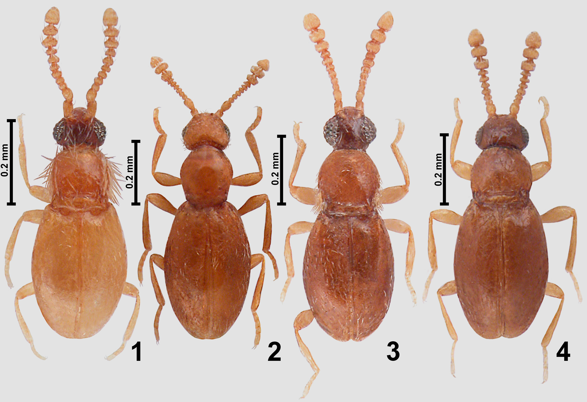
Redescription. Body of male ( Fig. 2 View FIGURES 1 – 4 ) strongly convex, elongate and slender, with moderately long appendages, BL 0.70–0.73 mm; cuticle glossy, light brown, vestiture fine.
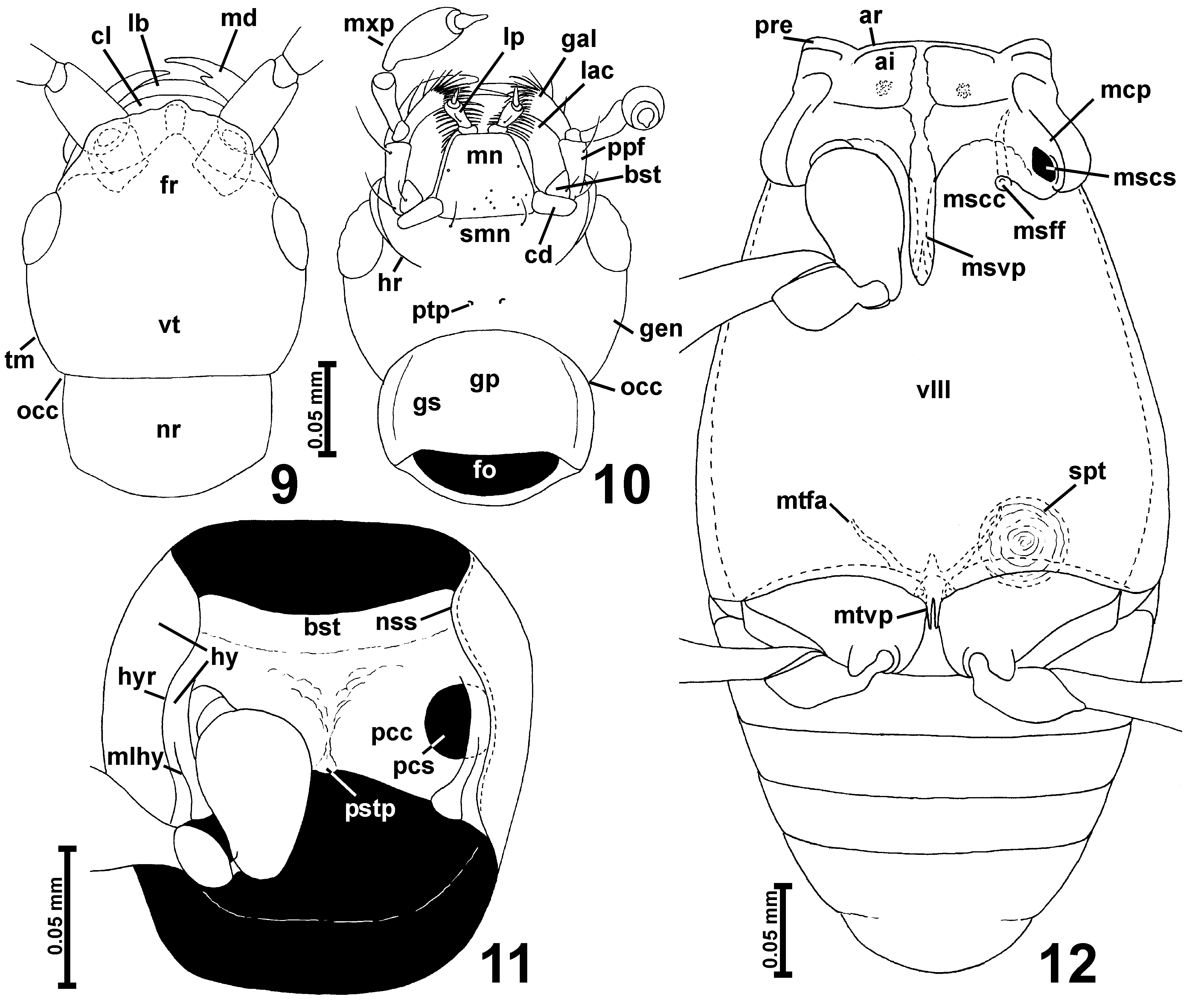
Head ( Figs. 2 View FIGURES 1 – 4 , 9–10 View FIGURES 9 – 12 ) with anterior part (in front of occipital constriction) about as long as broad, with moderately large eyes; occipital constriction ( Figs. 9–10 View FIGURES 9 – 12 ; occ) only slightly narrower than vertex; tempora ( Fig. 9 View FIGURES 9 – 12 ; tm) long and slightly convergent caudad, without bristles; vertex ( Fig. 9 View FIGURES 9 – 12 ; vt) broader than long, convex, not projecting dorso-caudad; frons ( Fig. 9 View FIGURES 9 – 12 ; fr) transverse and subtrapezoidal with anterior margin distinctly expanding in middle, posteriorly confluent with vertex; frontoclypeal groove absent; antennal insertions broadly separated.
Labrum transverse with rounded anterior margin. Mandibles ( Fig. 9 View FIGURES 9 – 12 ; md) symmetrical, with slender and curved distal parts, one mesal tooth located sub-apically; prostheca in the studied specimen not visible. Each maxilla ( Fig. 10 View FIGURES 9 – 12 ) with subtriangular basistipes ( Fig. 10 View FIGURES 9 – 12 ; bst), elongate galea ( Fig. 10 View FIGURES 9 – 12 ; gal) and lacinia ( Fig. 10 View FIGURES 9 – 12 ; lac) and moderately long maxillary palp ( Fig. 10 View FIGURES 9 – 12 ; mxp) composed of only slightly elongate palpomere I, strongly elongate, pedunculate palpomere II, elongate palpomere III broadest in distal third, and small, elongate, subconical and pointed palpomere IV with distinctly delimited apical part.
Labium ( Fig. 10 View FIGURES 9 – 12 ) with transverse submentum ( Fig. 10 View FIGURES 9 – 12 ; smn) not demarcated posteriorly and fused laterally with postcardinal parts of hypostomae; subtrapezoidal mentum ( Fig.10 View FIGURES 9 – 12 ; mn); and short prementum bearing narrowly separated at bases small 3-segmented labial palps ( Fig. 10 View FIGURES 9 – 12 ; lp). Hypostomal ridges ( Fig. 10 View FIGURES 9 – 12 ; hr) short and evenly arcuate.
Gular plate ( Fig. 10 View FIGURES 9 – 12 ; gp) large and indistinctly narrowing anterad; gular sutures ( Fig. 10 View FIGURES 9 – 12 ; gs) superficial; posterior tentorial pits ( Fig. 10 View FIGURES 9 – 12 ; ptp) small but distinct, located at base of submentum, anterior to transverse arcuate impression delimiting ventrally 'neck region' from anterior part of head.
Antennae ( Fig. 1 View FIGURES 1 – 4 ) short in relation to body, with distinctly delimited club composed of antennomeres IX–XI.
Pronotum ( Fig. 1 View FIGURES 1 – 4 ) in dorsal view oval with weakly arcuate anterior and posterior margins and sides rounded in anterior half and nearly straight in posterior third, anterior and posterior corners rounded and indistinct; marginal and sub-lateral carinae absent; base of pronotum with fine and arcuate transverse groove, without pits; sides of pronotum without bristles.
Prosternum ( Fig. 11 View FIGURES 9 – 12 ) with short basisternal part ( Fig. 11 View FIGURES 9 – 12 ; bst) indistinctly demarcated from procoxal cavities ( Fig. 11 View FIGURES 9 – 12 ; pcc) by fine arcuate carina; median part of sternum with weakly developed prosternal intercoxal process visible as a subtriangular slightly convex area between procoxal cavities; procoxal sockets ( Fig. 11 View FIGURES 9 – 12 ; pcs) closed by broad postero-lateral lobes of prosternum; hypomera ( Fig. 11 View FIGURES 9 – 12 ; hy) elongate, divided into broad lateral parts and narrow internal (adcoxal) parts, adcoxal parts of hypomera anteriorly fused with prosternum, so that pronotosternal sutures ( Fig. 11 View FIGURES 9 – 12 ; nss) visible only along sides of basisternal parts of prosternum; hypomeral ridges ( Fig. 11 View FIGURES 9 – 12 ; hyr) complete, anteriorly connected to pronotosternal sutures; internal parts of hypomera with long median longitudinal grooves ( Fig. 11 View FIGURES 9 – 12 ; mlhy).
Mesonotum very small, subtrapezoidal, with slightly concave lateral margins of subtriangular mesocutellum barely visible between bases of elytra; mesoscutoscutellar suture absent.
Mesoventrite ( Fig. 12 View FIGURES 9 – 12 ) with narrow anterior ridge ( Fig. 12 View FIGURES 9 – 12 ; ar); mesoventral intercoxal process ( Fig. 12 View FIGURES 9 – 12 ; msvp) narrow and moderately expanded ventrally, anteriorly connected with anterior ridge and with subtriangular posterior margin, without posterior arms; asetose lateral impressions ( Fig. 12 View FIGURES 9 – 12 ; ai) present, each with porous field near middle; mesanepisternum with short prepectus ( Fig. 12 View FIGURES 9 – 12 ; pre) and posterior part largely hidden in ventral view; mesepimeron not visible in ventral view; sides of mesothorax without foveae; mesocoxal projections ( Fig. 12 View FIGURES 9 – 12 ; mcp) with mesocoxal sockets ( Fig. 12 View FIGURES 9 – 12 ; mscs) located on their mesoventral surface, without posterior lobes.
Metaventrite ( Fig. 12 View FIGURES 9 – 12 ; vIII) slightly longer than wide, anteriorly fused with mesoventrite, posteriorly moderately shallowly bisinuate and with narrow median metaventral intercoxal process ( Fig. 12 View FIGURES 9 – 12 ; mtvp) composed of two long spines. Metanepisterna and metepimera narrow.
Metafurca ( Fig. 12 View FIGURES 9 – 12 ) with very short and broad stalk and divergent lateral furcal arms ( Fig. 12 View FIGURES 9 – 12 ; mtfa).
Elytra ( Fig. 2 View FIGURES 1 – 4 ) oval, each with two rudimentary and asetose basal foveae located in shallow basal impression; humeral calli well-marked and developed as longitudinal protuberances; elytral apices unmodified, separately rounded.
Hind wings well-developed, about twice as long as elytra.
Legs ( Figs. 2 View FIGURES 1 – 4 , 11–12 View FIGURES 9 – 12 ) moderately long and slender; procoxae subglobose, mesocoxae elongate, metacoxae transverse; all trochanters short; all femora weakly clavate; tibiae short and slightly thickening distally; tarsi short and stout.
Abdominal sternites ( Fig. 12 View FIGURES 9 – 12 ) unmodified, suture between VII and VIII barely marked.
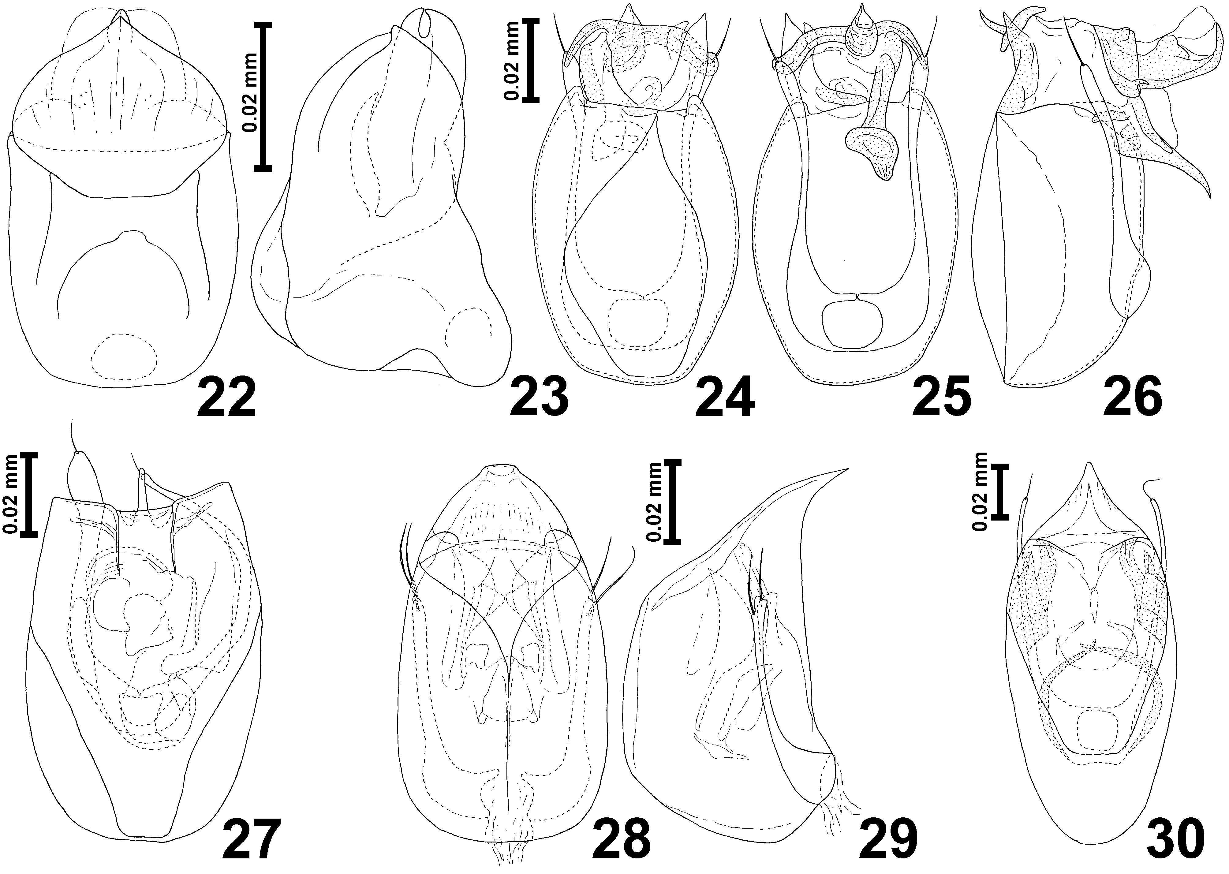
Aedeagus ( Figs. 24–27 View FIGURES 22 – 30 ) with symmetrical, lightly sclerotized and thin-walled median lobe, with complex and asymmetrical set of internal sclerites, parameres present, not fused with median lobe, with apical setae.
Spermatheca ( Fig. 12 View FIGURES 9 – 12 ; spt) globular, darkly sclerotized, located in metathorax.
Distribution and composition. Heteroscydmus is represented by a single species known from the central part of Bolivia ( Fig. 36 View FIGURES 35 – 38 ).
Remarks. Heteroscydmus in the first paragraph of Franz's description (Franz 1980) was compared to Pseudoeudesis , a genus of Scydmaenini , but in the further parts of the text it was correctly assigned to Cyrtoscydmini and stated to be closely similar to Microscydmus . One of characters that Franz discussed to place Heteroscydmus in Cyrtoscydmini and not Scydmaenini is the apical margin of the scape, which according to Franz is not emarginate. In fact, the distal margin of the scape in Heteroscydmus is relatively deeply emarginate dorsally ( Fig. 9 View FIGURES 9 – 12 ); this character as a feature to distinguish Scydmaenini (often with deeply emarginated or notched scape) from Cyrtoscydmini (usually with shallowly emarginate or straight apical margin) was recently discussed by Jałoszyński (2012). Interestingly, also in Microscydmus the apical margin of the scape is deeply emarginate. The shape of maxillary palpomere IV, ventral structures of the prothorax and the aedeagus suggest that the placement of Heteroscydmus in Cyrtoscydmini was correct. Similarly to Microscydmus and Microraphes , Heteroscydmus also has the internal parts of prothoracic hypomera anteriorly fused with the prosternum, so the pronotosternal sutures are visible only along sides of the basisternal part of prosternum, and in ventral view it seems like the notosternal sutures were continuous with the hypomeral ridges that separate the internal part of each hypomeron from the external part, which is merely a lateral deflexed part of the pronotum.
Heteroscydmus clearly differs from Microscydmus in the following characters: the posterior tentorial pits located distinctly anterior to transverse impression that ventrally demarcates the 'neck region' from the anterior part of head ( Fig. 10 View FIGURES 9 – 12 ) (pits in the impression in Microscydmus ); base of pronotum with short and narrow transverse groove (two pairs of pits in Microscydmus ); internal parts of prothoracic hypomera with long median longitudinal hypomeral grooves ( Fig. 11 View FIGURES 9 – 12 ) (grooves absent in Microscydmus ); lack of bristles on sides of pronotum (bristles present in Microscydmus ); the mesoventrite without setose impressions (present and fused in middle in Microscydmus ); the mesoventral intercoxal process connected anteriorly with the anterior ridge of mesoventrite (anterior margin of process distant from the anterior ridge in Microscydmus ); and each elytron with two asetose rudiments of basal foveae (one large and setose fovea in Microscydmus ). Major differences between Microscydmus and all genera treated in the present paper are compiled in Table 1.
Heteroscydmus yapacariensis Franz ( Figs. 2 View FIGURES 1 – 4 , 9–12 View FIGURES 9 – 12 , 24–27 View FIGURES 22 – 30 , 32 View FIGURES 31 – 34 , 36 View FIGURES 35 – 38 )
Heteroscydmus yapacariensis Franz, 1980: 208 , Fig. 195.
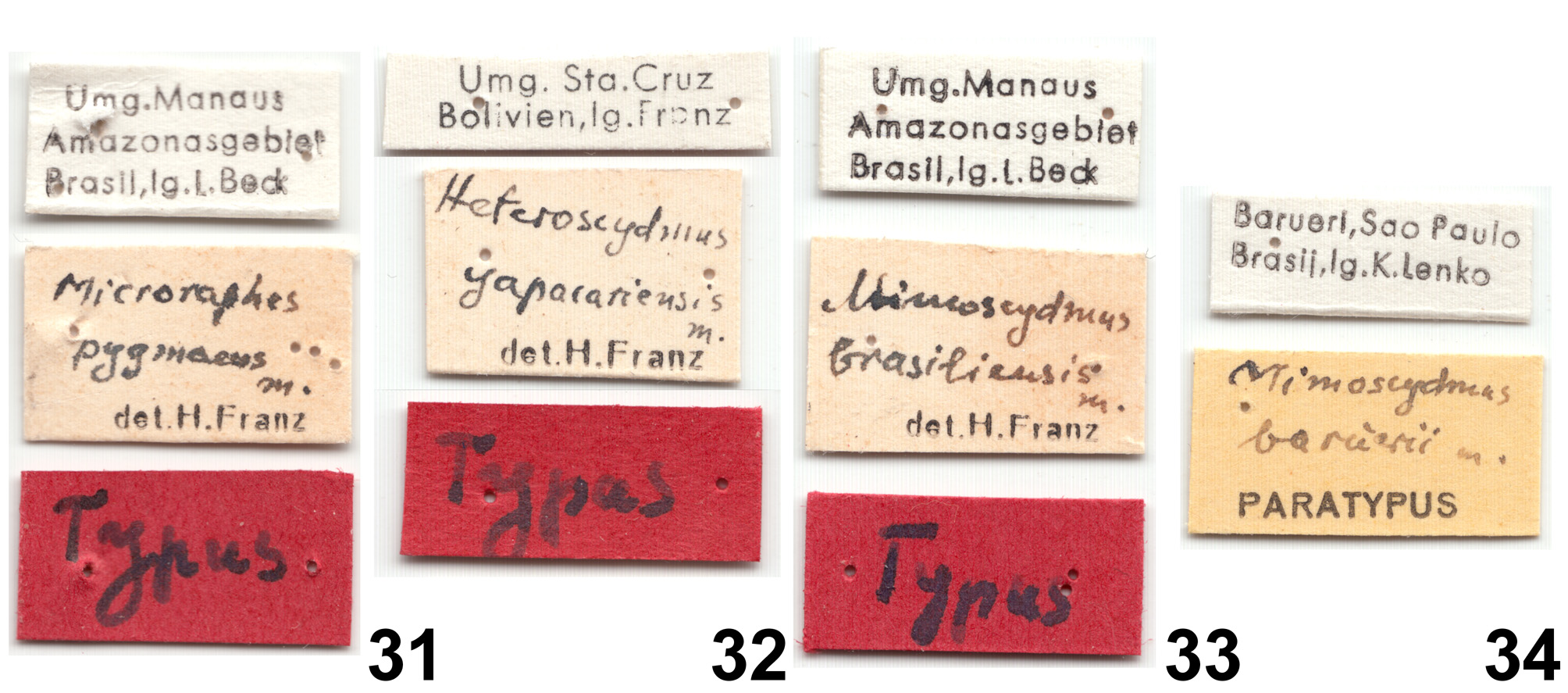
Material studied. Holotype: ♂: three labels ( Fig. 32 View FIGURES 31 – 34 ): "Umg. Sta. Cruz / Bolivien, lg. Franz" with "SA142" on the reverse side [white, printed; reverse handwritten], " Heteroscydmus / yapacariensis / m. / det. H. Franz" [white, handwritten and printed], " Typus " [red, handwritten] (NHMW). Paratypes: 1 ♀, 1 ♂: same data as for holotype, except for yellow " Paratypus " label with the female and a yellowish identification label with the male, without annotation stating the paratype status of this specimen (NHMW).
Diagnosis. This is the only known species of Heteroscydmus and can be identified on the basis of the generic characters and the aedeagus.
Redescription. Body of male ( Fig. 2 View FIGURES 1 – 4 ) strongly convex, elongate and slender, with moderately long appendages, BL 0.70 mm; cuticle glossy, body uniformly light brown, vestiture yellowish.
Head subtrapezoidal, broadest at eyes, HL 0.13 mm, HW 0.15 mm; tempora about as long as eyes, slightly convergent caudad; vertex and frons confluent, strongly convex and together about as long as broad; supraantennal tubercles indistinct. Punctures on head dorsum fine and sparse, inconspicuous; setae short, sparse and suberect. Antennae slender but strikingly short in relation to body ( Fig. 2 View FIGURES 1 – 4 ), with distinctly delimited club composed of antennomeres IX–XI, AnL 0.23 mm; antennomere I–II elongate, III–VIII each slightly transverse; IX–X strongly transverse; XI slightly broader than long, with subconical, blunt apex.
Pronotum in dorsal view oval, broadest near anterior third, PL 0.18 mm, PW 0.15 mm; anterior margin weakly arcuate; lateral margins strongly rounded in anterior half and nearly straight in posterior third, strongly convergent toward obtuse and blunt hind pronotal angles; posterior margin weakly arcuate; base of pronotum with short an narrow but distinct transverse ante-basal arcuate groove convex anteriorly. Punctures on pronotal disc fine and inconspicuous; setae sparse, thin, short and suberect.
Elytra oval, distinctly more convex than pronotum, broadest near middle, EL 0.40 mm, EW 0.28 mm, EI 1.45; humeral calli distinct; basal impressions shallow and short, basal foveae in a dry mount indiscernible; elytral apices separately rounded. Punctures on elytral disc slightly more distinct than those on pronotum but still fine and inconspicuous; setae short, sparse and suberect. Hind wings well-developed, about twice as long as elytra.
Legs moderately long and slender, without modifications.
Aedeagus ( Figs. 24–27 View FIGURES 22 – 30 ) elongate, AeL 0.05 mm, median lobe oval with truncate apex, internal sclerites asymmetrical and complex; parameres slender, each with single apical seta.
Female. Similar to male but differing in slightly smaller eyes, and in consequence tempora longer than eyes ( Fig. 9 View FIGURES 9 – 12 ). BL 0.73 mm; HL 0.13 mm, HW 0.15 mm, AnL 0.23 mm; PL 0.18 mm, PW 0.15 mm; EL 0.43 mm, EW 0.30 mm, EI 1.42.
Distribution ( Fig. 36 View FIGURES 35 – 38 ). Central Bolivia, Santa Cruz Department.
Remarks. The type series reported by Franz (1980) consisted of a holotype male and three paratypes of an unspecified sex. The type series preserved at NHMW consists of a male holotype, one female and two males, one of them incomplete and entirely embedded in euparal. The aedeagus of the holotype male is in an erected condition ( Figs. 24–26 View FIGURES 22 – 30 ) and was certainly not illustrated by Franz; his figure (1980, Fig. 195) was apparently based on the non-erected aedeagus of one of the paratype males ( Fig. 27 View FIGURES 22 – 30 ). The other male during the present study was found to belong to a different species and even genus than the holotype. It is a Microscydmus or Mimoscydmus , but its aedeagus is distorted to a degree that makes a species identification impossible, so ventral details of this specimen were not examined.
Franz named this species " yapacariensis " after the Rio Yapacani river in Bolivia, misspelled as "Yapacari" in his paper (Franz 1980). In the original description Franz (1980) gives further data related to the collecting site of the type series: a gallery forest along Rio Yapacani (as Yapacari), western Montero, forest litter and rotten wood of fallen trees.

No known copyright restrictions apply. See Agosti, D., Egloff, W., 2009. Taxonomic information exchange and copyright: the Plazi approach. BMC Research Notes 2009, 2:53 for further explanation.