
Itagonia medvedevi, Shi, Ai-Min, Yuan, Shi-Bin & Wu, Chun-Lian, 2010
|
publication ID |
https://doi.org/ 10.5281/zenodo.197440 |
|
DOI |
https://doi.org/10.5281/zenodo.6201091 |
|
persistent identifier |
https://treatment.plazi.org/id/03A3879B-CE32-2B4F-7C91-F8FB39ACFD0F |
|
treatment provided by |
Plazi |
|
scientific name |
Itagonia medvedevi |
| status |
sp. nov. |
Itagonia medvedevi , sp. nov.
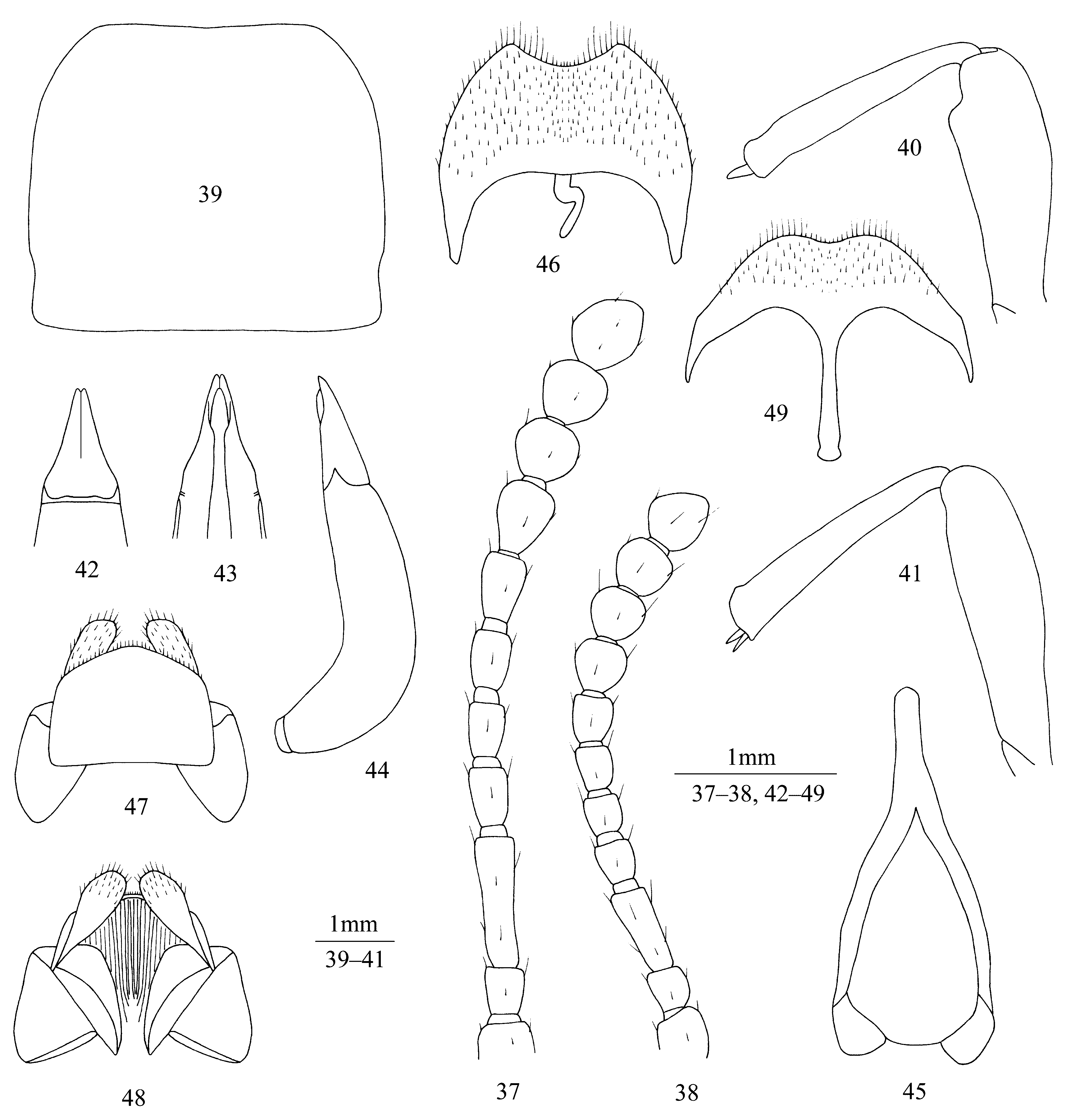
( Figs. 37–49 View FIGURES 37 – 49 , 61–62 View FIGURES 61 – 64. 61 – 62 )
Type material. Holotype: ɗ, CHINA: Sichuan, Maerkang, 31º53' N, 102º16' E, 2770 m, 30 Jul. 2009, collected by Ai-min Shi and Chun-lian Wu ( MCWNU). Paratypes: 1 ɗ and 1 Ψ, same data as the holotype ( MCWNU).
Diagnosis. The new species resembles Itagonia cordiformis Shi & Ren , with the following differences: Antennae long, when posteriorly extended, reaching pronotal base. Pronotum weakly transverse, 1.11–1.17 times as wide as long. Outer margin of epipleura visible from above in anterior 2/5 and apical part. Ventral surface of protarsomeres 1 to 3 with hair brushes. Parameres with outer margins sinuate in basal 1/3.
Etymology. This new species is named in honour of Gleb S. Medvedev, the great tenebrionidologist professor of the Zoological Institute, Russian Academy of Sciences, St. Petersburg.
Description. Body black, shining.
Male ( Figs 37, 39–46 View FIGURES 37 – 49 ). Anterior margin of clypeus weakly sinuate. Lateral margin of head with obtuseangled shallow incision above antennal base. Outer genal margin arcuately converging to clypeal base. Vertex with uniform punctures. Frontoclypeal suture moderately distinct. Antennae ( Fig. 37 View FIGURES 37 – 49 ) long, when posteriorly extended, reaching pronotal base. Length (width) ratio of antennomeres 2 to 11 as follows: 14 (12): 39(12): 17(12): 16(12): 17(12): 19(13): 21(17): 18(19): 18(19): 23(19).
Pronotum ( Fig. 39 View FIGURES 37 – 49 ) slightly transverse, 1.11–1.17 times as wide as long, 1.51–1.67 times as wide as head. Ratio of pronotal width at anterior margin to its maximum width and width at base (n=2) 0.59: 1.00: 0.96, on average. Lateral margins of pronotum arcuately converging to anterior margin in anterior 1/3, sinuate or nearly linear immediately before the base, finely bordered along entire length. Anterior margin of pronotum very weakly sinuate, bordered laterally; base straight, not bordered. Anterior angles of pronotum obtuse, rounded apically; posterior ones nearly rectangular. Pronotal surface between lateral margins convex, with punctures finer at disc than laterally, median depression clear. Propleura with longitudinal wrinkles and sparse granules. Prosternum in front of procoxae oblique; intercoxal process of prosternum with median depression, steeply sloping behind procoxae.
Elytra elongate-oval, 1.37–1.55 times as long as wide, widest before the middle, 1.44–1.52 times as wide as pronotum. Anterior 2/5 and apical part of outer margin of epipleura visible from above. Elytral surface between outer margin of epipleura and sutural margin convex, with shallow punctures and irregular wrinkles. Epipleura surface smooth, with fine wrinkles and very sparse granules. Visible abdominal sternites with punctures and brown setae, 1 to 3 visible abdominal sternites with longitudinal wrinkles.
Legs robust, length (width) ratio of pro-, meso- and metafemora 83(25): 89(22): 100(23); that of corresponding tibiae: 71(11): 69(14): 94(16). Upper edge of inner surface of profemur forming obtuse-angled prominence with rounded apex ( Fig. 40 View FIGURES 37 – 49 ). Protibiae with massive upper spur at apical margin, lower spur fine and pointed. Ventral surface of protarsomeres 1 to 3 with hair brushes, that of mesotarsomere 1 with apical tuft of pale hairs. Mesofemur and mesotibiae as in Fig. 41 View FIGURES 37 – 49 . Metatibiae weakly incurved. Length (width) ratio of metatarsomeres 1 to 4 as follows: 25(8.3): 15(8.2): 15(7.5): 25(7.5).
Aedeagus ( Figs 42–44 View FIGURES 37 – 49 ): length 2.31 mm, width 0.71 mm. Parameres 0.71 mm long and 0.47 mm wide, with outer margins sinuate in basal 1/3, and apical part regularly narrowing towards apex. Spiculum gastrale as in Fig. 45 View FIGURES 37 – 49 . Apical margin of abdominal inner sternite 8 sinuate ( Fig. 46 View FIGURES 37 – 49 ).
Female ( Figs 38, 47–49 View FIGURES 37 – 49 ). Body longer and wider. Antennae ( Fig. 38 View FIGURES 37 – 49 ) shorter than in male, when posteriorly extended, reaching beyond the middle of pronotum. Upper spur of fore tibiae massive and rounded apically; lower spur missing. Ventral surface of pro- and mesotarsomeres without hair brushes or apical tuft of pale hairs. Ovipositor as in Figs 47–48 View FIGURES 37 – 49 . Spiculum ventrale as in Fig. 49 View FIGURES 37 – 49 .
Measurements. Male body length 12.2–12.9 mm, width 5.5–6.2 mm; female body length 12.6 mm, width 6.6 mm.
Distribution. China: Sichuan (Maerkang).

No known copyright restrictions apply. See Agosti, D., Egloff, W., 2009. Taxonomic information exchange and copyright: the Plazi approach. BMC Research Notes 2009, 2:53 for further explanation.