
Kebira uteoides Row, 1909
|
publication ID |
https://doi.org/ 10.11646/zootaxa.4426.1.1 |
|
publication LSID |
lsid:zoobank.org:pub:18929E20-5296-4458-8A8A-4F5316A290FD |
|
DOI |
https://doi.org/10.5281/zenodo.5966772 |
|
persistent identifier |
https://treatment.plazi.org/id/386CC616-DCFB-A5BD-FF67-8C71FEA4FF4C |
|
treatment provided by |
Plazi |
|
scientific name |
Kebira uteoides Row, 1909 |
| status |
|
Kebira uteoides Row, 1909 View in CoL
Figs 85a–g View FIGURE 85 , 86a–e View FIGURE 86
Kebira uteoides Row, 1909: 210 View in CoL , pl. 20 figs 8–9, text-figs 7–8; Burton 1952: 164; Ilan & Vacelet 1993: 110, figs 2–4.
Material examined. RMNH Por. 9563, Saudi Arabia, Jeddah, near Thuwal, Al Fahal N, 22.300194°N 38.959583°E, depth 12 m, coll. N.J. de Voogd, scuba, field nr. THU04/JED052, 8 November 2014 GoogleMaps ; RMNH Por. 9664, Saudi Arabia, Jeddah , near Thuwal, Um Albalam, 22.193556°N 38.9475°E, depth 15 m, scuba, coll. N.J. de Voogd, field nr. THU10/JED168, 12 November 2014 GoogleMaps .
Description. Squat, laterally flattened, irregularly tubular individuals ( Figs 85a–d View FIGURE 85 ), attached to vertical walls and hanging down from the ceiling of cavities, with large apical or lateral oscules. In life ( Figs 85a,c View FIGURE 85 ), colors are shades of grey and pinkish white, on deck ( Figs 85b,d View FIGURE 85 ) and in preservation they become yellow. Size up to 4 cm high, and 5 x 3 cm in lateral expansion. Oscules 4–6 mm in diameter. Surface smooth. Consistency firm to hard.
Aquiferous system. Leuconoid.
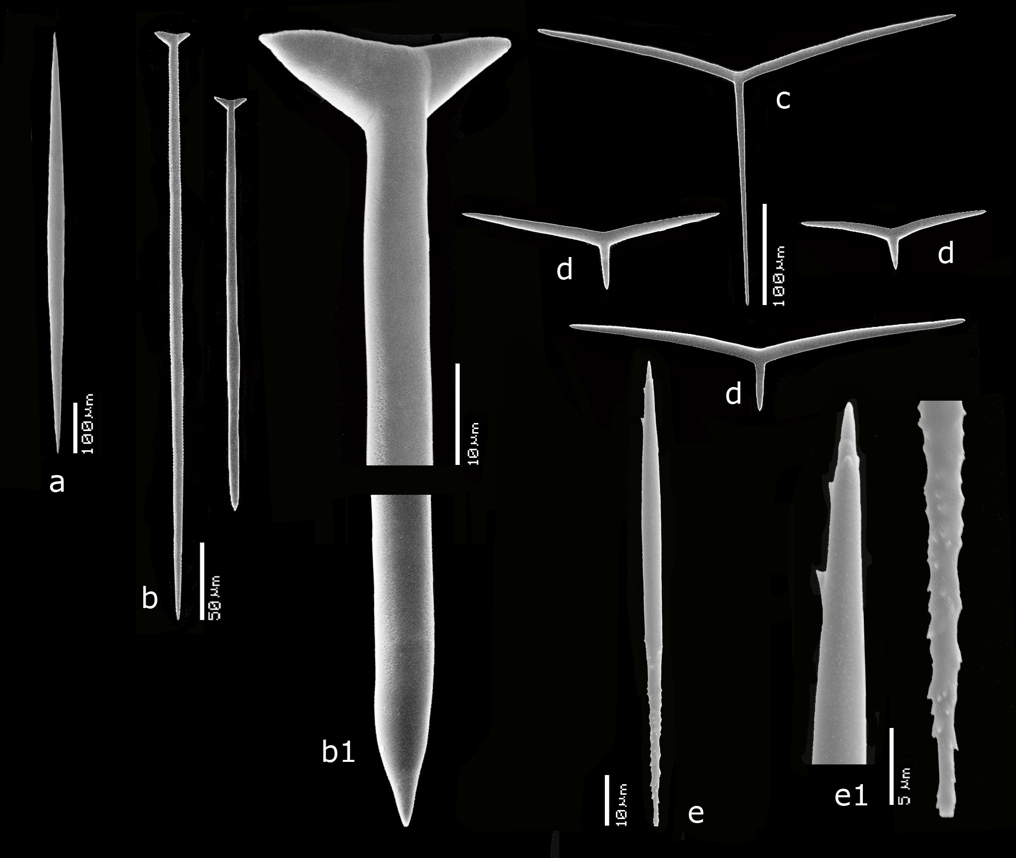
Skeleton. ( Figs 85e–g View FIGURE 85 ) In cross section ( Fig. 85e View FIGURE 85 ), there is a cortical skeleton of sagittal triactines, mostly with long unpaired actines ( Fig. 85f View FIGURE 85 ), carried by tangentially arranged giant diactines. Underneath, the choanosomal skeleton is formed by perpendicular giant diactines, tracts of nail-shaped triactines (arrows in Fig. 85e View FIGURE 85 ) and large sagittal triactines with long unpaired actines. The atrial skeleton ( Fig. 85g View FIGURE 85 ) is predominantly formed by sagittal triactines with short unpaired actines.
Spicules. ( Figs 86a–e View FIGURE 86 ) Giant diactines, nail-shaped triactines, sagittal triactines, microdiactines.
Giant and smaller diactines ( Fig. 86a View FIGURE 86 ), fusiform, symmetrical, sharply pointed, in a wide size range, 540– 1489 –2022 x 25– 79 –108 µm.
Nail-shaped triactines ( Figs 86b,b View FIGURE 86 1 View FIGURE 1 ), thinly fusiform, pointed at one end, at the opposite end provided with two conical actines arranged at a slight angle, in a large size range, unpaired actines 105– 317 –477 x 5.5– 8.5 –10 µm, paired actines 3–7 x 3 µm.
Large sagittal triactines ( Fig. 86c View FIGURE 86 ), with long unpaired actines, and flaring paired actines; unpaired actines 306– 371 –462 x 7 – 7.9 –8.5 µm, paired actines 204– 227 –248 x 9 – 10.4 –12 µm.
Atrial sagittal triactines ( Figs 86d View FIGURE 86 ) with short unpaired actines and straight paired actines; unpaired actines 57– 86 –121 x 8.5– 10.1 –12 µm, paired actines 179– 228 –282 x 11.5– 13.9 –17 µm.
Small thin diactines with tiny spines at both ends ( Figs 86e,e View FIGURE 86 1 View FIGURE 1 ), rare, not certainly proper to the sponge, but sufficiently characteristic to be a possible, previously unobserved, spicule type, 51–102 x 2–7 µm.
Distribution and ecology. Northern Red Sea: Gulf of Suez, Egypt (Sharm-al-Sheik), Saudi Arabia (Jeddah), hanging down in caves on shallow water reefs.
Remarks. The only noteworthy difference with Row’s description of the type specimen is that he confused the position of the cortical and the atrial sagittal triactines. It can be clearly seen in our Fig. 85g View FIGURE 85 , showing the atrial surface, that the predominant spicule type is the sagittal triactine with short unpaired actine. In Figs 85e View FIGURE 85 (upper part) and f, the dominant spicule type is the long-shafted sagittal triactine clearly visible. Our spicule sizes are more or less the same as in Row’s specimen, although we did not observe giant diactines of 4 mm length.
The small spined diactines may or may not be proper to the sponge.
Burton (1952) did not give a description other than mentioning that his specimen from Sharm al Sheik ( Egypt) was 2 cm high.
Ilan & Vacelet (1993) found giant diactines up to 4 mm, but omitted further spicule measurements, concentrating on a description of the soft parts.
We obtained partial 28SrRNA sequences for the two specimens. In our Phylogeny of Fig. 3 View FIGURE 3 , these grouped along with the new Kebira species described below in a larger clade together with Ute ampullacea , Paragrantia waguensis Hôzawa, 1940 , and Grantiopsis heroni Wörheide & Hooper, 2003 . These species were added to our analysis to confirm the affiliation of Kebira . The position of Ute ampullacea (cf. also above) in the group is surprising, but Kebira , Grantiopsis and also Paragrantia (see Van Soest et al. 2015) are likely members of the gamily Lelapiidae .


| RMNH |
National Museum of Natural History, Naturalis |
No known copyright restrictions apply. See Agosti, D., Egloff, W., 2009. Taxonomic information exchange and copyright: the Plazi approach. BMC Research Notes 2009, 2:53 for further explanation.
|
Kingdom |
|
|
Phylum |
|
|
Class |
|
|
Order |
|
|
Family |
|
|
Genus |
Kebira uteoides Row, 1909
| Van, Rob W. M. & De, Nicole J. 2018 |
Kebira uteoides
| Row, 1909 : 210 |
| Burton 1952 : 164 |
| Ilan & Vacelet 1993 : 110 |