
Macrostomum zhujiangensis Wang & Fang
|
publication ID |
https://doi.org/ 10.11646/zootaxa.4170.2.4 |
|
publication LSID |
lsid:zoobank.org:pub:666CFB09-4980-448A-9571-1DA0C34DBB00 |
|
DOI |
https://doi.org/10.5281/zenodo.5625949 |
|
persistent identifier |
https://treatment.plazi.org/id/039387E9-1D68-FFCB-4CD4-FE6DFB89FE30 |
|
treatment provided by |
Plazi |
|
scientific name |
Macrostomum zhujiangensis Wang & Fang |
| status |
sp. nov. |
Macrostomum zhujiangensis Wang & Fang View in CoL , n. sp.
( Figs. 2–4 View FIGURE 2 View FIGURE 3 View FIGURE 4 )
Material examined. Observations were made on several live and preserved speciemens. Holotype (PLA– Ma0070): permanent slides of stylet in polyvinyl-lactophenol. Paratype (PLA–Ma0071–76): six serially-sectioned specimen. Paratype (PLA–Ma0077–80): batches of animals in absolute ethanol. The type specimens were collected from a brackish fishpond in the Water-lands Resort in Shenzhen City, Guangdong Province, China (22°43′16″N, 113°46′03″E) ( Fig. 1 View FIGURE 1 ) on April 3rd and 30th, 2015. All specimens are deposited in IZCAS. GoogleMaps
Etymology. The name of this new species is derived from the name of the Zhujiang River Estuary in Guangdong Province, China.
Description. The body is dorsoventrally flattened and milk-white in color. The length and width of the body of active specimens is 1033 ± 76.7 µm (n=5) and 220 ± 41.7 µm (n=5), respectively ( Fig. 2 View FIGURE 2 A), while the length and width of the body of anaesthetized specimens is 912 ± 37.6 µm (n=5) and 257 ± 27.2 µm (n=5), respectively ( Fig. 2 View FIGURE 2 B). Its head and tail are rounded. When swimming, the body is broader anteriorly and narrower posteriorly. The thickness of epidermis is 8.6 ± 2.0 µm (n=5). Epidermal cilia are 8–10 µm (n=5) in length and distributed densely over the body surface. The body edge has tufts of rigid cilia that are 21–28 µm (n=5) in length. Rhabdites are 12–16 µm (n=5) in length and distributed along the dorsal body surface ( Figs. 3 View FIGURE 3 A, 4A–B). A pair of bean-shaped eyes (8.0 ± 0.9 µm in diameter, 25 ± 1.7 µm in eyes distance) is located at the anterior 12% position of the body. A lobeshaped pharynx is situated posterior to the eyes and surrounded by filiform pharyngeal glands, while the pharyngeal gland cell bodies are resided further back laterally on the ventral side of the animal. ( Figs. 2 View FIGURE 2 A–B, 4A). There are well developed adhesive glands at the posterior end of the body.
For the male reproductive system, a pair of testes, similar in size and shape, is located one at each side of the anterior 50% position of intestine. The testes of sexually mature individuals contain a large number of sperm ( Figs. 2 View FIGURE 2 A–B, 4A), which appear as dark regions in the center of the testis. Mature sperms are 130 ± 0.9 µm (n=10) in length. The lengths of feeler, body and shaft of the sperm are from 38–50 µm, 30–34 µm, and 45–48 µm, respectively. The sperm body has a pair of bristles, while a brush is not observed at the posterior end of the shaft ( Figs. 2 View FIGURE 2 E, 4E). The oval-shaped false vesicula seminalis is located at posterior 12% of the body length. The false vesicula seminalis connects to the spherically-shaped and muscular vesicula seminalis at its right edge, while the false vesicula seminalis connects to the vesicula granulorum on the right lower side ( Figs. 2 View FIGURE 2 B–C, 3A–E, 4A–C). The basal part of the C-shaped penis stylet connects to the posterior part of vesicula granulorum ( Figs. 2 View FIGURE 2 C, 4A–C). The curved-line (marked as ‘a’) and straight-line (marked as ‘b’) distances between the basal and distal ends of the penis stylet are 141.4 ± 3.4 µm (n=5) and 93.1 ± 2.4 µm (n=5), respectively ( Figs. 4 View FIGURE 4 D). Diameters of the basal and distal parts of the penis stylet are 13.9 ± 1.3 µm (n=5) and 5.2 ± 0.6 µm (n=5), respectively. The distal end of the penis stylet has neither spine nor thickening ( Figs. 2 View FIGURE 2 D, 4C–D), but has a beveled opening pointing at the male gonopore ( Figs. 2 View FIGURE 2 D, 4C).
For female reproductive system, a pair of ovaries is located one at each side of posterior region of the intestine. The light color or transparent ovary and dark color maturing or developing eggs are situated in an anterior to posterior sequence. ( Figs. 2 View FIGURE 2 A–B). The female antrum is situated at the mid-ventral line posterior to the intestine. The female gonopore, which is the opening of female antrum, is surrounded by cement glands ( Figs. 3 View FIGURE 3 B, 4A).
Phylogenetic analysis. The 18S rDNA phylogenetic analysis using neighbor joining (NJ) and maximum likelihood (ML) methods showed that three specimens of Macrostomum zhujiangensis n. sp. clustered together ( Figs. 5 View FIGURE 5. A , 6 View FIGURE 6. A ), forming a well-supported clade with the other 6 species of Macrostomum . Within the genera/species with sequences for analysis, this new species is more closely related to Bradynectes sterreri ( FJ715298 View Materials ) than the species from the families Dolichomacrostomidae , Microstomidae , or Haplopharyngidae .
Remarks. There are 12 previously described species that are similar to Macrostomum zhujiangensis n. sp. in the morphology of penis stylets (see Tab. 1 View TABLE 1 ). However, the distal ends of the penis stylets are evidently thickened in 8 species, including M. bicaudatum , M. clavituba , M. curvituba , M. gallicum , M. guttulatum , M. johni , M. lutheri and M. subterraneum ( Tab. 1 View TABLE 1 ). In M. bicurvistyla , M. semicirculatum and M. caprariae , although the distal ends of the penis stylets are non-thickened, the penis stylet openings are non-beveled. As for M. zhujiangensis n. sp., the wall of the distal region of penis stylet is not thickened and penis stylet opening is bevel-shaped, therefore the penis stylets of the above 8 species are easily distinguished from that of M. zhujiangensis n. sp.
M. obtusum is most similar to the M. zhujiangensis n. sp. in overall morphology of penis stylet. Their penis stylets both have a C-shape, without a thickening at the distal end. In addition, the distal openings of their penis stylet are similarly beveled. However, they showed obvious differences in their overall length, diameters of different regions of the penis stylets and bending angle. In M. obtusum , the length of penis stylet is 7 5–90 µm and the diameter of basal part of the penis stylet is approximately 18 µm. In contrast, the penis stylet of M. zhujiangensis n. sp. is about 140 µm in length and the diameter of basal part of penis stylet is approximately 14 µm. In M. obtusum , the diameter of the penis stylet changes abruptly in the anterior 33% position, while in M. zhujiangensis n. sp., diameter of the corresponding region is relatively constant. Furthermore, the bending angle of penis stylet in M. zhujiangensis n. sp. is clearly larger than that of M. obtusum . In addition, specimens of M. obtusum are much longer (2.0 mm) than M. zhujiangensis n. sp. (950 µm). Finally, M. obtusum lives in freshwater and M. zhujiangensis n. sp. lives in brackish-water. Together with the results of 18S rDNA phylogenetic analysis, it is evident that M. zhujiangensis n. sp. is a new species within the genus Macrostomum .
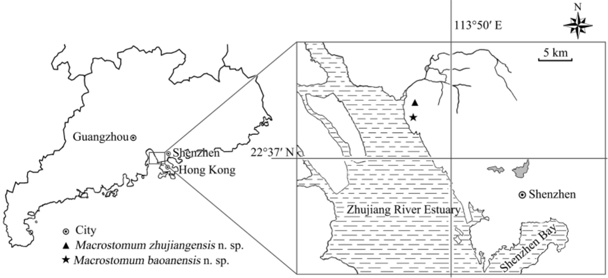
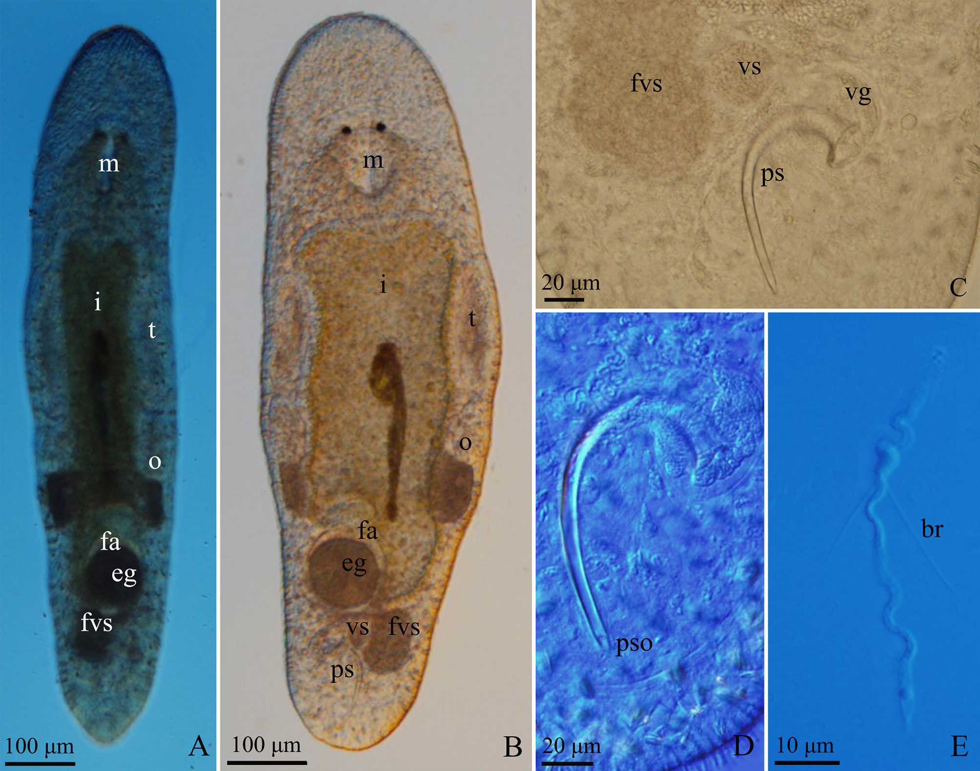

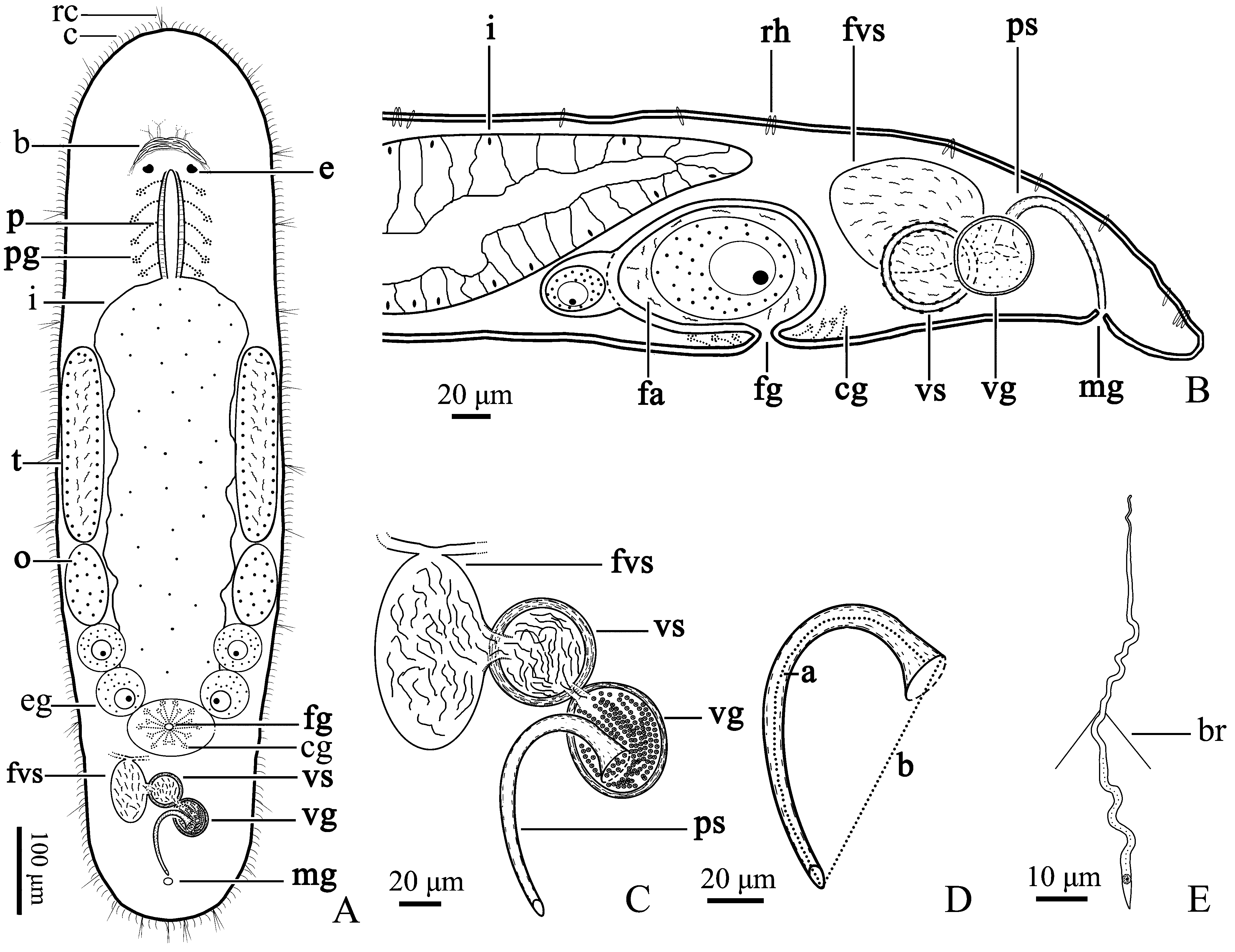


TABLE 1. Comparison of the stylets among Macrostomum zhujiangensis n. sp. and other 12 similar species in genus Macrostomum.
| Total Length of stylet (µm) | Diameter of basal stylet (µm) | Features of stylet distal opening | Habitat | Locality | Reference |
|---|---|---|---|---|---|
| M. zhujiangensis n. sp. 93.1 ± 2.4 | 13.9 ± 1.3 | Beveled Non-thickening | Brackish-water | China | This study |
| M. bicaudatum 160 | 11 | Beveled Lower margin thickened | Freshwater | China | Sun et al. 2015 |
| M. bicurvistyla 105–148 | 20–25 | Non-beveled Non-thickening | Brackish-water | Germany | Armonies & Hellwig 1987 |
| M. caprariae 106 | 38 | Non-beveled Non-thickening | Freshwater | Italy | Papi 1959 |
| M. clavituba 70–75 | 11–14 | Non-beveled Lower margin thickened | Brackish-water | France | Ax 2008 |
| M. curvituba 89–91 | 12–15 | Non-beveled Thickening | Brackish-water Freshwater | Finland | Luther 1947 |
| M. gallicum 65 | 10 | Non-beveled Thickened | Brackish-water | France | Ax 2008 |
| M. guttulatum 80–100 | 15–18 | Non-beveled Lower margin thickened | Brackish-water | Japan | Ax 2008 |
| M. johni 80–98 | 25–33 | Swollen with thickened wall | Freshwater | UK | Young 1972 |
| M. lutheri 210 | 50 | Beveled Lower margin thickened | Freshwater | Finland | Beklemischev 1927 |
| M. obtusum 75–90 | 18–27 | Beveled Non-thickening | Freshwater | Czechia | Vejdovsky 1895 |
| M. semicirculatum 80–100 | 20 | Non-beveled Non-thickening | Brackish-water | Japan | Ax 2008 |
| M. subterraneum 115 | 28 | Non-beveled Thickened | Freshwater | Germany | Rixen 1961 |
| IZCAS |
Institute of Zoology, Chinese Academy of Sciences |
No known copyright restrictions apply. See Agosti, D., Egloff, W., 2009. Taxonomic information exchange and copyright: the Plazi approach. BMC Research Notes 2009, 2:53 for further explanation.
|
Kingdom |
|
|
Phylum |
|
|
Class |
|
|
Order |
|
|
Family |
|
|
Genus |