
Magelona filiformis Wilson, 1959
|
publication ID |
https://doi.org/ 10.11646/zootaxa.4767.1.4 |
|
publication LSID |
urn:lsid:zoobank.org:pub:7B5EC00B-44DA-4A09-8B0A-3DCA78CD7C37 |
|
DOI |
https://doi.org/10.5281/zenodo.3799100 |
|
persistent identifier |
https://treatment.plazi.org/id/039487E1-FFD3-1567-FF37-41B918A13914 |
|
treatment provided by |
Carolina |
|
scientific name |
Magelona filiformis Wilson, 1959 |
| status |
|
Magelona filiformis Wilson, 1959 View in CoL
Figures 7–9 View FIGURE 7 View FIGURE 8 View FIGURE 9 , Table 1 View TABLE 1
Magelona filiformis minuta: Wilson (1959) View in CoL (juvenile M. filiformis View in CoL specimens, see Mills & Mortimer 2018 and Fig. 10 View FIGURE 10 )
Material examined. ENGLAND, Mill Bay , Salcombe : Holotype ( BMNH 1959.4.2.1 , c, slide mounted); paratypes ( BMNH 1959.4 .2.2, af, slide mounted; BMNH 1959.4.2.2-5, 4af, fluid preserved, poor preservation; BMNH 1959.4.2.6–10, 6af, fluid preserved). East Portlemouth, Mill Bay ( NMW.Z.2003.0.35.2003, c) 16.05.2003.
Diagnosis. A moderate species. Prostomium longer than wide, with a squared anterior margin. Chaetigers 1–8 with slender, smooth-edged, filiform lamellae, with superior dorsal lobes. Lamellae of chaetiger 9 triangular, without dorsal lobes. Thoracic chaetigers with capillary chaetae only. Abdominal lateral lamellae spatulate. Hooded hooks tridentate, in two groups, vis-à-vis.
Dimensions. Holotype complete (mounted on two slides: slide 1, majority of specimen; slide 2, posterior fragment of eight chaetigers and pygidium): prostomium 0.7 mm long (not possible to measure width of prostomium or thorax due to orientation on slide); thorax 4.5 mm long (including prostomium); abdomen 0.45 mm wide; 143 chaetigers. Slide mounted paratype (BMNH 1959.4.2.2): prostomium 0.75 mm long, 0.47 mm wide; thorax 0.36 mm wide, 5.45 mm long (including prostomium); abdomen 0.3 mm wide; 119 chaetigers long. Other paratypes 14–51.5 mm for 32–78 chaetigers (width measurements not including parapodia).
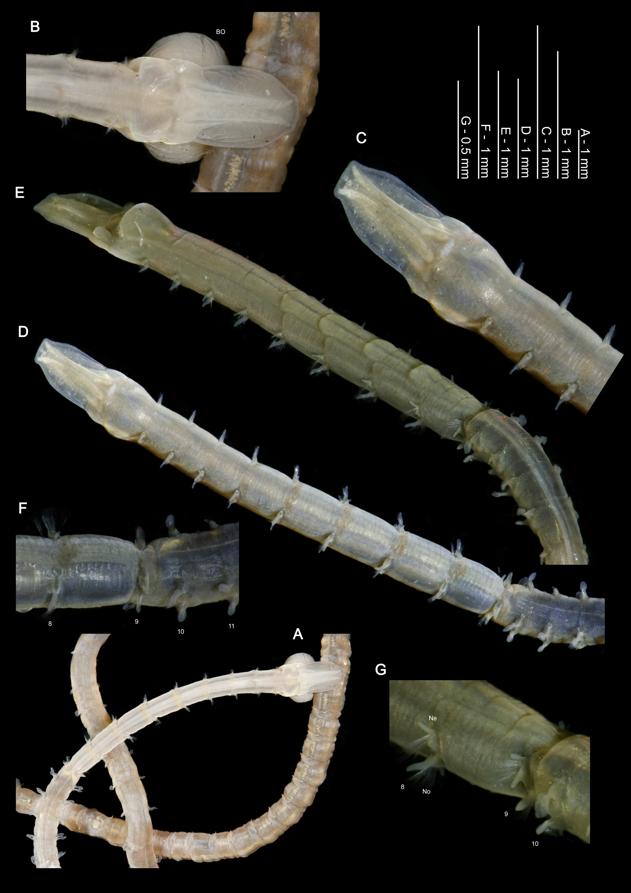
Description. A slender to moderate species, very long; thorax of similar width (when viewed dorsally, Figs 7A, D View FIGURE 7 ) but marginally thinner than abdomen (when viewed laterally, Figs 7E, G View FIGURE 7 ), difference between the two regions not marked ( Figs 7F, G View FIGURE 7 ).
Prostomium longer than wide (L:W ratio 1.6), with a squared anterior margin (often termed “rudimentary horns” but not as distinct as in other species), anterior margin smooth ( Figs 7 View FIGURE 7 A–E). Prostomium with two pairs of prominent longitudinal dorsal muscular ridges, relatively thin. Inner pair abutting medially but diverging at either end, outer pair less distinct, shorter and thinner, abutting inners for entire length. Faint markings present on either side of ridges ( Fig. 7B View FIGURE 7 ), indistinct, but generally present.
Burrowing organ either partially or fully everted on holotype ( Figs 8A, B View FIGURE 8 ) and five paratypes (BMNH 1959.4.2.6–10, Figs 7A, B View FIGURE 7 ), oval when partially everted, heart-shaped when fully everted. Longitudinally ridged inferiorly, appearing smooth superiorly. Both palps retained on holotype ( Figs 8A, B View FIGURE 8 ), long (approximately 5.2 and 7.5 mm in length) and slender ( Figs 8C, D View FIGURE 8 ). Palps of slide mounted paratype approximately 6.8–8.9 mm in length. Two further paratypes with palps retained. Papillae ampulliform ( Fig. 8D View FIGURE 8 ); proximally and medially in two rows either side of an inconspicuous ventral region, distally with 1–2 rows either side. Non-papillated region reaching chaetiger ~2 when folded backwards.
Achaetous region behind prostomium approximately one and a half times the size as chaetiger one ( Figs 7 View FIGURE 7 B–D). Chaetigers 1–8 similar ( Figs 7D, E View FIGURE 7 ; 8 View FIGURE 8 E–Q); parapodia biramous with low triangular notopodial prechaetal lamellae confluent with slender triangular lamellae, postchaetal to subchaetal in position. Postchaetal lamellae increase slightly in size along thorax, becoming more sinuous shaped by approximately chaetiger 6. Single, digitiform superior dorsal lobe present on all thoracic chaetigers except chaetiger 9; increasing slightly in size to chaetiger 8. Neuropodia with single slender triangular ventral lamella with pointed tip, directly below each chaetal bundle, marginally longer in mid-thoracic region. Ventral lamellae confluent with low pre- and postchaetal ridges, which encircle the chaetae cuff-like. Postchaetal lamellae of chaetiger 8 ( Fig. 7G View FIGURE 7 ) slightly expanded and triangular.
Notopodial postchaetal lamellae of chaetiger 9 ( Fig. 7F View FIGURE 7 ) triangular, slightly smaller than on preceding chaetiger. Postchaetal lamellae confluent with, and adjoining the low prechaetal lamellae toward distal tips, thus appearing almost lateral in position. Neuropodia ( Figs 7F, G View FIGURE 7 ) with triangular to digitiform postchaetal lamellae, pointing laterally, and appearing behind the upper part of the chaetal bundle, a smaller digitiform process below chaetal bundle on both sides. Thoracic chaetae simple, smooth-edged, winged capillaries, of similar size in both rami; those of the anterior thorax thinner. Distinct reniform ventral swellings, level with parapodia from chaetiger 5 ( Fig. 7E View FIGURE 7 ), paired.
Abdominal chaetigers with broad spatulate lateral lamellae, of about equal size in both rami, basally constricted and stalked ( Figs 7G View FIGURE 7 ; 9 View FIGURE 9 A–C). Lateral lamellae not extended behind chaetal rows ( Fig. 9H View FIGURE 9 ). Large digitiform processes present at inner margins of chaetal rows ( Fig. 9G View FIGURE 9 ), of a similar size to, or slightly shorter than abdominal chaetae in anterior abdomen.
Abdominal chaetae tridentate hooded hooks, superior two fangs parallel above main fang ( Fig. 9G View FIGURE 9 ). Hooks in two groups, main fangs vis-à-vis. Approximately 7–10 hooks per ramus initially, reducing to 5–6 on later chaetigers. Hooded hook adjacent to lateral lamellae smaller than rest ( Fig. 9H View FIGURE 9 ). No aciculae observed. Posteriorly opening pouches at extreme posterior end. Pouches, simple C-shaped and pocket-like, fairly convoluted, pattern irregular but essentially alternating from one side of the body to the other and on alternate chaetigers. Pouches evident on holotype on chaetigers 119 and 121, and on chaetigers 116L, 118R, 120L, 121R, 123L, 126R and 127L of a 135 chaetiger entire specimen (NMW.Z.2003.035.2003). Pygidium with two long, slender anal cirri ( Fig. 9F View FIGURE 9 ), with a small ventral anus.
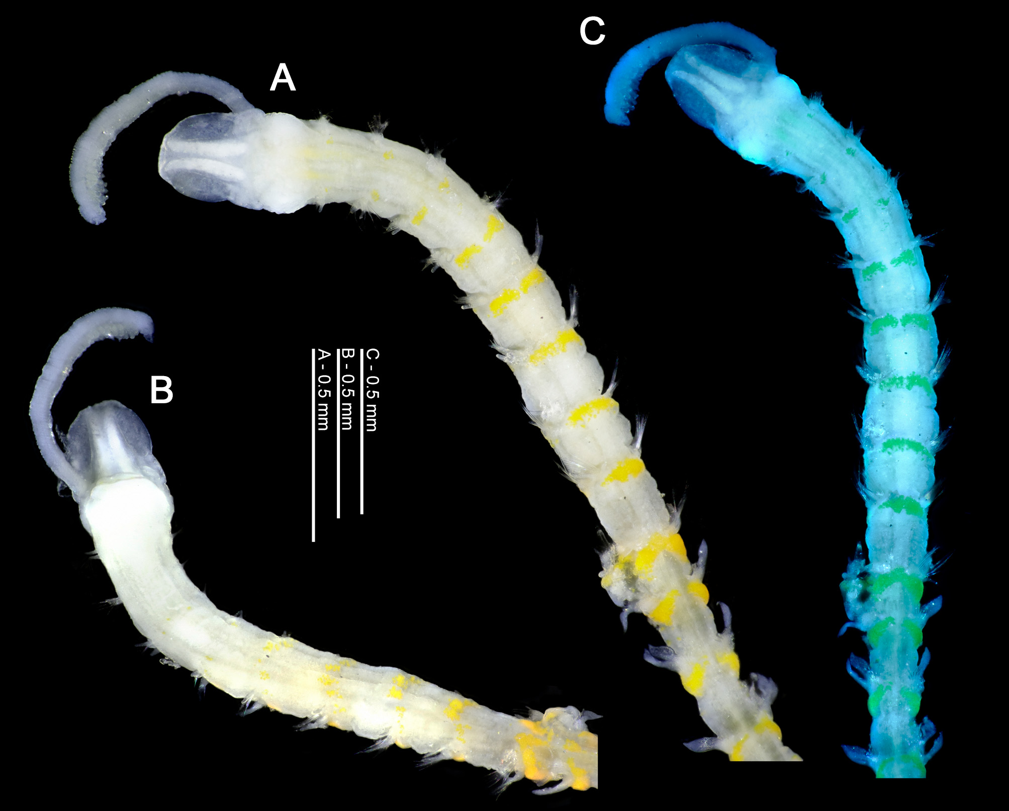
Colour. Live animals pinkish/white in colour, preserved specimens beige/white in alcohol ( Figs 7A, B View FIGURE 7 ). Paratypes (BMNH 1959.4.2.6–10) described as “pink” females ( Figs 7A, B View FIGURE 7 ) and “dark” males ( Figs 7 View FIGURE 7 C–G), although the label suggested “?cork staining” [sic], proposing the dark worms were possibly so because of leaching from the cork that had stoppered the vial. Dark colouration not observed in any other material. Distinct dorsal transverse bands just behind parapodia in the thorax, comprised of cream to yellow speckles, very characteristic of the species ( Figs 7A, D View FIGURE 7 ). On chaetigers 1–4, the band is medially interrupted, appearing as two separate circular areas. Behind the parapodia of chaetiger 9, the speckled areas are the largest and occur on almost half of the segment. Speckles continuing into anterior abdomen. Speckling also present on ventral surface, although not as conspicuous as those of the dorsal surface particularly in the anterior thorax. Interparapodial ‘glandular’ regions apparent as speckled areas ( Fig. 7A View FIGURE 7 ), relatively thin and appearing as a stripe in the posterior thorax. Staining with methyl green shows no clear pattern, although highlights the transverse bands as described above. The transverse bands of the thoracic region stain with Rose Bengal and can be a helpful identifier for this species in benthic samples (but note the similarity of the pattern in M. minuta View in CoL noted by Mills & Mortimer 2018). Dorsally, level with the parapodia of chaetiger 1, the stained buccal region can be seen as a large spot of stain through the epidermis of the specimen. Abdominally, staining behind the parapodia, and patches along the mid ventral line is present.
Habitat. Wilson (1959) stated that M. filiformis “lives in fragile tubes, which may be no more than the walls of burrows lined with a secretion to which sand grains adhere”. Observations of live animals within a laboratory setting by the first two authors, showed that whilst M. filiformis was the most inactive of the species under observation (see also Mortimer & Mackie 2014), as with other magelonid species, they move more or less continuously through the sediment. They do not inhabit burrows for any length of time and do not construct tubes such as is known for species such as M. alleni ( Mills & Mortimer 2019a) and M. equilamellae ( Fig. 4E View FIGURE 4 ). Meissner & Darr (2009) suggested the species in general has a preference for fine sands with low mud content (below 10%), and can be found from the intertidal zone to 45 m. Mortimer & Mackie (2014) collected large numbers of the species in fine sands of Oxwich Bay, South Wales.
Distribution. Distributed around the UK, Irish Sea, North Sea, English Channel, Bay of Biscay to Morocco, Mediterranean Sea.
Remarks. Several differences compared to the original description by Wilson are noted herein. Firstly, Wilson (1959) stated that the species had only one pair of prostomial ridges. However, there is an additional pair of thin out- er ridges alongside the inner pair previously reported. There are light markings either side of the ridges, not drawn or mentioned in the original description. Although indistinct, they are noticeable in the majority of specimens. Wilson, made no mention of the presence of lateral abdominal pouches and Fiege et al. (2000) later stated they were absent in this species. However, as detailed by Mortimer & Mackie (2014), posteriorly open pouches are present in the extreme posterior region of the species. Wilson described the neuropodium of chaetiger 9 as possessing “two fingerlike processes, the dorsal moderately long the ventral short, and 25–30 curved double-winged bristles arising from a setal sac anterior to the processes.” This implies that both lamellae are postchaetal. However, the lower lamella of this chaetiger is ventral in position, directly underneath the chaetal bundle. Furthermore, the “dorsal” neuropodial lamella, is more triangular, wider based than described or drawn by Wilson. The smaller abdominal hooded hook, adjacent to the lateral lamellae is a feature not previously reported for the species, although, hinted at in Wilson’s drawing of abdominal chaetigers.
As highlighted by Mills & Mortimer (2018), M. filiformis shares many morphological similarities with M. minuta (also originally described from north European waters), such as distinctive transverse bands of the thoracic region and a similar prostomial shape. However, they can be distinguished in terms of presence/absence of thoracic superior dorsal lobes (lacking in M. minuta ), dentition of abdominal hooded hooks (bidentate in M. minuta versus tridentate in M. filiformis ) and lamellar shape.
......continued on the next page
......continued on the next page
......continued on the next page
Fiege et al. (2000) stated that the abdominal hooded hooks of M. filiformis were in “one group in each ramus, all facing laterally”. The original description does not state their orientation, with the figure of the 13th parapodium showing the majority of hooks facing laterally, however, those nearest the lamellae, particularly in the neuropodium, appear twisted and facing anteriorly. The hooks of M. filiformis , as shown herein, for type and additional material are not unidirectional as stated by Fiege et al. (2000), they are vis-à-vis in two groups.
Wilson (1959) stated that the first nine pairs of parapodia carry only double-winged bristles, which he stated were most readily seen in formalin preserved material examined in water. His figure 1J showed two adjacent, distinctly bilimbate bristles, although not stating the chaetiger from which they were observed. As noted above the bristles of the anterior thorax are much thinner than those of the posterior thorax. Whilst the latter look distinctly bilimbate, as was noted by Wilson, the limbations of the anterior chaetae are much more difficult to observe and appear unilimbate. Variation in the thickness of chaetae is also evident in the posterior thorax, those toward the edge of the bundle being somewhat thinner. This is something which warrants further investigations, but it appears there is greater variation within the capillary chaetae than was previously reported.


| NMW |
Naturhistorisches Museum, Wien |
No known copyright restrictions apply. See Agosti, D., Egloff, W., 2009. Taxonomic information exchange and copyright: the Plazi approach. BMC Research Notes 2009, 2:53 for further explanation.
|
Kingdom |
|
|
Phylum |
|
|
Class |
|
|
Order |
|
|
Family |
|
|
Genus |
Magelona filiformis Wilson, 1959
| Mortimer, Kate, Mills, Kimberley, Jordana, Esther, Pinedo, Susana & Gil, João 2020 |
Magelona filiformis minuta: Wilson (1959 )
| Wilson (1959 |
| Mills & Mortimer 2018 |