
Magelona heteropoda Mohammed, 1973
|
publication ID |
https://doi.org/ 10.5281/zenodo.198268 |
|
DOI |
https://doi.org/10.5281/zenodo.6208086 |
|
persistent identifier |
https://treatment.plazi.org/id/03BDDD14-E465-5520-FF41-FF55BE8AFE5E |
|
treatment provided by |
Plazi |
|
scientific name |
Magelona heteropoda Mohammed, 1973 |
| status |
|
Magelona heteropoda Mohammed, 1973 View in CoL
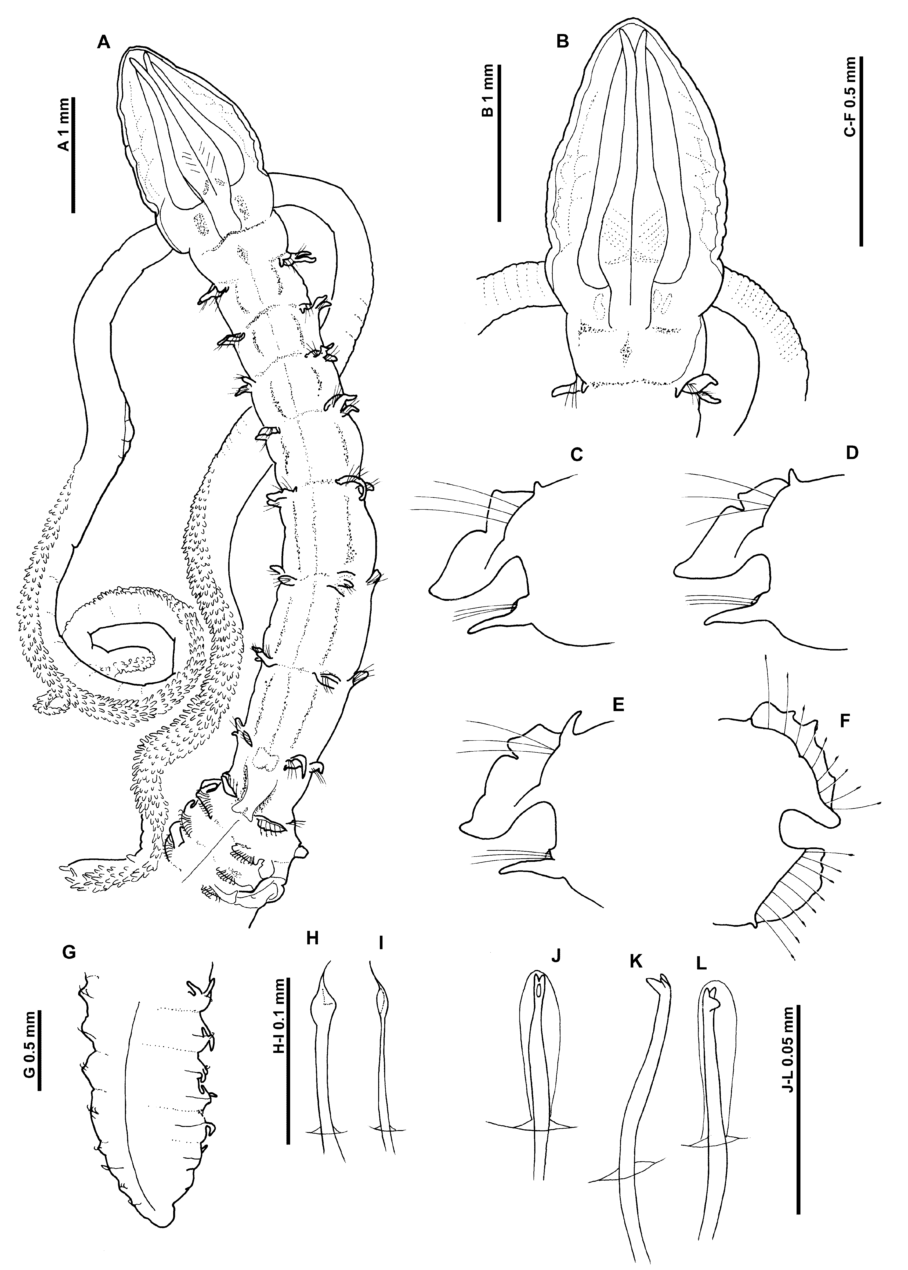
Figures 5–8 View FIGURE 5 View FIGURE 6 View FIGURE 7 View FIGURE 8
Magelona heteropoda Mohammed, 1973: 39 View in CoL –40, fig. 9
Material examined. Abu Halifa, Kuwait, Arabian Gulf (29°08’N, 48°07’E), in sand, intertidal, Holotype ( BMNH:ZB.1971.54, complete in 6 fragments), 26th September 1969, collected by Murad-B. M. Mohammed. (Note: type specimen originally described as being complete, presumably entire, although not stated), unfortunately the specimen fragmented during transit.
Description. A moderately stout species; abdomen marginally wider than thorax ( Fig. 5 View FIGURE 5 A). Holotype, complete (however, see below), extremely fragile (body almost detached at several segments). Anterior fragment, 35.3 mm for 63 chaetigers; prostomium 1.6 mm long, 1.1 mm wide; thorax (including prostomium) 7.1 mm long, 0.8 mm wide; abdomen 0.9 mm wide; four abdominal fragments (5.2 mm for 12 chaetigers, 2.6 mm for 4 chaetigers, 1.2 mm for 2 chaetigers and 4.3 mm for 8 chaetigers); posterior fragment 3.9 mm long for 16 chaetigers; total length 52.5 mm for 106 chaetigers (originally measured as 80 mm). Note: Mohammed (1973) stated “the holotype is complete and consists of two regions: a thorax of nine setigerous segments and an abdomen of about 100 segments”, although the specimen is now fragmented it is believed to be complete. Width measurements are less than those recorded by Mohammed, although Mohammed included the parapodia within his measurements. Chaetiger 3 on both sides, chaetigers 7 and 20 on the left, and the left neuropodium of chaetiger 9 have been previously dissected (presumably by Mohammed, as these are figured in the original description).
Prostomium elongate, much longer than wide (L:W ratio 1.45), slender triangular (less rounded and narrower than figured by Mohammed), without prostomial horns, anterior margin smooth (slightly folded over), rounded, lateral edges minutely undulating; eyes absent. Two pairs of prominent longitudinal dorsal muscular ridges, outer pair (slightly shorter) abutting inners for entire length, inner pair reaching the distal tip of prostomium, where they diverge only very slightly ( Figs 5 View FIGURE 5 A, 6A). Conspicuous, rounded oblong (muscular?) areas either side of ridges. Proboscis everted (not fully), heart–shaped; longitudinally ridged inferiorly, appearing smooth superiorly but with light transverse ridging (partially obscured as proboscis tip not everted). Palps arising ventrolaterally from base of prostomium, fairly robust, reaching around chaetiger 18, non–papillated region reaching chaetiger 3. Papillae short proximally, increasing in size, papillae long distally. Initially 3 rows of papillae either side of inconspicuous ventral groove, distally 2 rows.
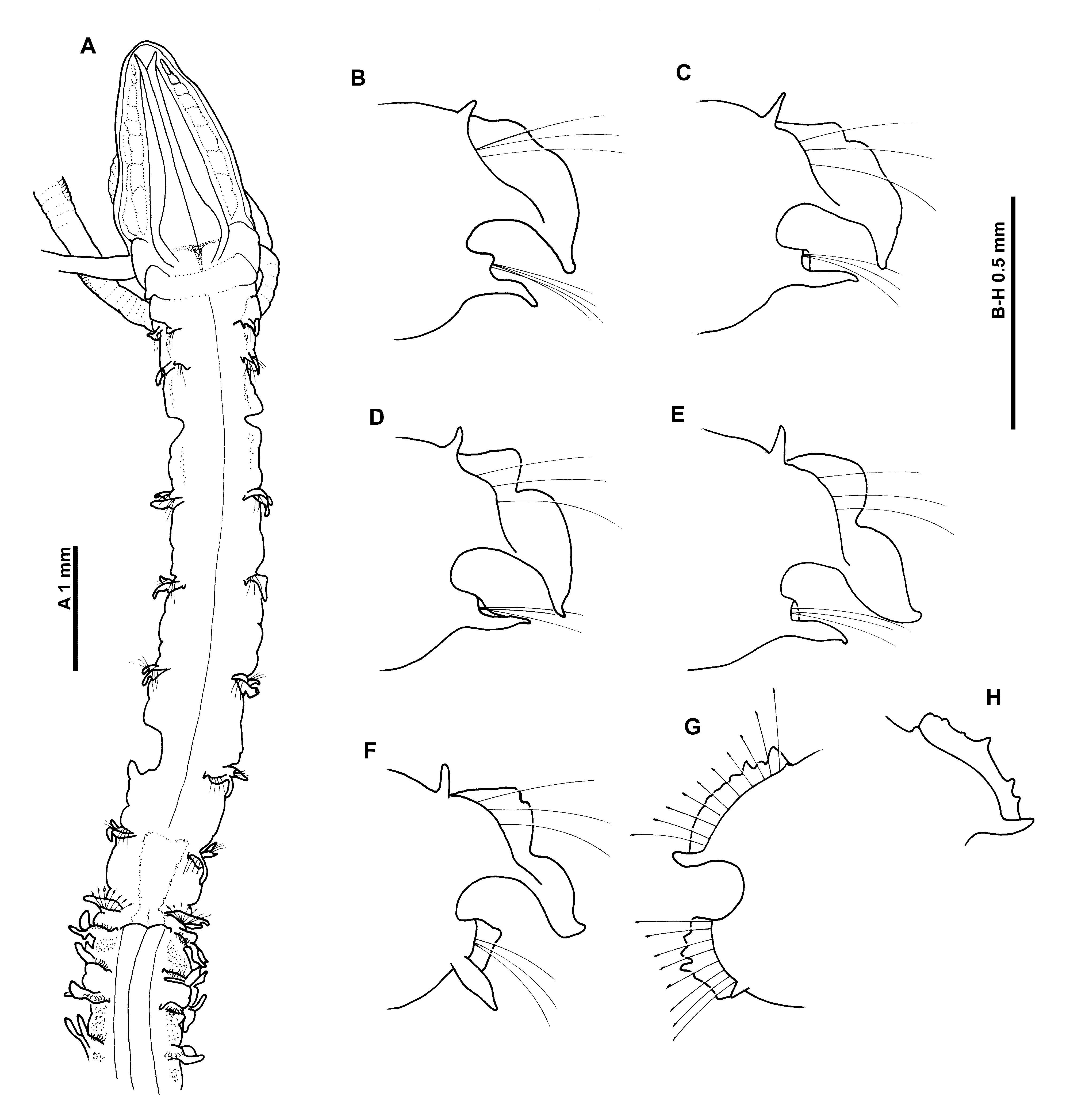
Peristomium achaetous, roughly twice the size of chaetiger 1. Chaetigers 1–7 similar; parapodia biramous with well–developed, thick notopodial prechaetal lamellae confluent with large delicate foliaceous postchaetal lamellae, latter of similar size throughout thorax ( Figs 5 View FIGURE 5 B–E). Upper edges of postchaetal lamellae lightly crenulated, a discrete notch present on fourth chaetiger ( Fig. 5 View FIGURE 5 C), developing into distinct bilobed lamellae by chaetiger 5 ( Fig. 5 View FIGURE 5 D) (note: chaetiger 3 could not be observed, but Mohammed’s original drawing of this chaetiger does not show any notch). Lateral portion of bilobed lamellae subtriangular with pointed tips, dorsalmost portions varying in shape from rounded to triangular. Single long, slender, tapering cirriform prechaetal superior process (DML) present on all thoracic chaetigers, gradually increasing in size to chaetiger 4 but decreasing towards posterior thorax.
Neuropodia of chaetigers 1–7 with single long slender cirriform processes ventrally (VNL), directly under chaetae but becoming distinctly prechaetal by chaetiger 7, distal tips pointed. Processes becoming shorter and broader, towards posterior thorax. Pre– and postchaetal lamellae well–developed, encircling chaetae cuff–like.
Chaetiger 8 ( Fig. 5 View FIGURE 5 F): Notopodial lamellae obviously bilobed, lateral portions subtriangular, dorsalmost portions triangular; apexes indented, prechaetal superior processes (DML) small. Neuropodial prechaetal processes, digitiform. Low prechaetal lamellae confluent with subtriangular postchaetal lamellae, encircling chaetae superiorly, cuff–like. Chaetae of chaetigers 1–8 simple winged capillaries.
Chaetiger 9 ( Fig. 5 View FIGURE 5 A): shorter than preceding chaetigers. Notopodial prechaetal lamellae low, broadly rounded, confluent with higher postchaetal lamellae, shorter than on preceding chaetigers, upper edge distinctly crenulated (degree of crenulation varying between rami on both sides of the body, see Figs 5 View FIGURE 5 G, H), lamellae encircling chaetae underneath as lateral expansions. Superior processes (DML) minute. Neuropodia similar to, but smaller than notopodia, without lateral expansions, small ventral prechaetal processes present (VML of some authors). Chaetae mucronate (not figured, in order to prevent further damage to specimen, although figured extensively by Mohammed 1973: figs 9G–L), arranged in arcs, chaetae longer towards margins of each fan.
A conspicuous, oval swelling present ventrally, level with the lamellae of chaetiger 8 ( Fig. 6 View FIGURE 6 E). Additional ventral swellings observed along thorax (visible from chaetiger 3), as two oblique anterior facing structures, increasing in development along thorax; those of chaetiger 7 more developed, cordiformis ( Fig. 6 View FIGURE 6 E). Swellings, with sporadic white speckles. A dorsal swelling, can be observed ( Fig. 5 View FIGURE 5 A) between the parapodia of chaetiger 8.
Abdominal chaetigers ( Fig. 6 View FIGURE 6 F) with broad, spatulate lateral lamellae, of about equal size in both rami, basally constricted, bluntly pointed. Lamellar shape shows some variation; rounded, subrectangular, oval with pointed tips. Extension of the lateral lamellae behind chaetal rows well–developed, especially in anterior abdomen, the apexes of which in some specimens form a distinct rounded tip. Small triangular processes (DML & VML) present at inner margins of chaetal rows. Lamellae of posterior fragment much reduced but initially still fairly broad, subtriangular with pointed tips, reducing to become cirriform.
Abdominal chaetae ( Fig. 6 View FIGURE 6 D) tridentate hooded hooks of similar size, superior two fangs parallel, above main fang. Hooks in two groups, main fangs vis–à–vis, group at outer margins of chaetal rows with fewer chaetae. Initially 14 hooks per rami, decreasing to approximately 10 hooks medially and 8 (evenly distributed between both groups) posteriorly.
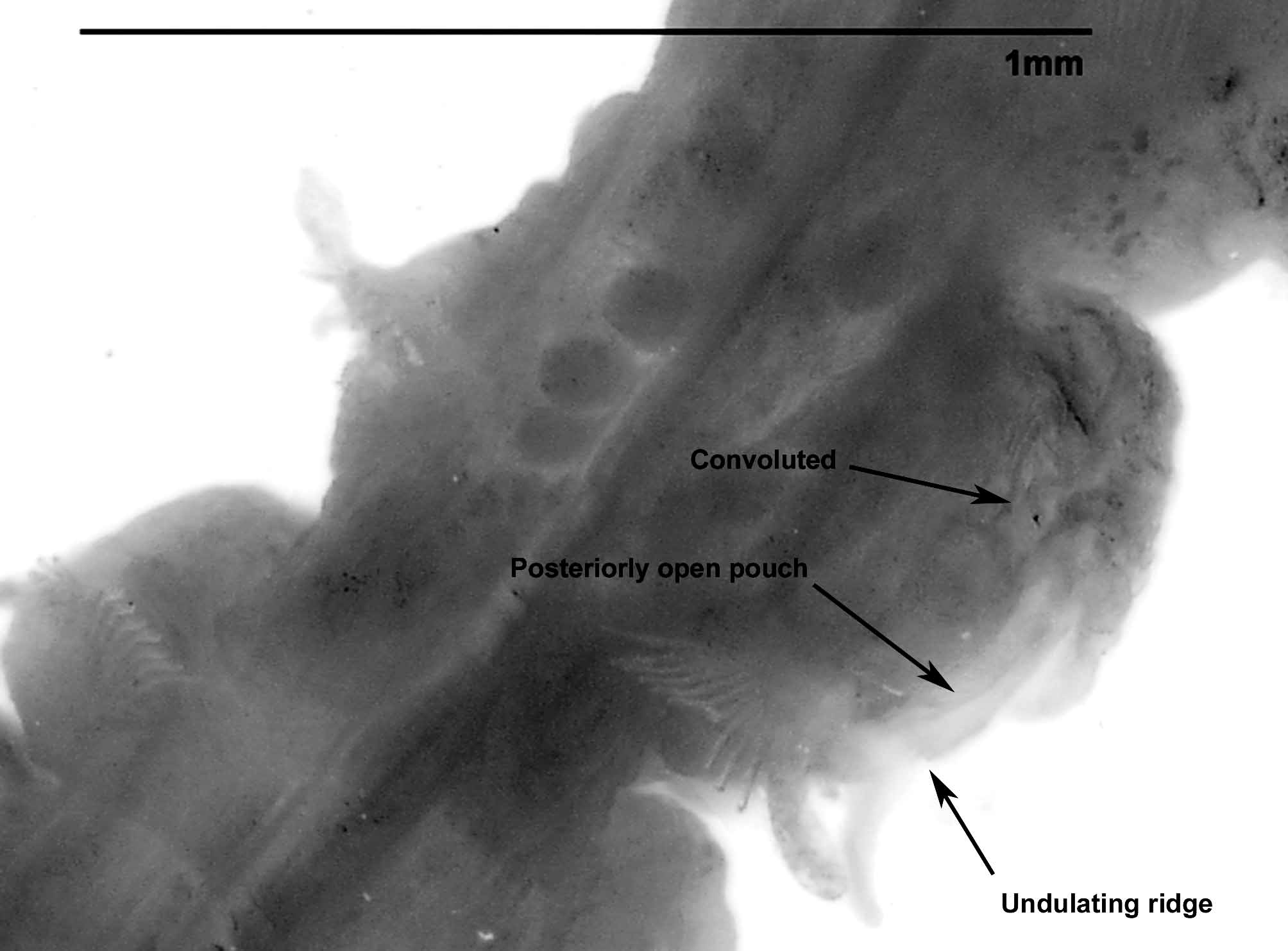
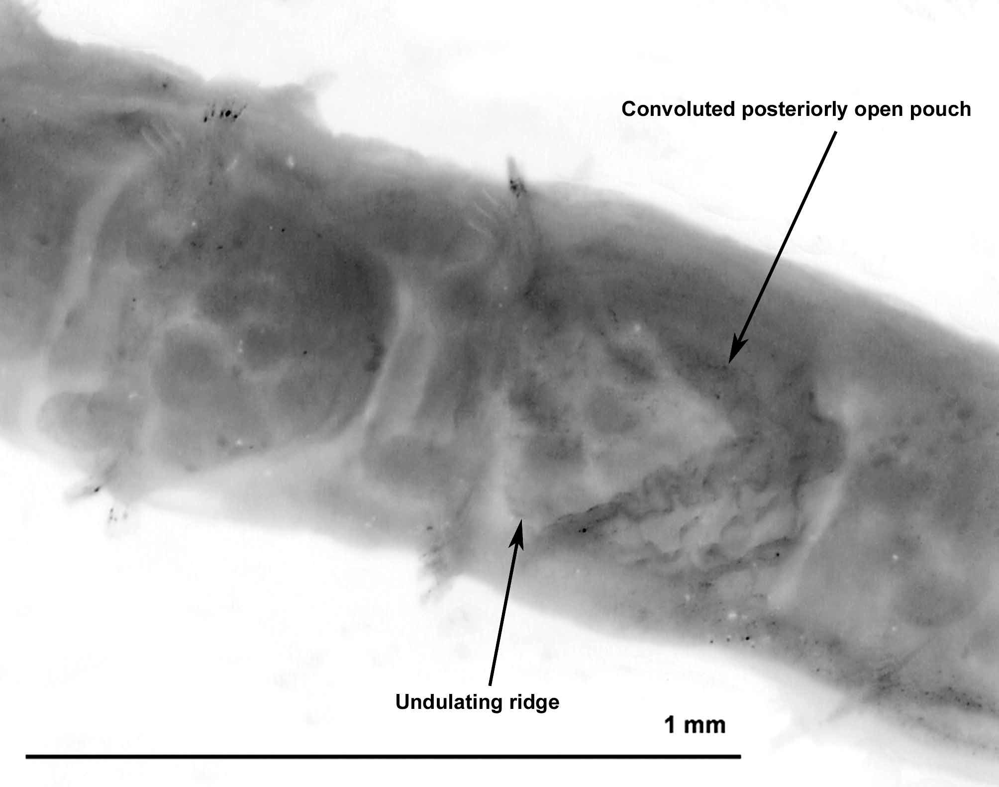
Paired anteriorly open pouches between chaetigers 11 and 12 (Σ configuration of Fiege et al. 2000 —see discussion) as two cuticular flaps (membrane absent on both sides, presumed missing). Unpaired posteriorly open pouches (C configuration of Fiege et al. 2000 —see discussion) present posteriorly, on alternate chaetigers, more or less regularly. Pouches differing from previously described C configuration pouches, often quite large, expanded more dorsally and ventrally, often convoluted ( Figs 7 View FIGURE 7 and 8 View FIGURE 8 ). Pouches observed on 11, 17L, 21L, 23R, 25L, 27R, 29L, 30R, 32L, 34R, 36L, 38R, 40L, 42R, 46L, 51R, 53L, 56R, 57L, 60R, 62L (on the anterior fragment), exact position of pouches difficult to discern due the fragility of the specimen. Posteriorly open pouches observed on posterior fragment (6th, 10th and 15th chaetiger from pygidium on left and the 4th, 8th and 12th on right). Pygidium rounded, left–hand anal cirri present ( Figs 6 View FIGURE 6 B–C), long, slender cirriform, right–hand missing (although original drawn by Mohammed).
Specimen ovigerous, eggs appearing within the body cavity, all of a similar size. The eggs appear to be separated from the pouch by a convoluted membrane.
Colour. The colouration originally described by Mohammed was uniformly white in life. The preserved material is yellowish/cream white, with darker glandular areas present inter–parapodially in the abdomen.
Staining with methyl green shows no clear pattern, but an overall diffuse stain. However, green speckled areas associated with the ventral swellings, and dorsally in the thorax (as seen in M. obockensis ) in particular between chaetigers 1–4 can be observed. Some staining evident abdominally between the parapodia.
Habitat. Specimens found in sand, intertidally at three locations: Hawalli, Salimiyah and Abu Halifa— Kuwait, Arabian Gulf. No other records of this species have been found.
Remarks. The holotype of M. heteropoda agrees very well with the syntypes of M. obockensis , in terms of prostomial shape, body size, the nature of the thoracic and abdominal lamellae, the presence of mucronate chaetae on the 9th chaetiger, the tridentate nature of the hooded hooks in two groups, and the presence of both anteriorly and posteriorly open pouches along the abdomen. However some variation between the material exists:
Prostomium: the figured syntype of M. obockensis appears to have a slightly broader prostomium than that of the holotype of M. heteropoda . However, the figured specimen of the former species is the broadest of all observed syntypes (variation between syntypes was observed) and the prostomia of other specimens were more akin to the slightly narrower prostomium of the latter species. As well as prostomial shape, conspicuous prostomial muscular areas either side of the dorsal longitudinal ridges were observed on the holotype of M. heteropoda . Such areas of similar shape and size were observed on the holotype of M. obockensis , although not as conspicuous ( Fig. 1 View FIGURE 1 B). This may be explained by the age and length of preservation of the M. obockensis material. However, they were conspicuous in the general collection specimens of M. obockensis .
Palps: The palps of the holotype of M. heteropoda , although robust were slightly more slender than those seen in the syntype material of M. obockensis , with only 3 rows of papillae present either side of the groove, proximally. However, variation in the size of the palps and number of papillae was also seen between the two specimens from the general collection (MNHN A 172), possibly showing variation caused by regeneration.
Thoracic lamellae: There is some variation in the figures of thoracic lamellae between those of M. heteropoda and M. obockensis . The upper edges appear more undulating in M. obockensis and smoother in M. heteropoda , and the nature of the bilobed lamellae (chaetiger where they first occur, and in the shape of the more dorsal portion differs a little). However, variation in lamellar shape between the syntypes of M. obockensis was observed ( Figs 1–2 View FIGURE 1 View FIGURE 2 ), some having more rounded dorsal portions, more akin to those seen in M. heteropoda , becoming more triangular on later chaetigers, and a degree of variation exists in the size of the notch on preceding chaetigers. The thoracic lamellae of the broadest specimen (39 chaetiger af, both palps retained, proboscis not everted) showed bilobed lamellae from chaetiger 1.
Chaetiger 9: the shape of the postchaetal lamellae of chaetiger 9 varies in the figures of the two species. However, the lamellar shape varies strongly between the notopodia on both sides of the body in M. heteropoda ( Figs 5 View FIGURE 5 G–H) (the variation between neuropodia could not be assessed on the holotype) and there is variation in the degree of crenulation between the syntype specimens of M. obockensis ( Figs 1 View FIGURE 1 F, 2J). Dorsal superior possesses of this chaetiger in M. heteropoda were minute and difficult to discern (Mohammed originally drew a minute process at the inner margin of the chaetae row), however, this was also true of several of the syntypes of M. obockensis .
Ventral and dorsal swellings: The presence of similar swellings on both the venter and dorsum in both species is an important characteristic. They are less well–developed in the holotype of M. heteropoda but are of similar size and shape, with white speckles being present on ventral swellings.
Thoracic/abdominal junction: the junction between the two body regions in M. heteropoda is not as clearly marked as it is in M. obockensis . However, the distinction between the two body regions will often depend on the degree of body constriction upon fixation. The holotype of M. heteropoda , appears to have been fixed in a very elongated state (possibly due to prior relaxation?), many of the segments are weakly connected, especially in the posterior region, adding to its fragility.
The variation seen between the two species is no more pronounced than the variation seen within the syntypes of M. obockensis . The morphological similarity between the two species is striking. Mohammed (1973) stated that…. “ M. heteropoda shows affinities with M. obockensis , but is readily distinguishable by its bilobed notopodial lamellae”, a feature now shown to be shared with M. obockensis . Consequently, with the corrections and additions to the original description of M. obockensis , I consider M. heteropoda to be a synonym of M. obockensis .

No known copyright restrictions apply. See Agosti, D., Egloff, W., 2009. Taxonomic information exchange and copyright: the Plazi approach. BMC Research Notes 2009, 2:53 for further explanation.
|
Kingdom |
|
|
Phylum |
|
|
Class |
|
|
Order |
|
|
Family |
|
|
Genus |
Magelona heteropoda Mohammed, 1973
| Mortimer, Kate 2010 |
Magelona heteropoda
| Mohammed 1973: 39 |