
Maritigrella makranica, Maghsoudlou, Abdolvahab & Rahimian, Hassan, 2014
|
publication ID |
https://doi.org/ 10.11646/zootaxa.3860.4.2 |
|
publication LSID |
lsid:zoobank.org:pub:3F59B0E9-F943-407C-A49B-6D05DBACCCEE |
|
DOI |
https://doi.org/10.5281/zenodo.6122982 |
|
persistent identifier |
https://treatment.plazi.org/id/DF12878D-FF91-FFBE-94A0-1B02F2D9F8D6 |
|
treatment provided by |
Plazi |
|
scientific name |
Maritigrella makranica |
| status |
sp. nov. |
Maritigrella makranica View in CoL sp. nov.
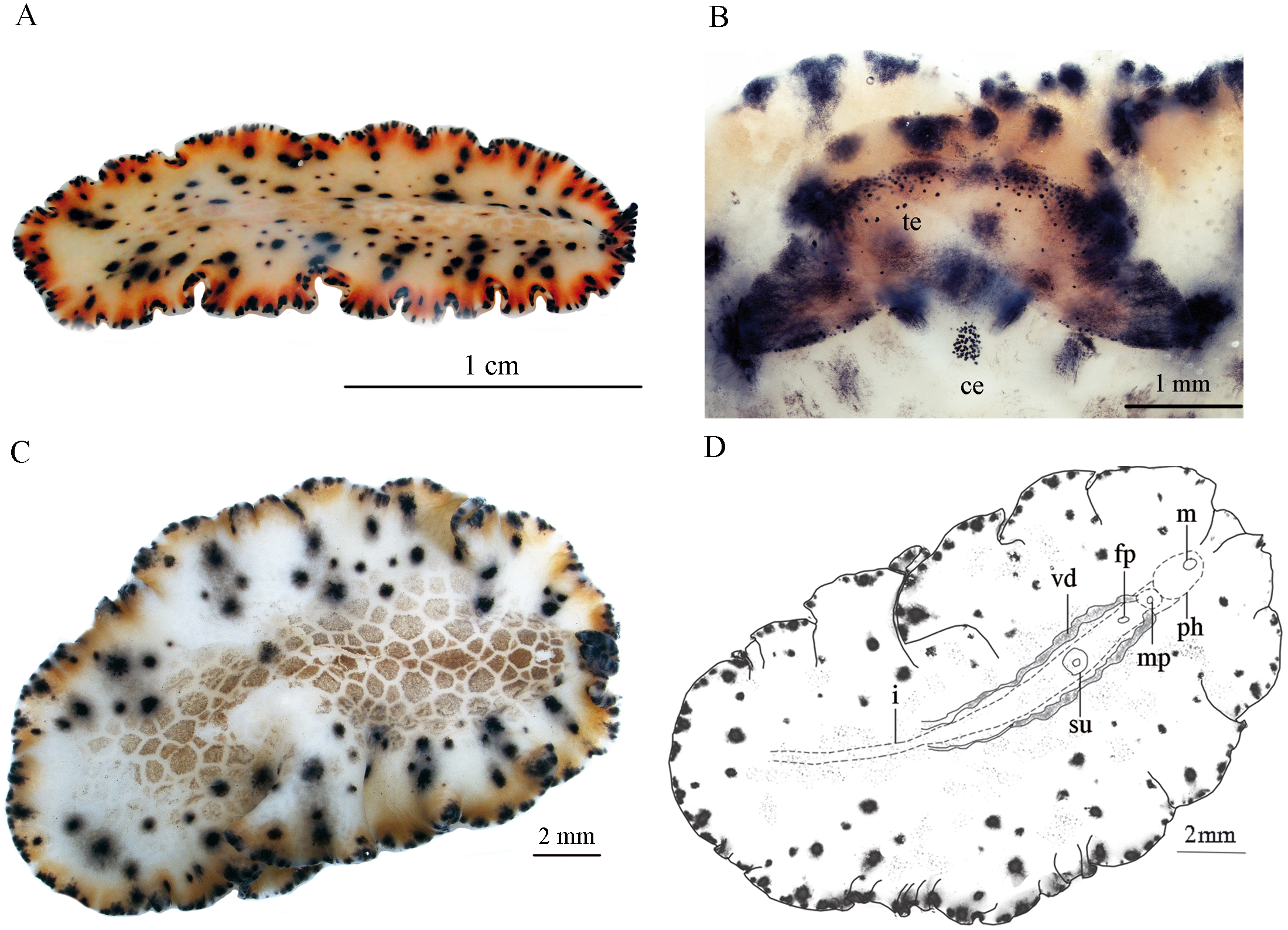
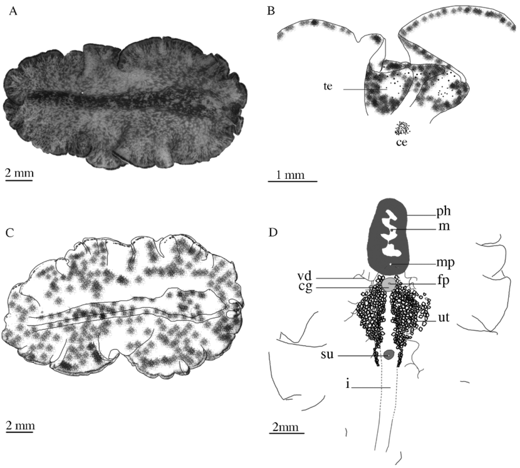
( Figures. 2 View FIGURE 2 , 3 View FIGURE 3 )
Material examined. Three specimens were collected in December 2010: one mature specimen provided serial sagittal sections, to elucidate the structure of the reproductive systems; two specimens, one damaged mature and one juvenile, were preserved in ETOH 70%, (Table 1).
Holotype. One mature specimen, ZUTC platy 1273 HS, with the posterior half preserved in ETOH 70%, and part of the anterior half containing the copulatory apparatus as series of sagittal sections (ten slides).
Paratypes. Two specimens, ZUTC platy 1274 S, one damaged mature and one juvenile preserved in ETOH 70%.
Etymology. The specific epithet refers to Makran area (Gulf of Oman) where the specimens were collected.
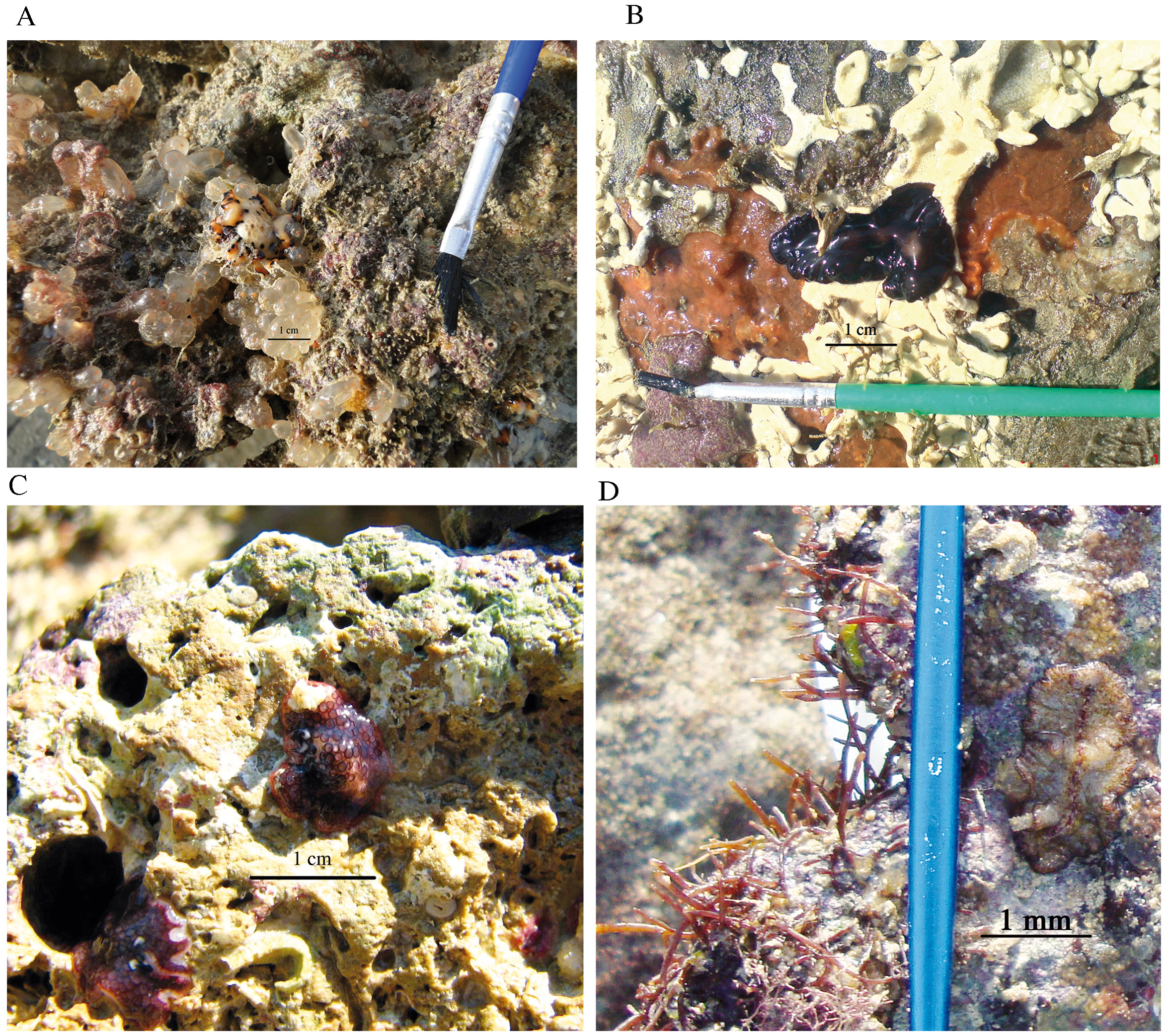
Type locality. All specimens were found together, associated with ecteinascidians, from intertidal rocky shores of the Gulf of Oman ( Fig. 1 View FIGURE 1 , station 38 and Table 1).
Type of the genus. Maritigrella eschara Newman and Cannon, 2000 .
Diagnostic characters. dorsal background with medial cream -white reticulated appearance containing pale orange spots in a honeycomb pattern, a distinct orange submarginal band around the entire body margin and between the marginal tentacles, black spots scattered around mid-dorsal surface, becoming more sparse on raised median region and towards body margin, surrounded by dark-grey halo around the body midline and orange-black halo towards margin.
Description. External morphology. Body soft and delicate, elongated, oval, slightly tapered posteriorly, elevated dorso-anteriorly only over the area of the pharynx, body margin with deep crenulated ruffles ( Fig. 2 View FIGURE 2 A), fixed specimens approximately 20 mm long × 12.5 mm wide ( Fig. 2 View FIGURE 2 C); 23 × 14 in mature, 12 × 10 in juvenile of ZUTC platy 1274. Dorsal background whitish cream, shades to orange toward the margin; an orange submarginal band is recognizable around the body margin and between the marginal tentacles, except on the tip of the tentacles where the color is black; large black spots exist on background, and in between marginal tentacles, more or less in transverse rows laterally ( Figs. 2 View FIGURE 2 A, B), each row containing 3–5 spots, smaller black spots, variable in size, scattered around mid-dorsal surface, becoming more sparse on raised median region and towards body margin ( Figs. 2 View FIGURE 2 A, C). A halo surrounds each of the black spots, dark gray around the spots of the median region, orangeblack towards margin, black spots disappear posterior to the body midline where pale orange honeycomb pattern also fades, dorsal surface with medial cream or white reticulated appearance containing pale orange spots in a honeycomb pattern; smaller black spots interspersed by white rims on the body margin appear as a black and white interrupted marginal band ( Fig 2 View FIGURE 2 A). Marginal tentacles long, slender and erect in living animals, having the same pattern as the body margin with black spots on the tentacles ( Fig. 2 View FIGURE 2 B). Dorsal and ventral eyespots scattered between tentacles, ventral eyespots more numerous ( Fig. 2 View FIGURE 2 B); cerebral eyes with 68–70 small spots, in two clusters, especially visible in living animals ( Fig. 2 View FIGURE 2 B). Ventral surface has the same pattern as dorsal one, but without orange honeycomb spots; pharynx small, 1.5 mm length, muscular and tubular, directed anteriorly; mouth anterior ( Fig. 2 View FIGURE 2 D), gut heavily branched; antero-ventral gonopores 1 mm apart, male gonopore immediately behind the pharynx, female gonopore located posterior to the male pore; sucker prominent, 1.6 mm posterior to the female pore ( Fig. 2 View FIGURE 2 D).
Reproductive structures. In ventral view, vasa deferentia as two parallel, un-branched strips besides the intestine ( Fig. 2 View FIGURE 2 D), extending upward joining each other and form a common vas deferens that eventually enters the posterior end of the seminal vesicle in sagittal view ( Fig. 3 View FIGURE 3 C). Seminal vesicle, 310 µm × 316 µm, muscular, oval ( Figs. 3 View FIGURE 3 A, C); prostatic vesicle more or less rounded-oval, 310 µm × 290 µm, highly muscular, prostatic lumen with smooth glandular lining ( Fig. 3 View FIGURE 3 C); ejaculatory duct straight joining the short prostatic duct at the base of penis; penis papillae with short stylet, 168 µm × 60 µm, stylet length to width ratio 1:2.8; deep ciliated male atrium pear-shaped ( Fig. 3 View FIGURE 3 C). Female atrium surrounded by well-developed cement glands, uterine vesicles not observed. Female gonopore located almost halfway between male pore and the sucker ( Fig. 3 View FIGURE 3 C).
Remarks. The presence of a tubular pharynx in euryleptids rather than the ruffled pharynx of pseudocerotids facilitates distinguishing these animals from each other ( Newman & Cannon 2000). The genus Maritigrella was erected recently within the Euryleptidae by Newman and Cannon (2000). The diagnostic characters for the genus include: a long body with a distinct striped color pattern, long marginal tentacles; two long clusters of cerebral eyes; small, muscular and tubular pharynx; short and pointed stylet; absence of uterine vesicles ( Newman & Cannon 2000).
To date, nine species of Maritigrella have been described ( Bolaños 2010): M. aurolineata (Verrill, 1901) Litvaitis, Bolaños and Quiroga 2010 , M. crozieri (Hyman, 1939) Newman, Norenburg and Reed 2000 , M. eschara Newman & Cannon, 2000 , M. fuscopunctata ( Prudhoe, 1977) , M. marygarsonae Newman & Cannon, 2000 , M. newmanae Bolaños, Quiroga and Litvaitis, 2007 , M. ocellata Newman & Cannon, 2000 , M. stellata Newman & Cannon, 2000 , M. virgulata Newman & Cannon, 2000 .
Species recognition within Maritigrella is mostly based on color patterns ( Newman and Cannon, 2000). M. fuscopunctata ( Prudhoe, 1977) and M. ocellata described by Newman and Cannon (2000) are closer together and to the species described here. Presence/absence of orange spots in a honeycomb pattern, number of black spots in transverse rows, and the halo color surrounding black spots are the major taxonomic characters with specific value that cause Newman and Cannon (2000) to distinguish M. fuscopunctata from M. ocellata . Our new species can be differentiated clearly from M. ocellata by having distinct medial orange spots in a honeycomb pattern, numbers of black spots in transverse rows (Figs. 12, 24 of Newman and Cannon, 2000), presence of distinct orange submarginal band over the entire body and between marginal tentacles, and the stylet length to width ratio (1:2.8 for M. makranica and 1: 3.2 for M. ocellata Newman & Cannon, 2000 ).
M. fuscopunctata View in CoL was originally described by Prudhoe (1977) as a pseudocerotid, Pseudoceros fuscopunctatus , from Australian waters. Based on the description by Prudhoe (1977), which indicates the presence of a tubular pharynx in his species, Newman & Cannon (2000) recognized that their specimens were similar to that of Prudhoe's description (1977), therefore they made a new combination as M. fuscopunctata ( Newman & Cannon 2000) View in CoL and described it as: dorsal background with large purple-black spots in transverse rows, black spots surrounded by purple-violet halo, with medial orange spots in a honycomb pattern (Fig. 22 of Newman and Cannon, 2000).
Our new species is therefore more similar to M. fuscopunctata View in CoL described by Newman & Cannon (2000); however, some differences can be seen with careful observation of the color pattern: M. fuscopunctata ( Prudhoe, 1977) View in CoL lacks black spots scattered on dorsal surface; unlike the species described here, these spots and larger transverse black spots have dark gray halo around the median region, orange-black halo towards margin in our new species, while in M. fuscopunctata View in CoL described by Newman and Cannon (2000) large transverse black spots are surrounded by purple halo; an orange submarginal band is present in M. makranica View in CoL but not in M. fuscopunctata ( Prudhoe, 1977) View in CoL (Fig. 22, Newman and Cannon, 2000).
Newman and Cannon also included other variants from Maldives, Indonesia and South Africa ( Newman and Cannon, 2003; p. 68, Figs 8 View FIGURE 8 –11 for M. fuscopunctata View in CoL of Newman and Cannon, 2005) as M. fuscopunctata View in CoL . All of these variants lack black spots scattered on the dorsal surface, the white reticulated network containing orange spots in a honey comb pattern also extends over the entire dorsal surface, while in M. makranica View in CoL sp. nov. black spots are scattered around middorsal surface, becoming more sparse on raised median region and towards body margin ( Fig. 2 View FIGURE 2 A); the cream or white reticulated appearance containing pale orange spots also located medially in a honeycomb pattern. Consequently, we introduced a new Maritigrella View in CoL species that inhabits the Iranian coast of the Gulf of Oman ( Fig.1 View FIGURE 1 Makran area) as M. makranica View in CoL . As a result it seems that M. fuscopunctata View in CoL previously reported from the West Indian Ocean ( Fig. 1 View FIGURE 1 k of Deepack and Reshma, 2011) is synonymous with our new described species.
Ascidians are the major prey of the pseudocerotid and euryleptid flatworms. Euryleptids rely more on muscular activity of their long tubular pharynx to extract tissues from zooids of colonial ascidians. Pseudocerotids, on the other hand, benefit from enzymatic digestion. They extend their ruffled pharynx on to several zooids simultaneously ( Newman & Cannon 1994). In this study all of the M. makranica View in CoL specimens were found in close association with ascidians E. turbinata ( Fig. 10 View FIGURE 10 A).
The present study introduces for the first time the presence of M. makranica View in CoL from the Gulf of Oman. Despite careful examination at all 38 stations, specimens of M. makranica View in CoL were observed only at one station ( Fig. 1 View FIGURE 1 , station 38) of the Gulf of Oman. Thus, it seems that the distribution of this species is restricted to the Gulf of Oman which may be due to the different physico-chemical environment of the Persian Gulf ( Banse 1997). Prudhoe (1985) believed that physical factors such as temperature restricted the distribution of polyclad flatworms.

No known copyright restrictions apply. See Agosti, D., Egloff, W., 2009. Taxonomic information exchange and copyright: the Plazi approach. BMC Research Notes 2009, 2:53 for further explanation.
|
Kingdom |
|
|
Phylum |
|
|
Class |
|
|
Order |
|
|
Family |
|
|
Genus |
Maritigrella makranica
| Maghsoudlou, Abdolvahab & Rahimian, Hassan 2014 |
M. fuscopunctata (
| Newman & Cannon 2000 |
M. fuscopunctata (
| Prudhoe 1977 |
M. fuscopunctata (
| Prudhoe 1977 |