
Mastigias papua ( Lesson, 1830 )
|
publication ID |
https://doi.org/ 10.11646/zootaxa.4457.4.2 |
|
publication LSID |
lsid:zoobank.org:pub:520C1691-5FD8-415A-BCB0-3C01CBBF5ECB |
|
DOI |
https://doi.org/10.5281/zenodo.5956951 |
|
persistent identifier |
https://treatment.plazi.org/id/03ABDD7D-FF86-127D-FF64-FAFFFAC1F8C6 |
|
treatment provided by |
Plazi |
|
scientific name |
Mastigias papua ( Lesson, 1830 ) |
| status |
|
Mastigias papua ( Lesson, 1830) View in CoL
( Figures 4–5 View FIGURE 4 View FIGURE 5 )
Mastigias View in CoL sp.: Cephea papua Lesson, 1830 ; Haeckel 1880; Vanhöffen 1888; Mastigias physophora Kishinouye, 1895 View in CoL ; Pseudorhiza thocambaui Agassiz & Mayer, 1899 ; Mastigias papua var. sibogae Maas, 1903 View in CoL ; Mayer 1910; Stiasny 1920, 1926; Uchida 1926; Kramp 1961; Hamner & Hauri 1981; Hamner 1982; Dawson et al. 2001; Dawson & Hamner 2003; Dawson 2005a, b; Swift et al. 2016.
Type Material. Smithsonian Museum USNM 1486941 (previously M0D001038-X of Dawson collection) neotype: Mastigias Papua Cove, Waigeo Island, West Papua (00 19.356’ S 130 34.277’ E), 21.xi.2007, M0D001038-X, 1 adult medusa (female) preserved in 4% formalin.
Other Material. Scripps Institution of Oceanography / UCSD PIC- 180420 -0001 -PT (previously M0D005690- V of Dawson collection): Mastigias Papua Cove , Waigeo Island , West Papua (00 19.35’ S 130 34.27’ E), 21.xi.2007, adult female medusa preserved in 4% formalin. GoogleMaps
California Academy of Sciences CASIZ 219752 (previously M0D005693-Y of Dawson collection), Danau Ctenophore , West Papua (0027.29’ S 13029.58’ E), 26.xi.2007, adult male medusa preserved in 4% formalin.
Michael N Dawson collection MOD05687-S, M0 D05688 View Materials -T, M0 D05689 View Materials -U, M0 D05691 View Materials -W, M0 D05692 View Materials -X, M0 D05694 View Materials -Z, M0 D05695 View Materials -A. Specimens preserved in 4% formaldehyde. Avaliable for examination upon request.
Type Locality: Waigeo Island, West Papua, Indonesia (00 19.356’ S 130 34.277’ E), in agreement with the original type locality reported in Lesson (1830, p 122): “chez les Papous de la Waigiou ( Lesson, 1830, p 122), i.e. the Coast of Waigeo Island,
Distribution: Western Pacific: West Papua, Palau and Marshall Islands.
Diagnosis. Five to ten velar lappets per octant (mean 9.7 ± 1.17 s.d.), 10.8 ±2.3 adradial origins at Gastrovascular Cavity (GVC) per quadrant, oral arms 0.3 ±0.05 times the size of bell diameter (bd; winged part of oral arm 0.21 ±0.05 bd; unwinged part of oral arm 0.10 ±0.03 bd). Thymine at positions 2, 79, 244, 292, 496, 559 and 575; guanine at positions 77, 209, 259, 332 and 409; cytosine at positions 103, 106, 109, 190, 344, 460 and 530 and adenine at positions 94, 199, 202, 205, 424, 512, 547, 550 and 625 of COI (Appendix 1).
Habitat. Medusae planktonic in coastal waters and marine lakes; occasionally oceanic. Usually present near surface (to several meters depth) during the day, deeper (tens of meters) at night. Abundance may vary through the year at and between locations ( Dawson 2005a).
Description of Neotype Specimen of Mastigias papua . Medusa is yellow-brown with white spots of various sizes on the exumbrella ( Fig. 4A, B View FIGURE 4 , 5A View FIGURE 5 ). Bell diameter 122.2 mm. Ring canal about twice the diameter of the oral disc (respectively 104 mm and 49 mm). Mesoglea of the bell flexible, thicker around the center portion, thinning towards bell margin. Velar lappets round ( Fig. 5E View FIGURE 5 ), 80 in the whole medusa (number in each octant: 9, 10, 10, 11), with furrows often between adjacent pairs of lappets. Mean (±s.d.) oral arm length is 43.37± 0.51 mm, with winged portion about 1.5 times longer than unwinged part (respectively 26.65± 1.27 mm and 16.7± 1.07 mm). Four perradial and four interradial rhopalia, each between two pointed rhopaliar lappets. Brood filaments present at the base of the oral arms and oral disc (mature female specimen). Terminal clubs about 1.4 the size of bell diameter (bd; mean 175.42± 6.65 mm) and shaped like a tricorn in cross-section ( Fig. 5B View FIGURE 5 ). Oral pillars 15.05± 0.07 mm wide, 3.68 ± 0.12 mm long and 2.8± 0.14 mm deep. Genital ostia 35.5 ± 0.28 mm wide. Gastrovascular cavity (GVC) with one perradial, one interadial and 7±0.5 adradial origins per octant ( Fig. 5D View FIGURE 5 ). Two perradial, two interadial and 27.5±0.68 adradial anastomoses per octant. One gastrovascular and one adradial sinus, no perradial nor interadial sinuses, per quadrant. One ring canal sinus present in each of 2 quadrants.
Palau and West Papua samples: variation from the type specimen. Ocean samples from West Papua presented terminal club mean 0.86±0.24 bd; interradial diameter of the GVC mean 0.52±0.08 bd, perradial diameter of the GVC 0.21±0.03 bd; 6.5±0.5 mean adradial origins at GVC and 27.2±4.05 mean adradial anastomoses per octant. When compared to lake samples from West Papua, the features presented similar values: terminal club mean 0.83±0.05 bd; interradial diameter of the GVC mean 0.54±0.09 bd, approximately two-and-aquarter times the length of the perradial diameter of the GVC; 6.37±0.58 mean adradial origins at GVC and 23.93± 1.85 mean adradial anastomoses per octant.
Ocean samples from Palau presented terminal club mean 0.67±0.16 bd; interradial diameter of the GVC mean 0.55±0.03 bd, perradial diameter of the GVC mean 0.25±0.04 bd; 6.65±0.33 mean adradial origins at GVC and 48.64±16 mean adradial anastomoses at GVC per octant. Samples from marine lakes from Palau presented interradial diameter of GVC mean 0.54±0.04 bd and perradial diameter of the GVC mean 0.26±0.05 bd; 5±1.02 adradial origins at GVC and 19.36±17.2 adradial anastomoses per octant; and differed from samples from ocean locations in having shorter terminal clubs (terminal club length 0.17±0.11 bd; t = -16.649, df = 155, p-value <0.0001).
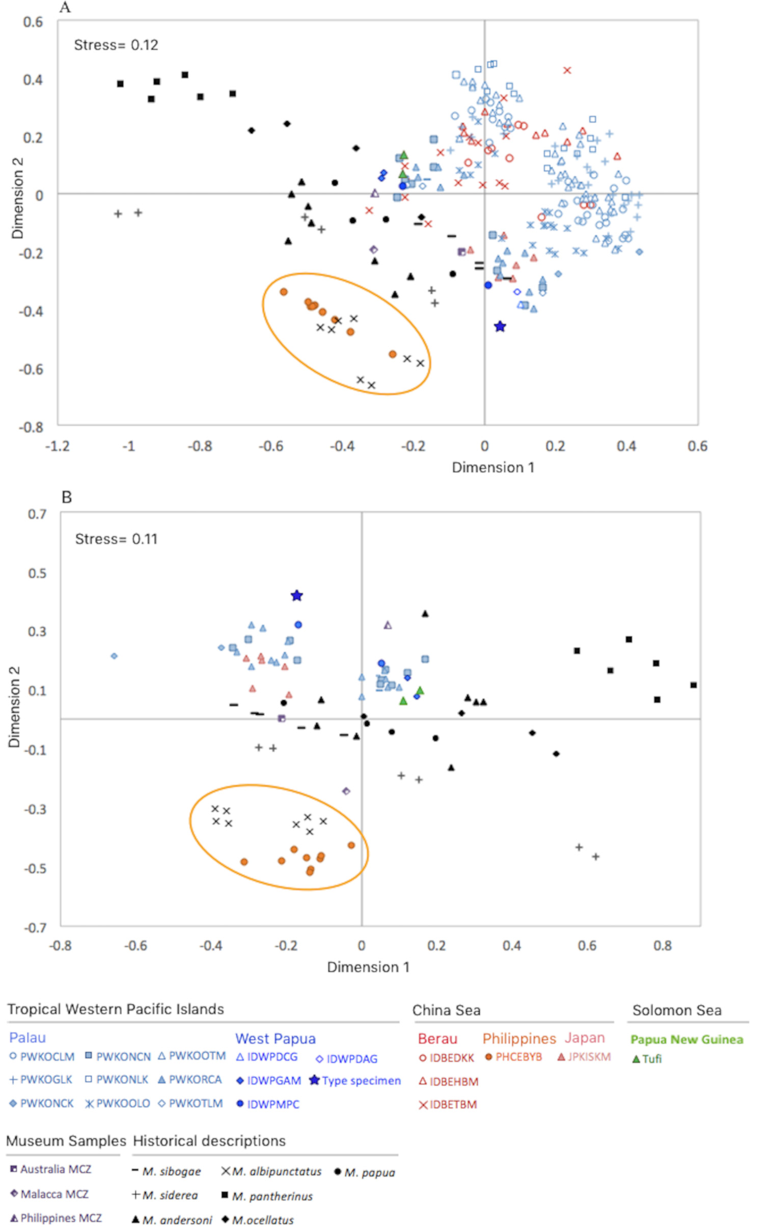
Comparison with historical descriptions. Both nMDS analyses visually represent the data reasonably well (stress = 0.11–0.12; Fig. 6 View FIGURE 6 ). In both representations, the new samples from Philippines were close to the historical description for M. albipunctatus ; although the museum sample from Philippines was not close in this representation to the other samples from Philippines. The museum sample from Australia was close in both representations to the historical description of M. sibogae . Samples from Tufi were most similar to the historical descriptions of M. papua , M. ocellatus and M. andersoni ( Fig. 6A View FIGURE 6 ). The geographically distant Mastigias pantherinus , from Samoa Islands, is distinct morphologically from all other samples in both representations.


| USNM |
Smithsonian Institution, National Museum of Natural History |
No known copyright restrictions apply. See Agosti, D., Egloff, W., 2009. Taxonomic information exchange and copyright: the Plazi approach. BMC Research Notes 2009, 2:53 for further explanation.
|
Kingdom |
|
|
Phylum |
|
|
Class |
|
|
Order |
|
|
Family |
|
|
Genus |
Mastigias papua ( Lesson, 1830 )
| Souza, Mariana Rocha De 2018 |
Mastigias papua var. sibogae
| Maas 1903 |
Pseudorhiza thocambaui
| Agassiz & Mayer 1899 |
Mastigias physophora
| Kishinouye 1895 |
Mastigias
| L. Agassiz 1862 |
Cephea papua
| Lesson 1830 |