
Merobruchus bIcolorIpes ( Pic, 1927 )
|
publication ID |
https://doi.org/ 10.11646/zootaxa.4078.1.25 |
|
publication LSID |
lsid:zoobank.org:pub:78037DEE-A4F0-4E28-8135-EFD57552C3D0 |
|
DOI |
https://doi.org/10.5281/zenodo.6057382 |
|
persistent identifier |
https://treatment.plazi.org/id/039487A5-0178-FF82-FF3B-FE3C67A4F929 |
|
treatment provided by |
Plazi |
|
scientific name |
Merobruchus bIcolorIpes ( Pic, 1927 ) |
| status |
|
Merobruchus bIcolorIpes ( Pic, 1927)
( Figures 4 View FIGURES 1 – 8 , 9 View FIGURES 9 – 17 , 34 View FIGURES 27 – 34 , 38 View FIGURES 35 – 40 , 41–42 View FIGURES 41 – 46 , 56 View FIGURES 49 – 56 , 81 View FIGURES 81 – 92 , 105 View FIGURES 105 – 109 , 131 View FIGURES 129 – 133 )
Pseudopachymerus bicoloripes Pic, 1927: 32 (original description, type locality: "Pernambuco, Brazil ", type depository: MNHN).
Caryedes bicoloripes: Blackwelder, 1946: 757 (distribution); Udayagiri & Wadhi, 1989: 71 (distribution).
Merobruchus bicoloripes: Terán & Muruaga de L'Argentier, 1981: 61, 74–83 (new combination, biology, description, host plant, distribution); Muruaga de L’Argentier & Dipierri, 1983: 87 (host plant); Kingsolver, 1988: 1 –5, 63 (distribution, host plant, taxonomy); Macêdo et al., 1992: 331, 333 (distribution, host plant); Link & Costa, 1995: 113 –114, 116–117, 120–121 (host plant); Johnson & Siemens, 1997: 14, 16 (host plant); Johnson & Romero-Nápoles, 2004: 404 (behavior); Kingsolver & Muruaga de L’Argentier, 2004: 87–88 (distribution, host plant).
Pseudopachymerus ruficornis var. subuniformis Pic, 1938: 22 (original description; synonymized by Kingsolver, 1988: 2).
Caryedes ruficornis var. subuniformis: Blackwelder, 1946: 758 (distribution).
Material Examined. Type material. Pseudopachymerus bicoloripes Pic, 1927 . Holotype deposited in MNHN, male ( Fig. 155): (1) “Pernambuco/( Brésil)” [Handwritten] (2) “ Type ” [Orange label, handwritten in black] (3) “ bicoloripes /n sp” [Handwritten] (4) “ HOLOTYPE ” (5) “ bicoloripes /Pic/det.70/J.M.Kingsolver” (6) “ Merobruchus / bicoloripes ( Pic, 1927) /Ribeiro-Costa, C.S. det. 2007”. Additional material. BRAZIL: Pernambuco: 7, Pesqueira, Fazenda São Francisco, Serra do Ororubá, 12.i.2005, Iannuzzi L. col. Enterolobium contortisiliquum ( DZUP). Mato Grosso: 2, Cuiabá, Marques M. col., Enterolobium contortisiliquum ( DZUP); 1, Salobra, 18–29.x.1938, Lane F. col. ( MZSP). Distrito Federal: 3, Brasília, 04.ix.1995, Ramos F.A. col. ( DZUP). Minas Gerais: 23, Montes Claros, Parque Estadual da Lapa Grande, 13.viii.2010, Moreira P.A. col., Enterolobium contortisiliquum ( DZUP); 9, Paraopeba, Heringer col. ( USNM); 2, Montes Claros, Parque Estadual da Lapa Grande, Moreira P.A. col. ( FIOC); 7, Viçosa, 18.vii.1979, Santana P.C. col., orelha de negro ( DZUP). São Paulo: 2, Teodoro Sampaio, R.E. Morro do Diabo, 22°27’S 52°10’W, viii.1993, Teixeira, E.O. col., Tamboril ( DZUP); 2, Teodoro Sampaio, R.E. Morro do Diabo, 28.viii.1986, Teixeira, E.O. col., Enterolobium contortisiliquum ( DZUP); 1, Itú, Faz. Pau d’Alho, viii.1960, Martins U. col. ( MZSP); 20, Botucatu, Fazenda experimental Edgardia-FCA- UNESP, 22°48” S 48°24”31.56’W 577m, 15.v.2009, Rodrigues L.M.S. col, Enterolobium contortisiliquum ( DZUP). Paraná: 1, Capitão Leônidas Marques, 4.vi.1999, Timbaúva ( DZUP); 9, Curitiba, UFPR Campus Jardim Botânico, 12.ix.2009, Ribeiro-Costa C.S. col., Enterolobium sp. ( DZUP); 43, Curitiba, UFPR Campus Jardim Botânico, 25°44”58.66’ S 49°24” 31.56’W 27.viii.2010, Ribeiro-Costa C.S., Viana J.H. & Andrade B.V. col. ( DZUP); 27, Curitiba, UFPR Campus Jardim Botânico, 25°44”58.66’ S 49°24” 31.56’W, 27.viii.2010, Ribeiro- Costa C.S. & Viana J.H. col. ( DZUP); 8, Curitiba, UFPR Campus Jardim Botânico, 25°26”45.18’ S 49°14” 20.86’W 929m, 30.v.2011, Viana J.H., Albuquerque F.P., Manfio D. & Ribeiro-Costa C.S. col. ( DZUP); 6, Lunardelli, 11.viii.1983, Penteado col., Enterolobium contortisiliquum ( USNM). Rio Grande do Sul: 4, Passo Fundo, 9.ix.2010, Savaris M. col., Enterolobium contortisiliquum ( DZUP); 1, Pelotas, 25.xii.1995, Moura L. col. ( DZUP); 4, São Sepé, 10.vii.1983, Costa E.C. & Link D. col., Enterolobium contortisiliquum ( USNM); 1, Porto Alegre, 7.x.1936, Timbaúva ( FIOC); 5, Santa Maria, 6.viii.1971, Link D. col., Enterolobium timbouva ( USNM); 3, Santa Maria, 12.ix.2012, Garlet J. col., Enterolobium contortisiliquum ( DZUP); 12, Santa Maria, 07.v.13, Dorneles D. U. col. ( DZUP). PARAGUAY: Cordillera: 2, Caacupé, 7 km W, 11.x.1968, O’Brien C.W. & O’Brien L. col. ( USNM).
Redescription, holotype. Body length: 4.36 mm; width: 2.84 mm.
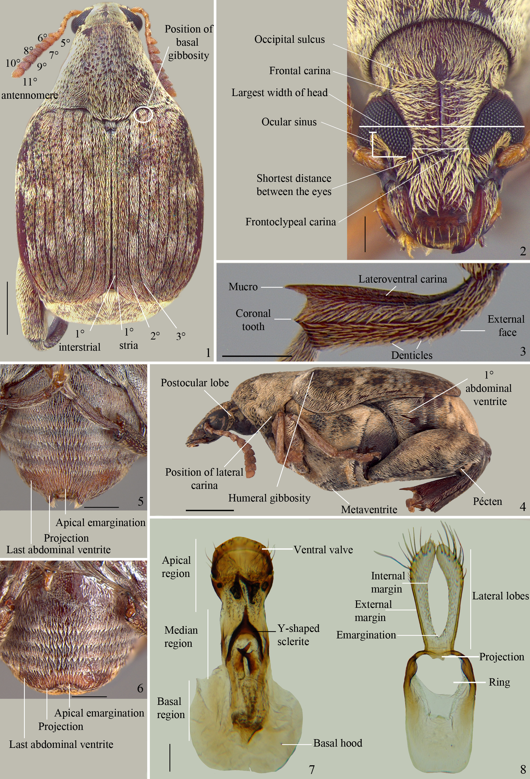
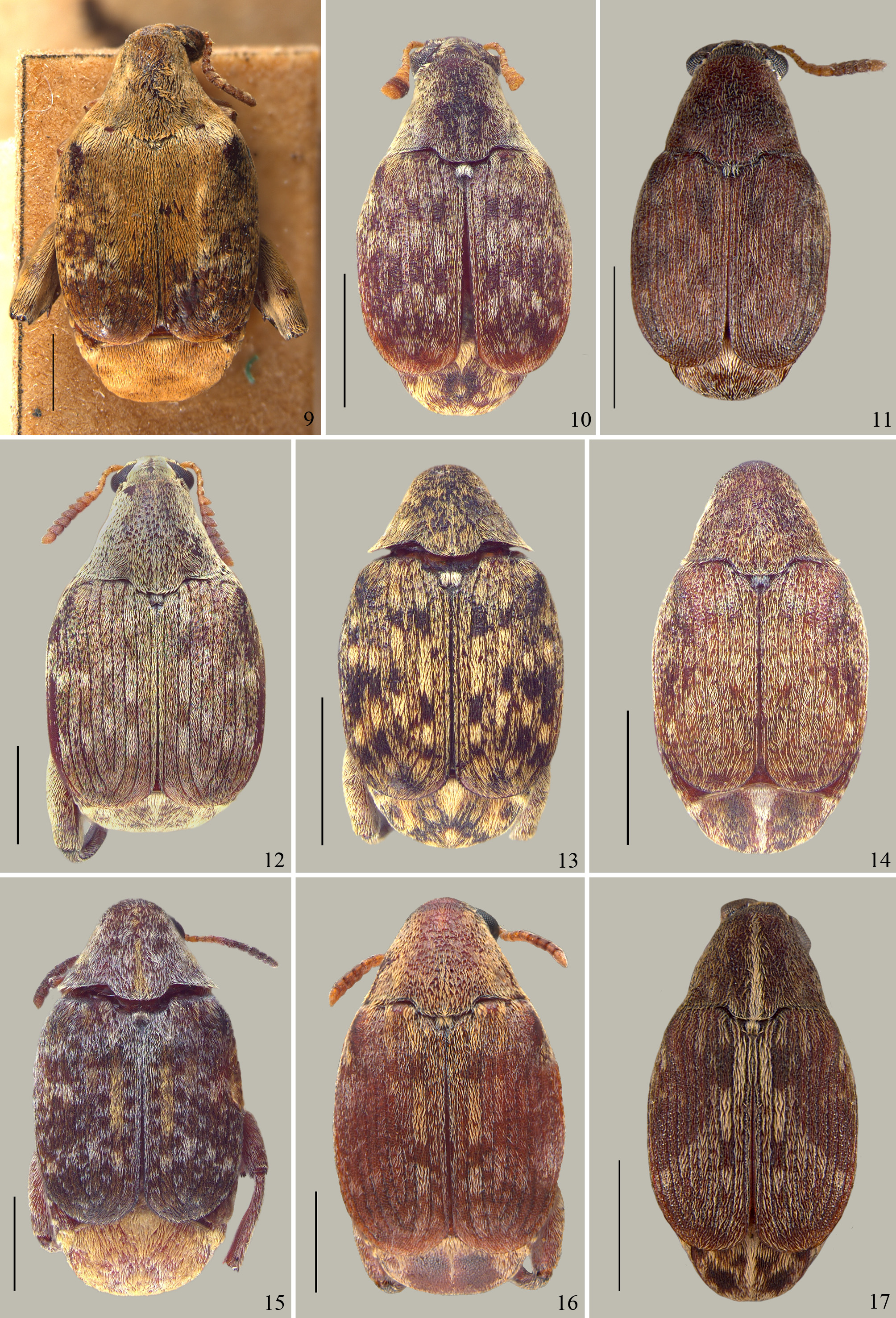
Integument. Dorsum ( Fig. 9 View FIGURES 9 – 17 ): reddish brown and dark brown. Antenna: pale brown; antennomeres 8–10 slightly darker ( Fig. 4 View FIGURES 1 – 8 ). Ventral region: reddish brown and dark brown except anterior and middle legs pale brown ( Fig. 4 View FIGURES 1 – 8 ). Pygidium : reddish brown ( Fig. 81 View FIGURES 81 – 92 ).
Pubescence. Dorsum ( Fig. 9 View FIGURES 9 – 17 ): pronotum homogeneous and dense, yellowish gray, pale brown and coppery; scutellum yellowish gray; elytron variegate, yellowish gray, white, pale brown and coppery, except for pale brown pubescence forming a dense subrectangulate area from anterior to submedian region of elytra. Ventral region: yellowish gray, pale brown and white ( Fig. 4 View FIGURES 1 – 8 ); abdominal ventrites 1–2 with glabrous and polished lateral areas ( Fig. 4 View FIGURES 1 – 8 ). Pygidium : homogeneous and dense, pale brown ( Fig. 56 View FIGURES 49 – 56 ).
Body. Subrectangulate body.
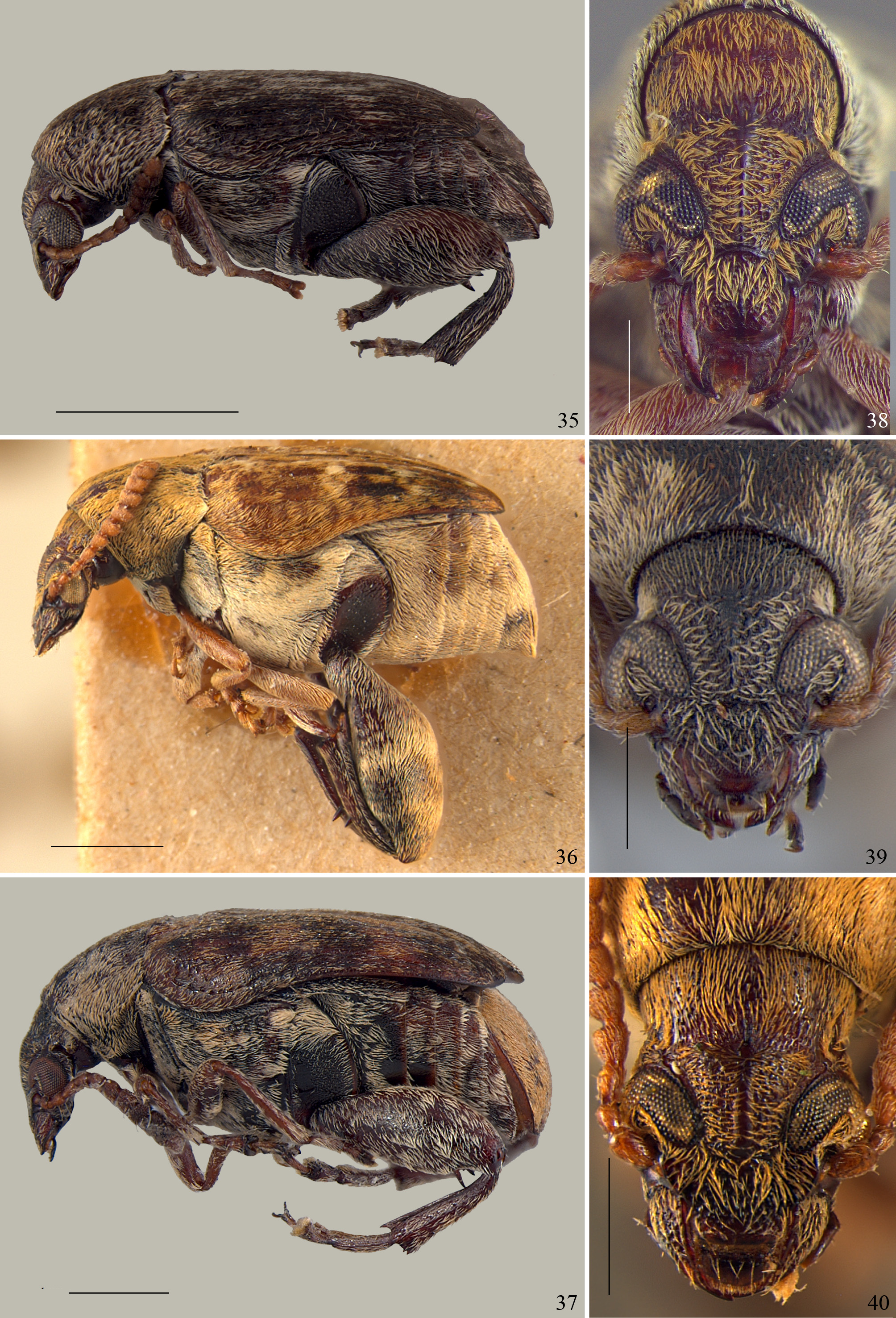
Head: without occipital sulcus ( Figs 2 View FIGURES 1 – 8 , 38 View FIGURES 35 – 40 ); ocular index 3.1 mm; ocular sinus 0.17 mm ( Figs 2 View FIGURES 1 – 8 , 38 View FIGURES 35 – 40 ); long postocular lobe ( Fig. 4 View FIGURES 1 – 8 ); frons slightly elevated ( Fig. 38 View FIGURES 35 – 40 ); conspicuous frontal carina, micropunctate, with regular borders, not enlarged at base ( Figs 2 View FIGURES 1 – 8 , 38 View FIGURES 35 – 40 ); frontoclypeal carina distinct ( Figs 2 View FIGURES 1 – 8 , 38 View FIGURES 35 – 40 ). Antenna: slightly clavate from antennomere 5 ( Fig. 4 View FIGURES 1 – 8 ); 6–10 wider than long; 11 longer than wide. Pronotum: lateral margins concave in dorsal view ( Fig. 9 View FIGURES 9 – 17 ); basal lobe with straight margin; median gibbosity absent ( Fig. 4 View FIGURES 1 – 8 ); pronotum flat at posterior region, without longitudinal median sulcus ( Fig. 9 View FIGURES 9 – 17 ); lateral gibbosities absent, only slightly depressed on lateral corners ( Fig. 9 View FIGURES 9 – 17 ); sparse moderate and shallow punctation intermixed with fine punctation all over pronotum; lateral carina absent. Scutellum: subquadrate, as wide as long ( Fig. 9 View FIGURES 9 – 17 ); bidentate. Elytron: humeral gibbosity strongly conspicuous ( Figs 4 View FIGURES 1 – 8 , 9 View FIGURES 9 – 17 ); basal gibbosity absent ( Figs 9 View FIGURES 9 – 17 , 41–42 View FIGURES 41 – 46 ); striae regular in course, without basal denticles, free apically and visibly impressed, except 4–6 formed only by isolated punctation ( Figs 41–42 View FIGURES 41 – 46 ). Mesoventrite: mesoventral process rounded apically; postmesocoxal sulcus rounded, following the curvature of the coxa ( Fig. 47 View FIGURES 47 – 48 ). Metaventrite: protuberant in lateral view ( Fig. 4 View FIGURES 1 – 8 ); metanepisternum with sparse coarse punctation; dorsal carina incomplete, not limiting posterior corner and margin. Hind leg: femur in lateral view projects beyond pygidium ( Fig. 4 View FIGURES 1 – 8 ); pecten with 4 teeth; internal margin without denticles at anterior region before pecten. Tibia, external face smooth, not microserrated; lateroventral carina incomplete; mucro longer than width of tibia apically; coronal tooth absent; curvature of the tibia at external margin reaching half length of tibia. Abdomen: last ventrite with median apical border strongly emarginate, with short lateral projections ( Fig. 55 View FIGURES 49 – 56 ). Pygidium : entirely convex; median lateral tubercles slightly elevated; apical margin rounded.
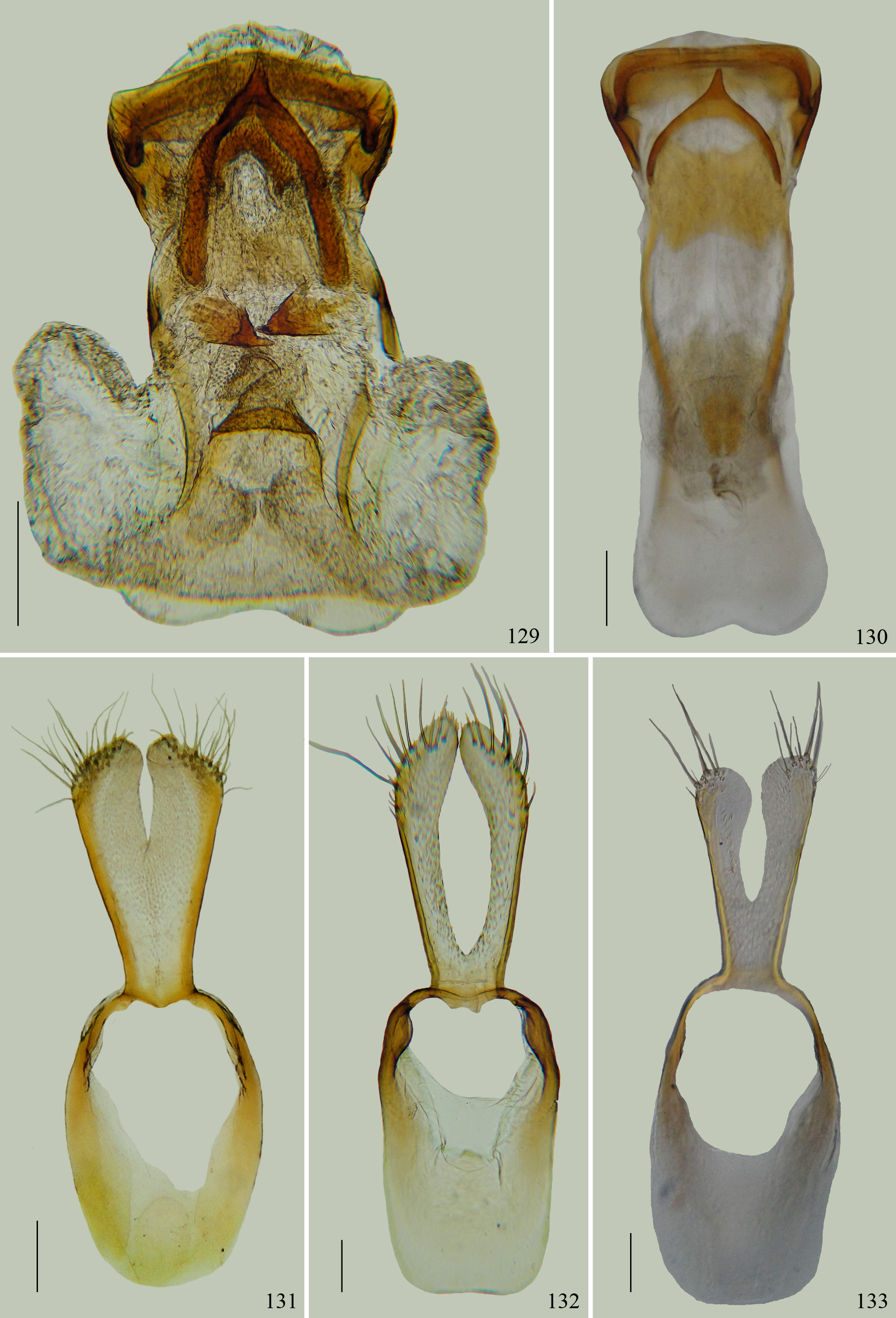
Male genitalia. Median lobe ( Fig. 105 View FIGURES 105 – 109 ): length about 3.5 x the maximum width at base of ventral valve. Ventral valve arcuate, wider than long and narrower than median lobe apically. Internal sac, apical region without spines, spicules or denticles; median region with a large Y-shaped sclerite, rounded apically; basal region with dense small and thin spicules, a few sparse setae, a pair of small spine-shaped sclerite with narrow base and short apex. Basal hood bilobate, 2 x wider than largest width at base of ventral valve. Tegmen ( Fig. 131 View FIGURES 129 – 133 ): lateral lobes slightly deep emargination, not reaching half of its length; external and internal margins straight; truncate and oblique apex, not convergent, wider than basal region of lobes; without projections toward the ring.
Variability. Body length (n = 10): 3.15–4.33 mm; width: 2.16–2.85 mm.
Pubescence. Pygidium : yellowish gray and white or yellowish gray ( Fig. 81 View FIGURES 81 – 92 ).
Body. Head: ocular index 3.06–3.57 mm; ocular sinus 0.13–0.18 mm. Hind leg: femur, pecten with 5 teeth.
Female. Abdomen: last ventrite, median apical border with arcuate emargination ( Fig. 52 View FIGURES 49 – 56 ), with long lateral projections forming digitate process laterally ( Figs 52 View FIGURES 49 – 56 , 81 View FIGURES 81 – 92 ). Pygidium : almost straight in female.
Diagnosis. This species differs from M. pickeli mainly by the long postocular lobe, pronotum concave laterally, elytron without basal denticles.
Distribution. Brazil (Pernambuco, Mato Grosso, Distrito Federal, Minas Gerais, São Paulo, Paraná, Rio Grande do Sul), Argentina (Jujuy, Salta, Formosa, Tucumán, Misiones, La Rioja, Santa Fé, Entre Rios, Santa Cruz), Paraguay (Cordillera).
Host plants. Fabaceae , Mimosoideae , Ingeae : Enterolobium contortisiliquum (Vell.) Morong. , Enterolobium timbouva Mart.
Remarks. When Terán & Muruaga de L'Argentier (1981) proposed a new combination to Merobruchus bicoloripes ( Pic, 1927) they listed the literature related to Bruchus bicoloripes Pic, 1930 erroneously because they must list the literature related to Pseudopachymerus bicoloripes Pic, 1927 .


No known copyright restrictions apply. See Agosti, D., Egloff, W., 2009. Taxonomic information exchange and copyright: the Plazi approach. BMC Research Notes 2009, 2:53 for further explanation.
|
Kingdom |
|
|
Phylum |
|
|
Class |
|
|
Order |
|
|
Family |
|
|
Genus |
Merobruchus bIcolorIpes ( Pic, 1927 )
| Manfio, Daiara & Ribeiro-Costa, Cibele Stramare 2016 |
Merobruchus bicoloripes: Terán & Muruaga
| Johnson 1997: 14 |
| Link 1995: 113 |
| Macedo 1992: 331 |
| Kingsolver 1988: 1 |
| Dipierri 1983: 87 |
Caryedes bicoloripes:
| Udayagiri 1989: 71 |
| Blackwelder 1946: 757 |
Caryedes ruficornis
| Blackwelder 1946: 758 |
Pseudopachymerus ruficornis
| Kingsolver 1988: 2 |
| Pic 1938: 22 |
Pseudopachymerus bicoloripes
| Pic 1927: 32 |