
Mesabolivar spinosus (González-Sponga, 2005)
|
publication ID |
https://doi.org/ 10.11646/zootaxa.4395.1.1 |
|
publication LSID |
lsid:zoobank.org:pub:B43C234D-45C4-4A6D-9836-8A7524A5B291 |
|
DOI |
https://doi.org/10.5281/zenodo.5950523 |
|
persistent identifier |
https://treatment.plazi.org/id/160AC713-C66A-FF9A-2A9C-9BE637F07D1C |
|
treatment provided by |
Plazi |
|
scientific name |
Mesabolivar spinosus (González-Sponga, 2005) |
| status |
|
Mesabolivar spinosus (González-Sponga, 2005) View in CoL
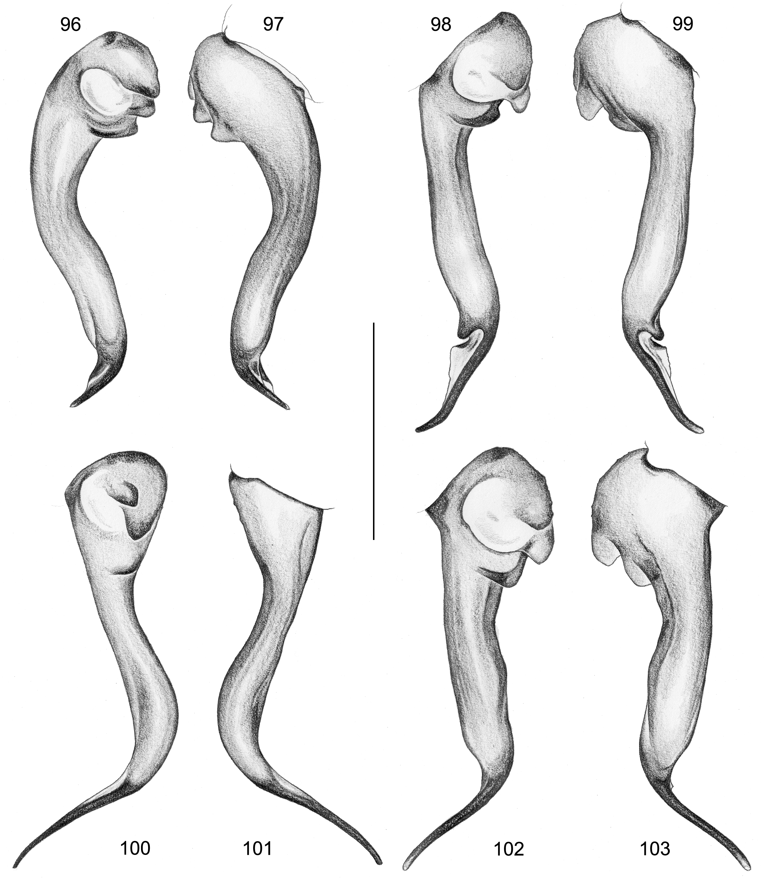
Figs 102–103 View FIGURES96–103 , 110–114 View FIGURES 104–114 , 130–132 View FIGURES 128–137
Rioparaguanus spinosus González-Sponga, 2005: 104 View in CoL , pl. 3, figs 1–9 (♂♀, Venezuela). Mesabolivar spinosus: Huber et al. 2014a: 418 View in CoL .
Diagnosis. Distinguished from most similar known relative ( M. cyaneus ) by wider and shorter procursus (compare Figs 101 and 103 View FIGURES96–103 ), by bulbal process with complex distal structures (compare Figs 109 and 111 View FIGURES 104–114 ), and by female external and internal genitalia (epigynum wider; relatively larger internal sclerotized structure, compare Figs 112, 114 View FIGURES 104–114 and Huber 2000: figs 799–800); from M. aurantiacus also by much wider palpal femur, by absence of ventral process on procursus, and by simple epigynal pocket (not extending anteriorly into long furrow).
Type material. VENEZUELA: Bolívar: 4♂ 2♀ 2 juvs types (see Note below), MIZA ( MAGS 1176), Municipio Heres, Río Paragua, base of Cerro Guaiquinima (5.800°N, 63.533°W), 920–1344 m a.s.l., 5.ii.1990 and 21.ii.1990 (E. Toro, L. Jaspe, M.A. González-Sponga); examined.
Redescription. Male (type, see Note below)
MEASUREMENTS. Total body length 3.8, carapace width 1.5. Distance PME-PME 160 µm, diameter PME 140 µm, distance PME-ALE 100 µm, distance AME-AME 30 µm, diameter AME 60 µm. Sternum width/length: 1.0/ 0.65. Leg 1: 56.0 (13.9 + 0.7 + 13.5 + 25.1 + 2.8), tibia 2: 9.2, tibia 3: 7.2, tibia 4: 9.1. Femora 1–4 width (at half length): 0.18, 0.18, 0.29, 0.19.
COLOR (in ethanol). Carapace ochre-orange with dark median line, ocular area light brown; sternum pale ochre-orange, margins brown; legs brown, tips of femora and tibiae lighter (pale ochre-yellow), without dark rings; abdomen greenish gray, dorsally and laterally densely covered with dark internal marks, ventrally with indistinct plate in front of gonopore.
BODY. Habitus similar to M. aurantiacus (cf. Figs 78, 80 View FIGURES 78–89 ); ocular area raised; carapace with distinct median furrow; clypeus unmodified; sternum unmodified.
CHELICERAE. With one pair of frontal apophyses close to median line, similar to M. aurantiacus (cf. Huber 2000: fig. 807) but slightly shorter.
PALPS. In general similar to M. aurantiacus (cf. Huber 2000: figs 805–806) but with much wider palpal femur; coxa and trochanter apparently identical to M. aurantiacus , femur proximal retrolateral apophysis also as in M. aurantiacus , but femur distally much wider; procursus as in Figs 102–103 View FIGURES96–103 , similar M. cyaneus , without ventral process (in contrast to M. aurantiacus ); bulbal process with complex distal elements ( Figs 110–111 View FIGURES 104–114 ).
LEGS. Without spines, without curved hairs, few vertical hairs; retrolateral trichobothrium on tibia 1 at 2.5%; prolateral trichobothrium present on tibia 1; tarsus 1 with>40 pseudosegments, distally distinct.
Male (variation). Tibia 1 in three other males: 12.4, 14.1, 16.3. Femur 3 in smallest male barely wider than other femora.
Female. In general similar to male but all leg femora approximately same width. Total body length 3.0, 4.5. Tibiae 1 missing in both females. Epigynum as in Figs 112–113 View FIGURES 104–114 , 130–131 View FIGURES 128–137 ; anterior plate wider than long, with median pocket, round internal sclerotized structure clearly visible through cuticle. Internal genitalia as in Figs 114 View FIGURES 104–114 , 132 View FIGURES 128–137 , with pair of small pore-plates.
Distribution. Known from type locality in Venezuela only ( Fig. 725 View FIGURES 724–725 ).
Notes. In the original description, González-Sponga (2005) mentions 2♂ 2♀ (♂ holotype “1176a”, 1♀ paratype “1176b”, and 1♂ 1♀ paratypes without explicit number). The MIZA collection has two vials, both labeled “1176”, one containing a single male, the other 3♂ 2♀ 2 juvs. While there is no reasonable doubt that this is the type material, it remains unclear why there are more specimens than reported in the original description, and which specimen is the holotype.
The separated male does not seem to be the holotype. Its tibia 1 is much longer (16.3) than that reported by González-Sponga (2005) for the holotype (12.9). Two of the other three males come close to this measurement (12.4, 13.5). The two vials possibly represent the two collecting dates. Since the conspecificity of all these specimens is beyond doubt, the males are here simply all considered “ types ”. There is no need at this point to treat these specimens as syntypes and to select a lectotype.
The total body lengths reported in the original description are clearly wrong (♂ 5.70, ♀ 6.65).


| MIZA |
Museo del Instituto de Zoologia Agricola Francisco Fernandez Yepez |
No known copyright restrictions apply. See Agosti, D., Egloff, W., 2009. Taxonomic information exchange and copyright: the Plazi approach. BMC Research Notes 2009, 2:53 for further explanation.
|
Kingdom |
|
|
Phylum |
|
|
Class |
|
|
Order |
|
|
Family |
|
|
Genus |
Mesabolivar spinosus (González-Sponga, 2005)
| Huber, Bernhard A. 2018 |
Mesabolivar spinosus:
| Huber et al. 2014: 418 |
spinosus González-Sponga, 2005: 104
| Gonzalez-Sponga 2005: 104 |