
Mesocoelium crossophorum, Perez Veigueras, 1942
|
publication ID |
https://doi.org/ 10.11646/zootaxa.3387.1.1 |
|
DOI |
https://doi.org/10.5281/zenodo.5256037 |
|
persistent identifier |
https://treatment.plazi.org/id/F21487DC-D309-056A-FAD7-8AC5C973F8FD |
|
treatment provided by |
Felipe |
|
scientific name |
Mesocoelium crossophorum |
| status |
|
Mesocoelium crossophorum View in CoL
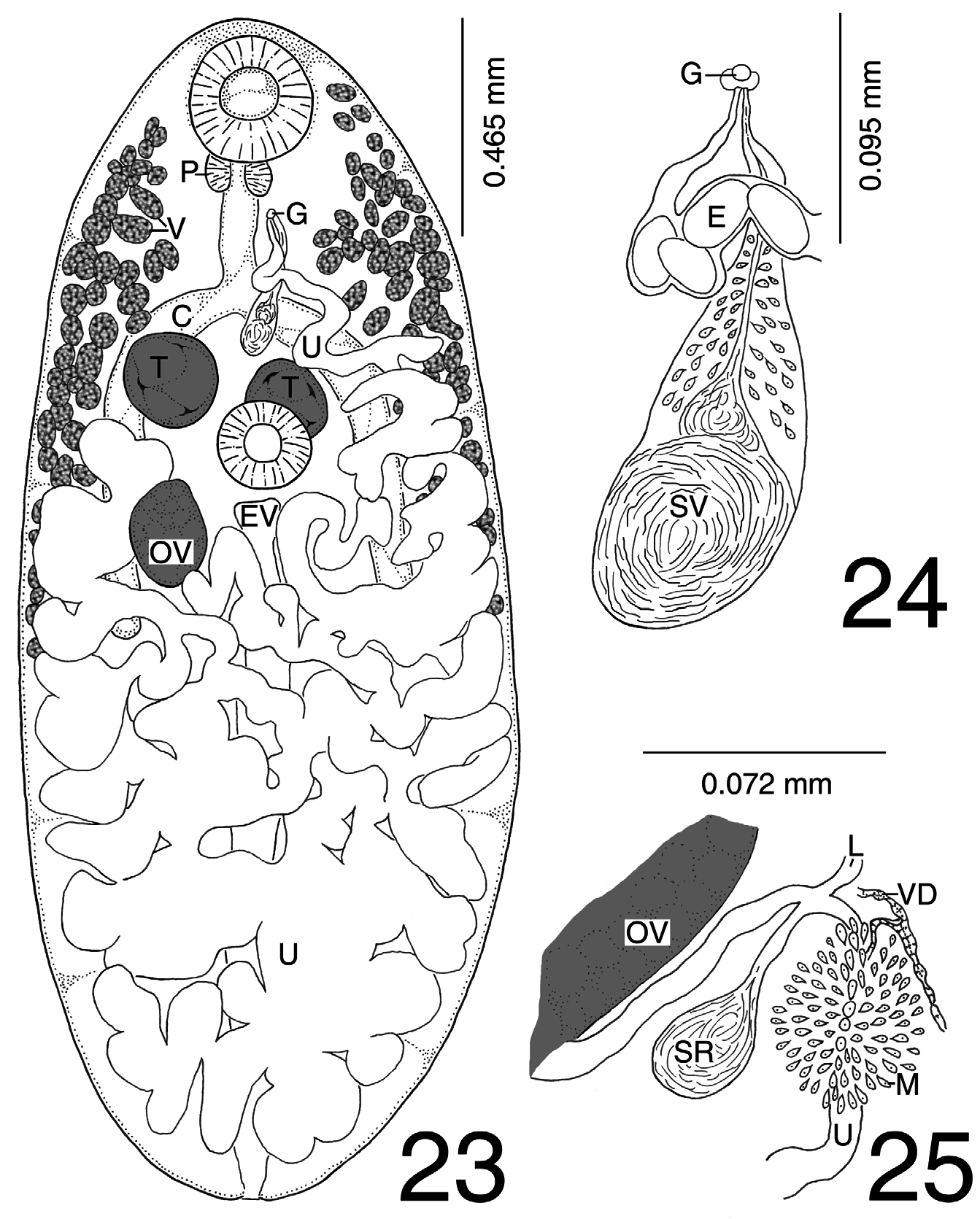
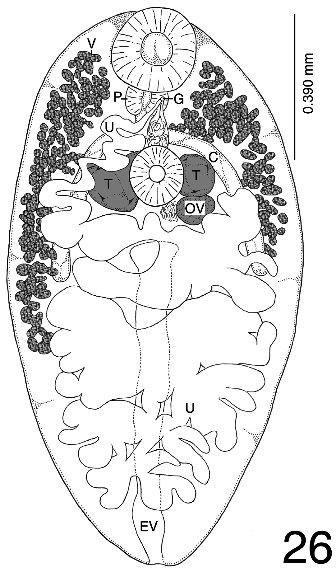
( Figures 23–26 View FIGURES 23–25 View FIGURE 26 ; Table 14)
Definitive hosts: Mochlus fernandi Mertens , African fire or Fernando’s skink ( Squamata : Scincidae ); Bufo marinus Linnaeus , the cane toad ( Anura : Bufonidae ).
Localities: Uganda; Honolulu, USA.
Site: Intestine.
Specimens examined: USNPC 045302.00 ( Uganda); 045639.00 (Honolulu).
Description of specimens: Based on four specimens. With characteristics of genus. Body monas type, small, oval, spinose, 2,263 (1,763 –2,750) by 1,054 (900–1,200), body spines 10–18 long; forebody 680 (500–870) long, 24–32% of body length. Mouth slightly subterminal; oral sucker spherical to subspherical, 302 (270–320) by 295 (275–320); prepharynx short; pharynx subspherical to spherical, wider than long, 101 (90–113) by 119 (105–110); esophagus 127 (38–210) long; cecal bifurcation near midlevel of forebody; ceca surpassing ovary posteriorly, terminating about ⅓ distance down hindbody, occupying 10–17% of postovarian space. Ratio of widths of oral sucker and pharynx 1:2.6 (1:2.1–1:2.8). Ventral sucker located anterior to midlevel of body, smaller than oral sucker, 227 (200–255) by 223 (190–255). Ratio of sucker widths 1:1.4 (1:1.1–1:1.6).
Testes smooth, side by side to diagonal, situated at level of ventral sucker. Right testis 175 (113–250) by 180 (165–200); left testis 168 (85–230) by 182 (148–205). Cirrus sac situated between pharynx and ventral sucker, enclosing short cirrus, reduced pars prostatica, short ejaculatory duct surrounded by prostate cells, and bipartite seminal vesicle, 207 (175–225, 6–11% of body length) by 74 (69–78). Genital pore near posterior margin of pharynx to slightly more posterior, prebifurcal, submedian.
Ovary smooth, posttesticular, situated short distance posterior to right or left testis, 156 (105–250) by 159 (148–170), removed from posterior end by some distance; postovarian space 1,250 (1,000 –1,425) long, 51–61% of body length. Ratio of width of ovary to mean width of testes 1:1.1 (1:1.0–1:1.2). Seminal receptacle spherical, located immediately sinistral and slightly posterior to ovary. Laurer’s canal present, opening not observed. Vitelline fields distributed along ceca from level of oral sucker posteriorly to about ⅓ distance down hindbody or slightly more posterior, terminating near to, or surpassing cecal ends; vitelline follicles 79 (24–104) by 55 (24–148) wide (n = 25). Uterus largely postacetabular, filling most of hindbody. Eggs operculate, 37 (35–40) by 23 (21–25) (n = 30).
Excretory vesicle I-shaped, reaching anteriorly to just above midbody, terminating near posterior margin of ovary; excretory pore subterminal.
Remarks: These specimens (USNPC 045302.00, 045639.00) have moderately long ceca, and a genital pore that is prebifurcal and submedian, placing them in the monas body type. The posterior extent of the vitelline fields terminate near to, or surpass the cecal ends posteriorly; the gonads overlap the area of the ventral sucker; the body is oval, widest from midlevel of body to the level of ventral sucker; the genital pore is immediately post pharyngeal; the ceca are relatively short, occupying 10–17% of the postovarian space and the eggs have a maximum size of 45 by 30, placing them in M. crossophorum . These specimens conform in all diagnostic characteristics to M. crossophorum (Table 14), but it should be noted that the body spines of those from Uganda (USNPC 045302.00) are 10–11 long, but are 14–18 from the specimens from Honolulu (USNPC 045639.00). Although it is possible that the specimens from Uganda represent a separate species from those from Honolulu, we do not feel that the length of body spines from a small sample is sufficient reason to separate these specimens. The original description of M. crossophorum by Pérez Veigueras (1942) distinguished it largely based on the presence of “cuticular, digitiform prolongations” on the lateral and anterior aspects of the oral sucker. As explained under “Characteristics unique to species” above, we believe these structures to be an artifact and did not consider them in our evaluation of these specimens. The specimen represented in Figure 4 View FIGURES 4–6 of the original description and those we examined from Uganda were more contracted than the specimens we examined from Honolulu, which we feel resulted in the genital pore being displaced further anterior (from the midlevel of the pharynx in the very contracted specimens from Uganda to immediately postpharyngeal from Figure 4 View FIGURES 4–6 of the original decription rather than being immediately anterior to the midlevel of the esophagus in those from Honolulu).

No known copyright restrictions apply. See Agosti, D., Egloff, W., 2009. Taxonomic information exchange and copyright: the Plazi approach. BMC Research Notes 2009, 2:53 for further explanation.
|
Kingdom |
|
|
Phylum |
|
|
Class |
|
|
Order |
|
|
Family |
|
|
Genus |