
Moina hemanti, Padhye, Sameer M & Dumont, Henri J, 2014
|
publication ID |
https://doi.org/ 10.11646/zootaxa.3860.6.4 |
|
publication LSID |
lsid:zoobank.org:pub:7143E18A-20D0-402F-A4F9-2EF986BD54BA |
|
DOI |
https://doi.org/10.5281/zenodo.5248435 |
|
persistent identifier |
https://treatment.plazi.org/id/9A174742-577C-5B30-C5F2-FC3DFEAEF805 |
|
treatment provided by |
Felipe |
|
scientific name |
Moina hemanti |
| status |
sp. nov. |
Moina hemanti sp. nov
( Figs. 1–5 View FIGURE 1 View FIGURE 2 View FIGURE 3 View FIGURE 4 View FIGURE 5 )
Etymology. The species is dedicated to Dr. Hemant Ghate, a well known zoologist from Pune, India who has studied a range of insects and has helped many Indian students in their entomological studies.
Type locality. Moina hemanti was seen only in one pool in the studied site, but consistently over three years (2010 – 2013). The diameter of the pool was about 90 cm. The depth was about 10 cm with no aquatic vegetation. The substrate consisted of mud with dead leaves fallen from the canopy. Its pH of ranged between 7–7.5 and temperature between 26–28°C.
Type material. Holotype A parthenogenetic female mounted on a glass slide in Polyvinyllactophenol ( PVLP) (Registration number: C 6196/2). It has been submitted at Zoological Survery of India ( ZSI) Kolkata.
Paratypes. Ten parthenogenetic females (in 70% ethanol) have been deposited as paratypes (Registration number: C 6197/2) to ZSI. Nine parthenogenetic females and one male (in 5% formalin) have been deposited as paratypes to Wildlife Information Liaison Development society (Registration number: WILD-14-CRU-001)
Collector for Holotype and paratype material: Dr. Yugandhar Shinde.
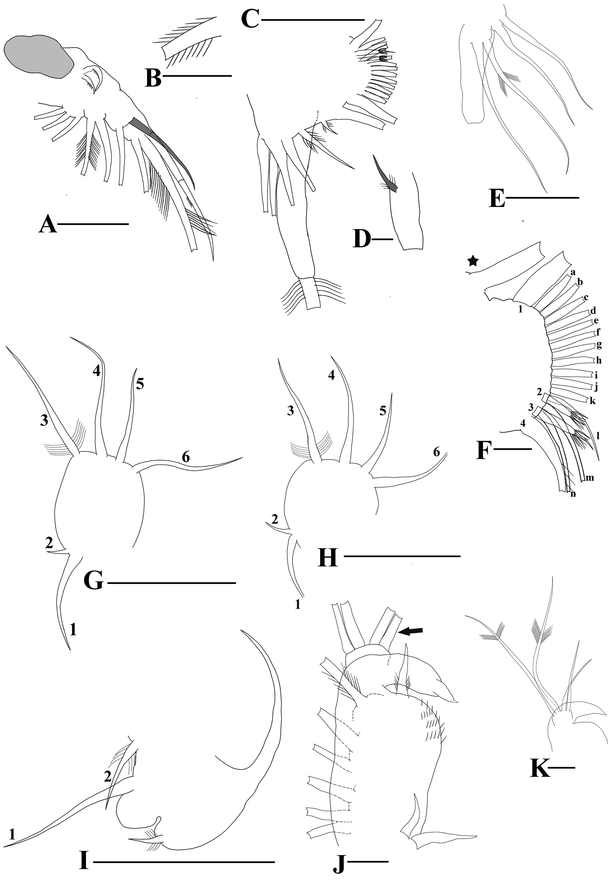
Diagnosis. Parthenogenetic female oval in shape with height/length ratio 0.6–0.74. Dorsal valve convex, valves striated. Rostrum slightly developed with varying rostral tip. Large ocellus present. Antenna 1 slender and lined with fine setae in groups on the posterior face; second antennae with two sensory setae of unequal length, small spine at base, projecting outwards. Abdomen with one protuberance. Postabdomen with 5–10 setulated teeth and asymmetric bident tooth, 4–5 basal spines on postabdominal claw.
Male with long antennule and 4 stout hooks at the tip. IDL of P1 with four setae, two long and setulated, two short and naked.
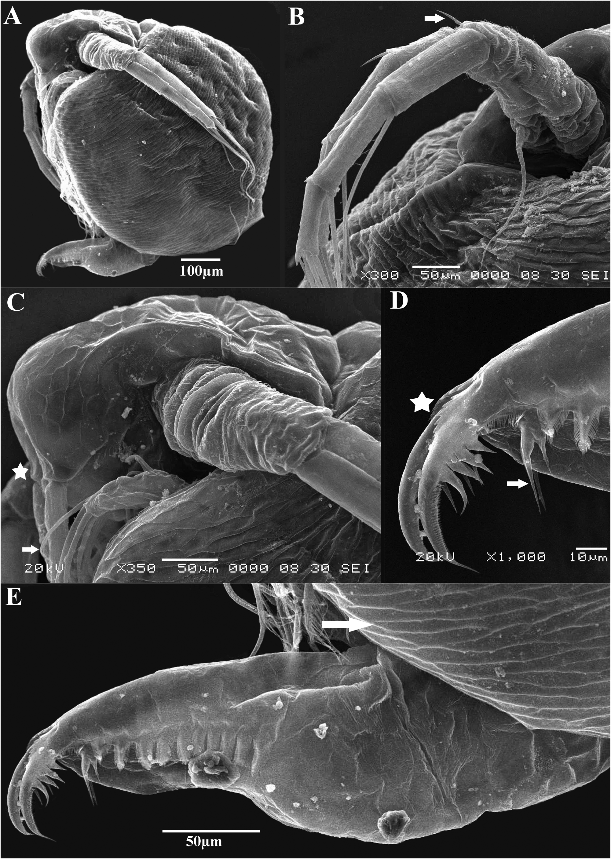
Description. Parthenogenetic female. General. Body oval and with height/length ratio (0.60–0.74) ( Fig.1 View FIGURE 1 : C, Fig.5 View FIGURE 5 : A).
Head of small size, length less than 4 times the total body length ( Figs.1C View FIGURE 1 , 2A View FIGURE 2 , 5C View FIGURE 5 ), supra-ocular depression present but not prominent ( Figs. 2A View FIGURE 2 , 5C View FIGURE 5 ). Rostrum well visible, its tip slightly upturned ( Figs. 2A View FIGURE 2 ‘arrow’, 4C ‘star’) but few specimens had a straight tip with no curvature ( Fig. 1C View FIGURE 1 ). Compound eye large ( Figs. 2A View FIGURE 2 , 5C View FIGURE 5 ), small ocellus located beneath the compound eye ( Figs. 1C View FIGURE 1 , 2A View FIGURE 2 ). No hairs present on the head ( Fig. 5C View FIGURE 5 ).
Labrum. Main body with a slightly convex dorsal margin, basal segment roughly cylindrical in shape, apical segment tip quadrate in shape, slightly upturned (Daphnid type, Dumont et al. 2013 and references therein) and lined with setae gradually decreasing in size posteriorly. ( Fig. 2C View FIGURE 2 ).
Valves. Dorsal margin high under head with a slight depression between head and rest of body ( Fig. 1C View FIGURE 1 ), highly arched when carrying embryos ( Fig. 5A View FIGURE 5 ) but laterally compressed and almost straight posteriorly otherwise ( Fig. 1C View FIGURE 1 ). Valves ornamented with elongated cells ( Fig. 5E View FIGURE 5 ‘arrow’). Ventral spinules ordered in two groups with initial larger spinules present on nearly the entire ventral margin, distance between each of these spinules roughly half their length; smaller spinules following the larger ones also arranged with longest in the first position followed by shorter spinules decreasing in length ( Fig. 2B View FIGURE 2 ); setulated hook seen at the dorso-posterior region of the valves, slightly re-curved and pointing inward ( Fig.2M View FIGURE 2 ).
Antennule slender, cylindrical, lined with setules on the posterior face and short spinules on the anterior face. Sensory seta located closer to base than to mid length and slightly more in length than the antennule base. Nine aesthetascs located at the tip Fig. 2D View FIGURE 2 ).
Antenna long, two setae located at the base of unequal length ( Fig. 2F View FIGURE 2 ) with length slightly more than the length of coxal segment. Long sensory seta, attached to the base ( Fig. 2E View FIGURE 2 ), extending up to middle of the third antennal segment. Basal segment with a short spine projecting outwards ( Figs. 2E View FIGURE 2 , 5B View FIGURE 5 “arrow”) Antennal setal formula: 0-0-1-3/1-1-3 and spines formula: 0-1-0-1/0-0-1. Each segment lined with transverse rows of small spinules ( Fig. 5B View FIGURE 5 ) with plumose setulation; lateral seta of basal segment of endopod asymmetrically setulated with one side having long plumose setulation while the other side having very short setules ( Fig.2 View FIGURE 2 : G).
Abdomen with a small single abdominal projection ( Fig. 2H View FIGURE 2 shaded gray).
Postabdomen well extended and narrowing distally ( Figs. 2I View FIGURE 2 , 5E View FIGURE 5 ); ventral margin straight but slightly variable ( Figs.2J & K View FIGURE 2 ) and with slightly convex border. Margin lined with rows of setules. Six-ten triangular plumose teeth laterally ( Figs. 2I View FIGURE 2 , 5E View FIGURE 5 ). Bident tooth with highly unequal branches, ( Figs. 2L View FIGURE 2 , 5D View FIGURE 5 ‘arrow’) with distal tooth at least twice as long as proximal tooth and first few plumose denticles. A fine row of spinules present anterior to the plumose teeth ( Fig. 5 D View FIGURE 5 ). Large postabdominal claw, bowed and with an acute and projecting apex, lined with fine pecten dorsally ( Fig. 5D View FIGURE 5 ). Usually four robust basal spines present at the proximal end of the claw ( Figs. 2I View FIGURE 2 , 5D View FIGURE 5 ); 5 spines also seen but 3 spines not observed. Ventral margin at the base of claw also lined with 3–6 stout denticles ( Fig.5 View FIGURE 5 : D ‘star’).
Limb 1 with 10 setae, ejector hooks well developed, unequal in size ( Fig. 3A View FIGURE 3 ). IDL with 3 setae, the anterior most with fine setulation. Endite 3 with two setae, one relatively short anterior seta slightly more chitinized, curved and lined with undersized setulation ( Figs. 3A View FIGURE 3 shaded gray, 3B, 5C ‘arrow’) and one posterior seta with long setules. Endite 2 with 2 long setae and endite 1 with 3 setae and plumose setulation ( Fig. 3A View FIGURE 3 ).
Limb 2. Exopodite with long seta with long plumose setulation ( Fig. 3C View FIGURE 3 ), small seta present at its base, slightly longer than the base of exopodite ( Fig. 3D View FIGURE 3 shaded gray). Three endites having 4 setae with plumose setulation ( Fig. 3E View FIGURE 3 ). Two setae present in the intermediate zone of P2 with disproportional lengths, the longer seta (adjacent to endite 1) at least 3 times longer than the shorter one. 'Beating seta' (as given by Kotov et al. 2005) present adjoining the gnathobase ( Fig. 3F View FIGURE 3 ‘star’). Gnathobase with one long anterior inner row seta bordering the 'beating seta' ( Fig. 3F View FIGURE 3 ‘1’) and plumose setulation; 3 setae in the posterior inner row ( Fig. 3F View FIGURE 3 ‘2–4’). Seta 2 and seta 3 are of dissimilar size, seta 2 always smaller and usually 2/3 rd the length of seta 3. Both the setae lined with concentrically arranged thick setules; seta 4 lined on one side with long thick setules interspersed between slender setules ( Fig. 3F View FIGURE 3 ). Twelve - thirteen setae ( Fig. 3F View FIGURE 3 ‘a-n’) present on the filtering plate with the last two more chitinized than the rest.
Limb 3. Exopodite sub-rectangular with 6 setae, bilaterally setulated, setal length: 3>4>6>1~5>2 ( Fig.3G View FIGURE 3 ). Four endital setae and 38–42 gnathobasic filter setae present.
Limb 4. Exopodite rounded with 6 setae, bilaterally setulated, setal lengths similar to exopodite of limb3 ( Fig.3 View FIGURE 3 : H).Two endital setae and 30–34 gnathobasic filtering setae present.
Limb 5. Exopodite with 2 setae, highly unequal size, longer seta at least 1.2 times the length of the exopodite body. Endopod with ovoid lobe and 2 setae, one nearly as long as the 1st exopodite seta, 2nd seta 1/3rd of the length of the first. All setae bilaterally setulated ( Fig.3 View FIGURE 3 : I).
Male. Rostrum reduced, eye large ( Fig. 4A View FIGURE 4 ). Postabdomen similar to female, gonopores tending to open on the ventral face of postabdomen ( Fig. 4C View FIGURE 4 ). Long, slender antennule, slightly bent; two small setae present nearer to the base, sensory seta being longer of the two ( Fig. 4A View FIGURE 4 ); nine aesthetascs present alongside four ejector hooks at the antennal tip, small spinules also present lining the tip adjacent to the hooks ( Fig. 4B View FIGURE 4 ). Four setae present in IDL of P1 ( Fig. 3K View FIGURE 3 ), endite 3 with two setae, highly uneven in size. Copulatory hook short with undulating margin and a pointed apex, base opposite the hook lined with spinules, two ejector hooks situated at the base of the corm ( Fig.3J View FIGURE 3 ).
Ephippial female. In spite of intensive search, we were not able to find any sexual females or ephippia either.
Size. Females ranging from 640 to 850 µm (n=15). Males ranging from 510 to 620 µm (n=5).
Distribution and ecology: Known only from type locality. It was found in a stream-fed ephemeral pool. The pool was extremely labile with regards to its duration. It used to get turbid in few days (with distinct pungent smell) if the rains stopped and became dry soon after, suggesting little Dissolved Oxygen. Therefore, all animals collected were characteristically red in color ( Fig.1C View FIGURE 1 ). The pool was visited by cattle regularly and hence dispersal via these carriers would be likely but no specimens were seen in any other pools that formed in that habitat nor in any localities nearby.


No known copyright restrictions apply. See Agosti, D., Egloff, W., 2009. Taxonomic information exchange and copyright: the Plazi approach. BMC Research Notes 2009, 2:53 for further explanation.