
Montesinus monnei, Grossi, Paschoal Coelho, 2016
|
publication ID |
https://doi.org/ 10.11646/zootaxa.4078.1.20 |
|
publication LSID |
lsid:zoobank.org:pub:836FA2B8-7871-4DED-9646-BD39A4BAA031 |
|
DOI |
https://doi.org/10.5281/zenodo.6073654 |
|
persistent identifier |
https://treatment.plazi.org/id/0395E107-FF9D-680E-FF60-FF2C3622FC01 |
|
treatment provided by |
Plazi |
|
scientific name |
Montesinus monnei |
| status |
sp. nov. |
Montesinus monnei View in CoL sp. nov.
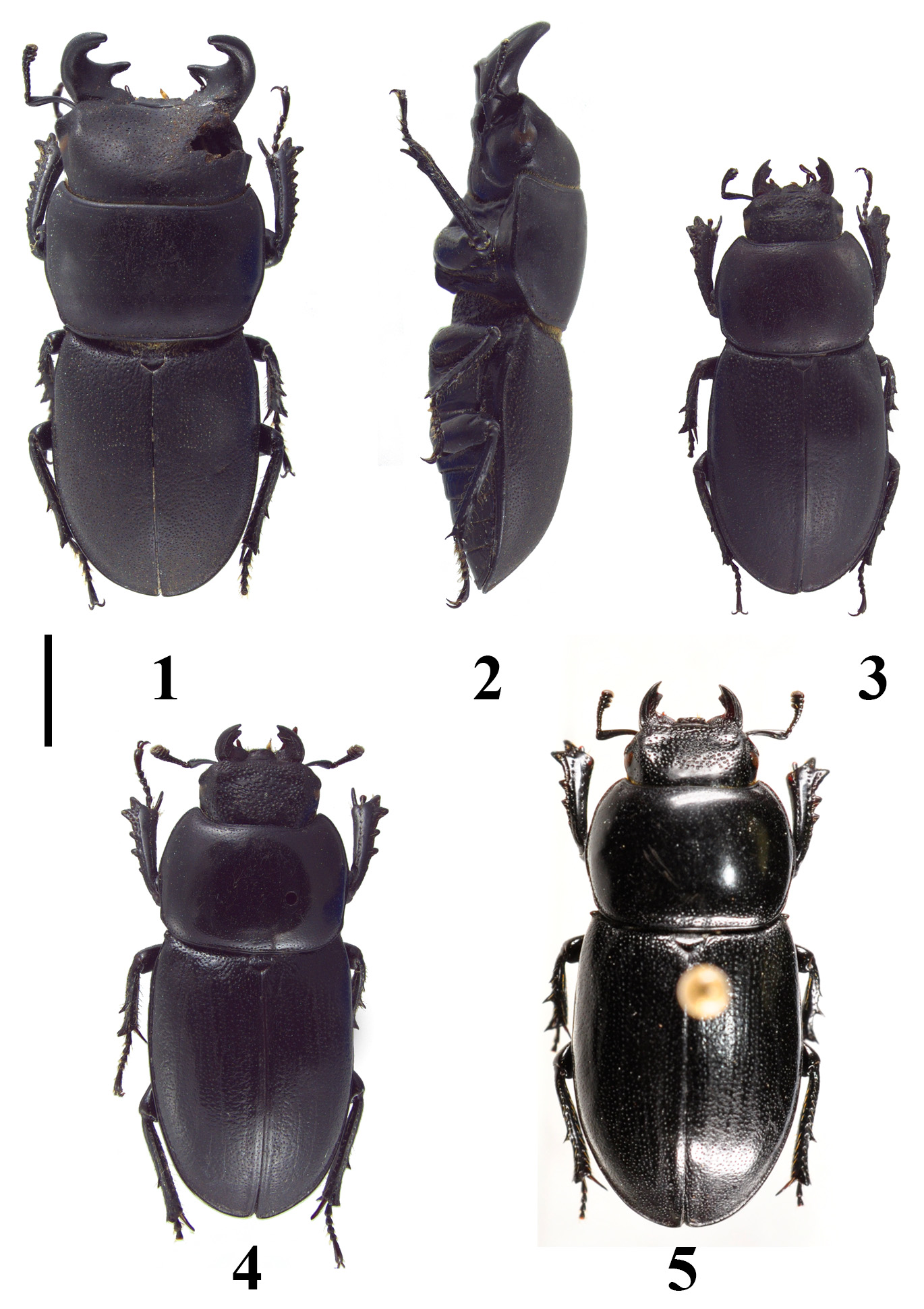
( Figures 1–3 View FIGURES 1 – 5 , 6–19 View FIGURES 6 – 12 View FIGURES 13 – 17 View FIGURES 18 – 21 , 22 View FIGURES 22 – 27 )
Material examined. Holotype male (dissected) labeled as follows: Handwritten white label, a) “BRASIL-Esp. Santo/Parque Nac. Caparaó/ Próx. Pico da Bandeira/ (2500 m) 13.IV.2001 / Fabio Oliveira Correia”; b) “ Montesinus monnei n. sp. /♂ HOLOTYPUS / P. C. Grossi det. 2015”. Allotype female labeled as holotype except for the female symbol and the ALLOTYPUS label. Holotype and allotype deposited at MNRJ.
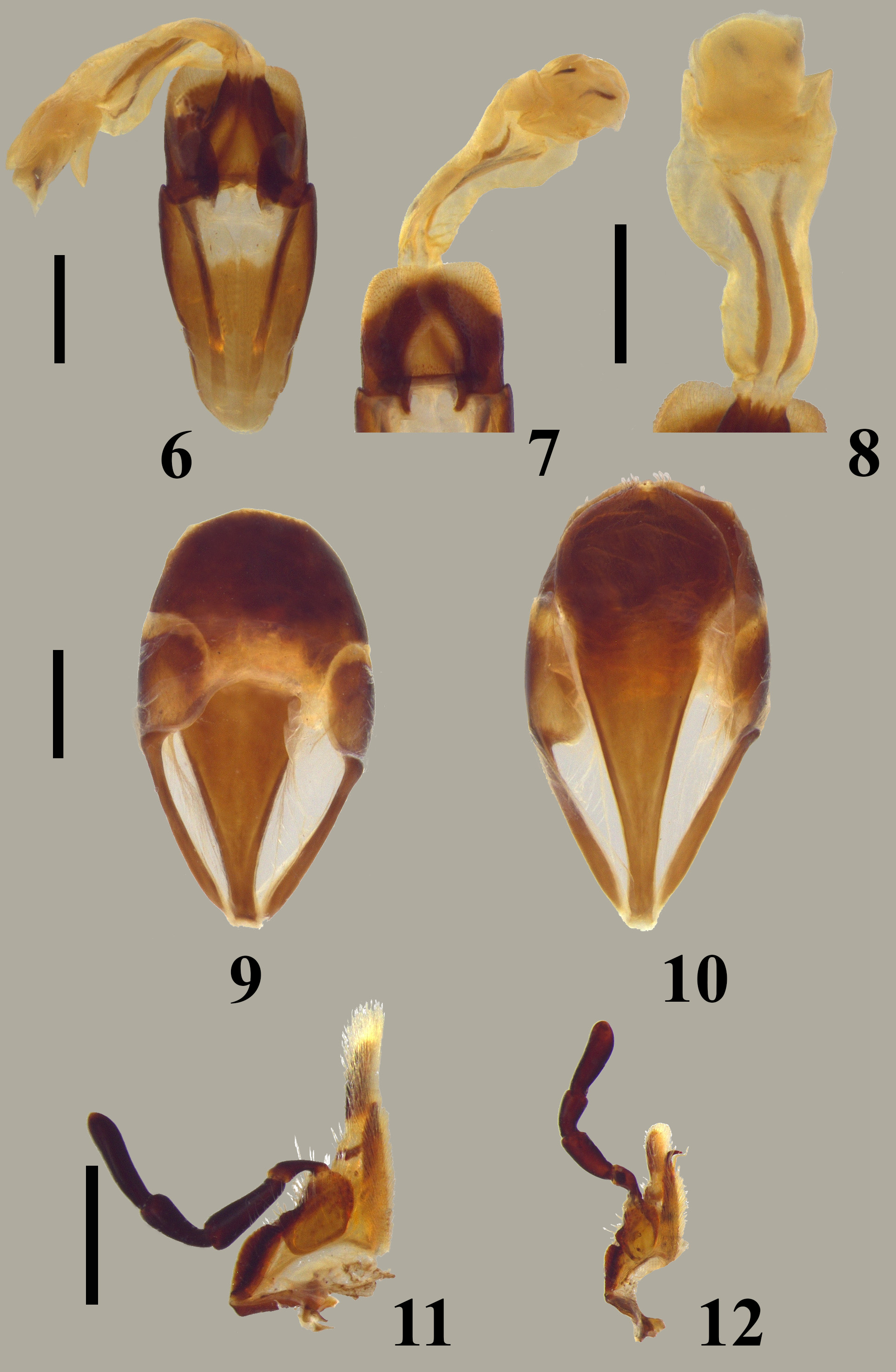
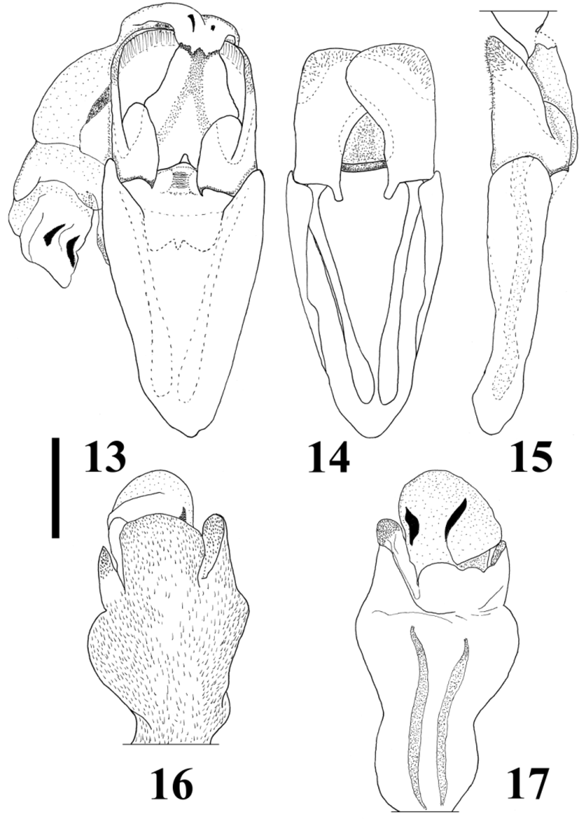
Holotype description. Total length, including mandibles, 19.0 mm. Greatest pronotal width 7.7 mm. Color: Dorsal surface dull black, pronotum and legs somewhat opaque ventrally with gular area, femora and trochanter reddish dark. Mouthparts: Mandibles symmetrical, cylindrical in cross section, abruptly upturned; apex slightly incurved, acutely pointed and with an obscure internal tooth; integument finely reticulated; dorsal base with a convex process; basal tooth conical, slightly emarginate at posterior apex ( Figs 1–2 View FIGURES 1 – 5 ). Ventrally near basal tooth with a subtriangular punctate area. Labrum trapezoidal, with 3 anterior rounded processes; punctures concentrated at edge; anterior margin and anterior angles with yellowish setae. Mentum with sparse large punctures from middle to anterior edge. Maxillae with lacinia simple, not curved, and with no hooklets; palpi slender with 2–4 almost with the same length ( Figs 11–18 View FIGURES 6 – 12 View FIGURES 13 – 17 View FIGURES 18 – 21 ). Head: Shape transverse, as wide as elytra; vertex broadly convex and scarcely punctate, punctures fine; disc depressed until anterior margin, more punctate, punctures fine to moderate. Canthi subtriangular in shape, externally projected. Anteriorly and above eyes with a convex process each side. Temporal process lacking, instead with a small convex area. Gena deeper than gula, disc convex and sparsely punctate; punctures fine. Gula anteriorly with integument microreticulate and sparse to moderately punctate; punctures fine to moderate; posteriorly with a subrectangular smooth area limited by two subparallel sutures. Prothorax: Pronotum slightly convex, sparsely punctate elsewhere; punctures fine, more punctate near anterior angles; along posterior margin with moderate punctures; all pronotal angles rounded. Scutellum: Shape subtriangular, very sparsely and finely punctate; surface anteriorly depressed. Elytra: Surface totally punctate; punctures fine to large (mostly moderate); near suture punctures fine, anteriorly larger. Each puncture with internal lateral wall, visible floor, and single setae. Punctures on elytral declivity with elongated setae, longer than puncture diameter. Humeri externally projected and rounded distally. Legs: Protibiae distally with 2 external strong teeth; medially to base with 6–8 smaller ones, 2 of these more developed. Mesotibiae with 3 external teeth, distal one much longer than the others; right tibia with the distal longer and left tibia with the proximal longer. Metatibiae with 1 external middle tooth. Genitalia: Genital capsule simple, rounded and sclerotized. Tergite with lateral paired plates quadrate, discal area more strongly sclerotized; between disc and lateral plates less membranous. Sternite setose at middle until discal area and with some scattered setae at posterior margin. Genitalia with simple genital capsule ( Figs 9–10 View FIGURES 6 – 12 ). Aedeagus symmetrical, instead internal sac apex ( Figs 6–8 View FIGURES 6 – 12 , 13–17 View FIGURES 13 – 17 ); basal piece strongly sclerotized ventrally with lateral apical sides directed backward being mostly membranous dorsally and slightly curved ( Figs 6 View FIGURES 6 – 12 , 13 View FIGURES 13 – 17 ); paired struts present, as long as basal piece length with apex wider and less sclerotized. Parameres large, leaf-like shaped and apically broad, 1.3x shorter than basal piece; ventrally in its curvature with a two-folded flange ( Figs 6–7 View FIGURES 6 – 12 , 13–15 View FIGURES 13 – 17 ); internal ventral base with an outcurved process; dorsally with internal base projected ( Figs 6 View FIGURES 6 – 12 , 13, 15 View FIGURES 13 – 17 ); external surface near apex with small, dense setae. Median lobe trapezoidal, strongly sclerotized and weakly, irregularly emarginate at apex; base with one symmetrical emargination. Dorsal cross bar transverse, straight. Internal sac permanently everted, shorter than tegmen (basal piece + parameres), almost membranous and partly coated by dense short setae; middle expanded laterally and apex with two asymmetrical, lateral projections covered by minute sclerotized spicules; internally with 4 paired sclerites, 2 elongate from base until base of apical part, and two subtriangularly shaped located at apex ( Figs 6–8 View FIGURES 6 – 12 , 13, 16–17 View FIGURES 13 – 17 ).
Female ( Figs 3 View FIGURES 1 – 5 , 12 View FIGURES 6 – 12 , 19 View FIGURES 18 – 21 , 22 View FIGURES 22 – 27 ). As male in general aspect, except by the following characters. Total length 15.0 mm. Greatest pronotal width 5.6 mm. Mouthparts: Mandibles shorter than head, incurved; basal teeth lacking, apex acute with an internal concavity; each mandible with a single forward projected tooth, the left tooth somewhat sinuous, posteriorly to it abruptly curved; internal dorsal carina present, above it concave; external dorsal carina present only at mandibular base, incomplete and making with internal carina a weak depression. Labrum trapezoidal, irregularly sinuous in anterior margin, sparsely punctate and setose only anteriorly. Maxillae with lacinia hook-shaped right angled, apex slender, and with one spiral-shaped hooklet ( Figs 12 View FIGURES 6 – 12 , 19 View FIGURES 18 – 21 ). Mentum less transverse, anteriorly slightly sinuous; each lateral convex; moderately punctate, punctures large to coalescent, each puncture with a single elongate setae, more evident near margins. Head: Smaller in size, ½ width of male’s head; broader than 1 elytron width. Disc at each side with concave impression, almost totally punctate; punctures large to coarse. Vertex posteriorly impunctate, anteriorly convex and strongly punctate; punctures large to coarse. Canthi not externally projected, just rounded, intruding eyes less than a half of eye length. Eyes closer to anterior pronotal margin. Gena not convex and more punctate, posteriorly setose and reddish. Gula broader with semicircular punctures, a transverse deep suture and reddish brown coloration. Prothorax: Pronotum not as transverse, rounded at lateral edges, slightly convex and almost reddish except near lateral margins, black just before anterior and posterior angles; surface moderately punctate; punctures fine. Scutellum : smaller, less punctate and surface nearly smooth. Elytra: Shape oblong and more elongate. Surface totally punctate; punctures smaller and shallow. Legs: Shape smaller. Protibiae wider, with 2 distal and only 2 proximal teeth. Mesotibiae less dentate, left tibia with only one external tooth, and right tibia with two teeth, the proximal tooth smaller. Metatibiae with teeth less acute, apex almost truncate.
Etymology. The specific name is an homage to my friend Dr. Miguel Angel Monné Barrios, one of the greatest longhorn beetles specialist, and who kindly showed me the specimens here described, that were among the miscellaneous Coleoptera at MNRJ.
Remarks. The specimens described here are the only two specimens known, and were apparently found dead, as the male has the right part of head, including canthus, eye and antennae, missing, as is the last tarsomere of the right metatarsus. The female is also damaged, since the right antennae, four last tarsomeres of left protarsus and two last tarsomeres of left mesotarsus are missing. This species can be distinguished from the other species by its smaller body size, opaque body surface and deeper, more distinct elytral punctures. It was also found near the highest place in the Serra do Caparaó, called Pico da Bandeira, while the second species described here is from another formation (Serra do Espinhaço), and the third from a lower place at Caparaó.


| MNRJ |
Museu Nacional/Universidade Federal de Rio de Janeiro |
No known copyright restrictions apply. See Agosti, D., Egloff, W., 2009. Taxonomic information exchange and copyright: the Plazi approach. BMC Research Notes 2009, 2:53 for further explanation.