
Moschoneura, Butler, 1870
|
publication ID |
https://doi.org/ 10.11646/zootaxa.4347.3.1 |
|
publication LSID |
lsid:zoobank.org:pub:610C16FC-0583-4325-B264-6D768E48BC88 |
|
DOI |
https://doi.org/10.5281/zenodo.6002171 |
|
persistent identifier |
https://treatment.plazi.org/id/4F018817-FF99-144D-FF09-F8FDDABAF948 |
|
treatment provided by |
Plazi |
|
scientific name |
Moschoneura |
| status |
|
Moschoneura View in CoL View at ENA
We studied only males of the following subspecies of Moschoneura : M. ela xanthella , M. ithomia ithomia , M. pinthous patricia , and M. pinthous monica (see the appendix).
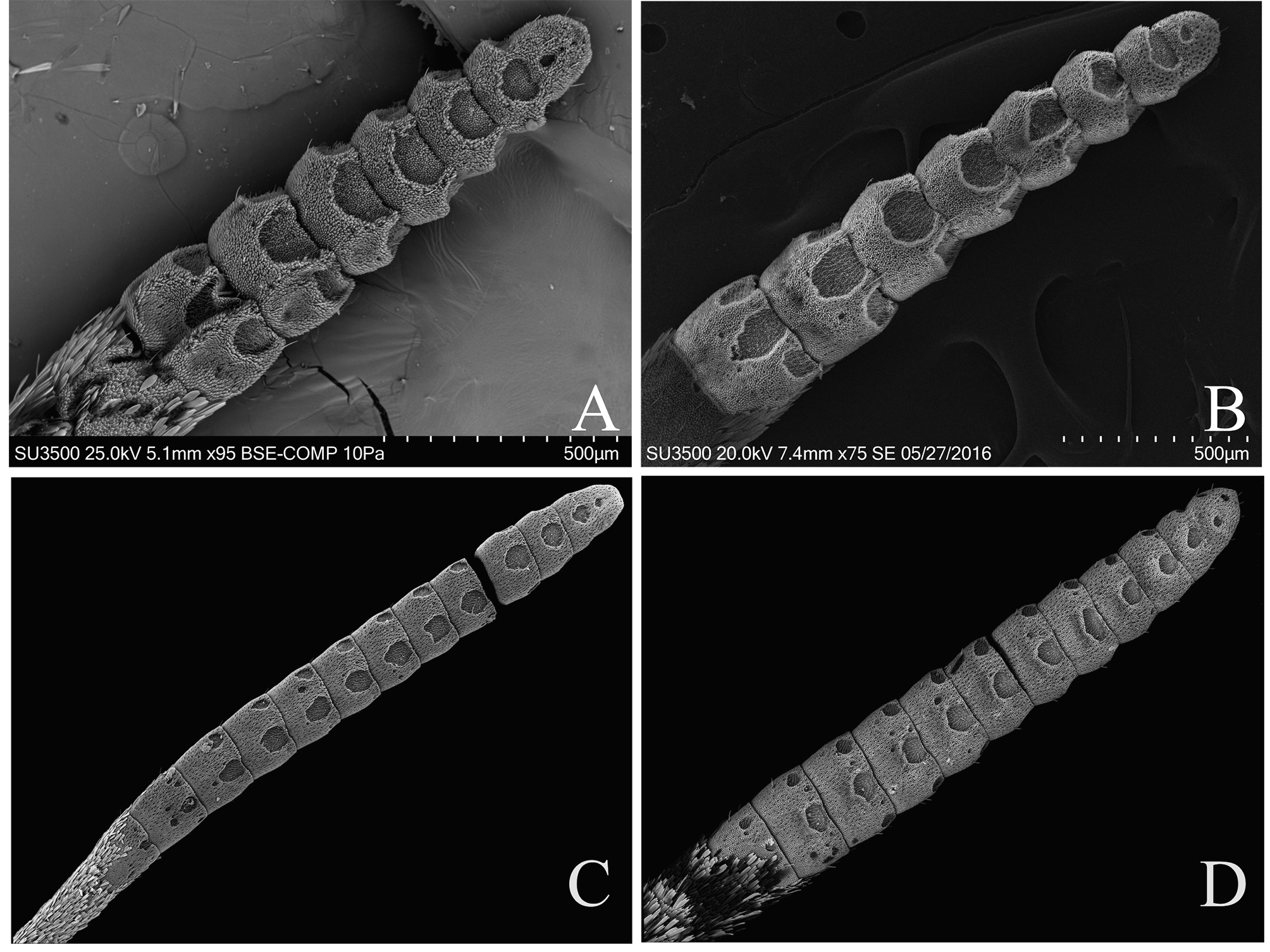
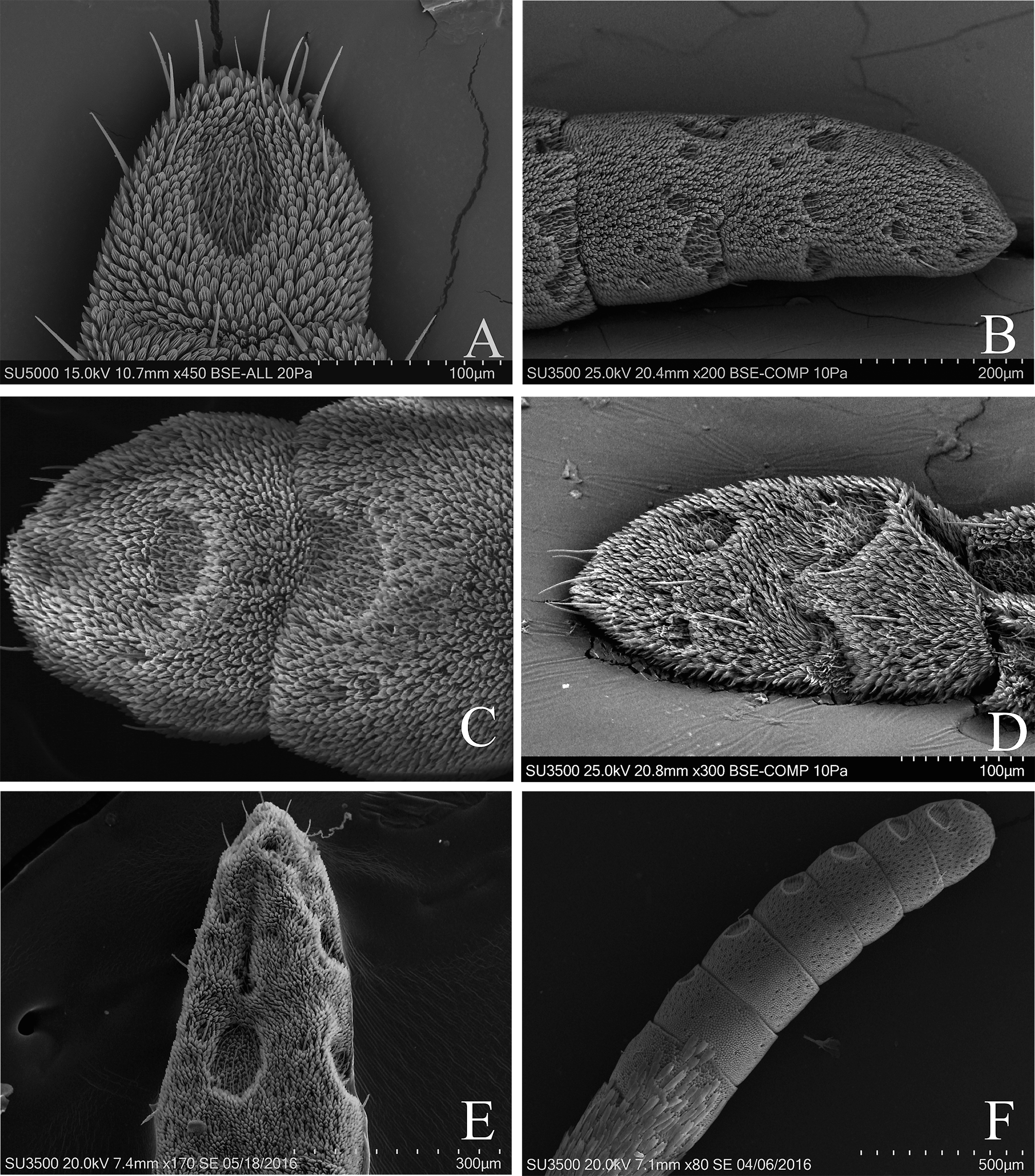
ANTENNAL CLUB: The antenna is greater than one-half the length of the costal margin of the forewing; it is clavate. The scaleless antennal club ranges from 1280–1700 µm ( Fig. 8B View FIGURE 8 ), with an average of 1.4 mm (1410 µm). M. e. xanthella has the longest club and M. p. monica the shortest.
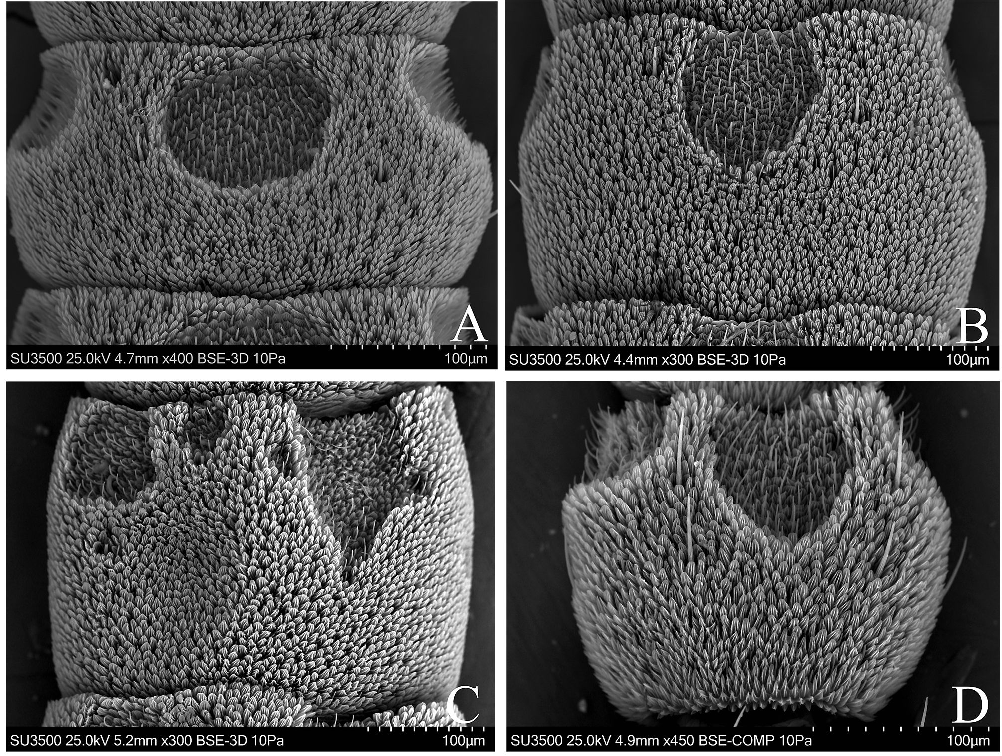
ANTENNOMERES: There are usually seven to eight scaleless antennomeres: seven in M. i. ithomia and M. p. monica ; eight in M. e. xanthella and M. p. patricia . The first scaleless antennomere is cylindrical, and the following have a hexagonal outline and are depressed (their amplitude is 1.5 times its length) ( Fig. 10D View FIGURE 10 ). The distal antennomere is digitiform with a slight constriction in the middle ( Fig. 8B View FIGURE 8 ). The first antennomere is scaled on the dorsal surface and there are a few scales laterally; however, M. p. monica has scales on the mesial-ventral surface but not at the base of the antennomere. Usually the scaled area on the dorsal surface occurs on the first and sometimes the second antennomere ( M. p. monica ). The distal antennomere corresponds to two fused and is a little less than twice in length than preceding ( Fig. 11D View FIGURE 11 ). Sometimes between the distal and penultimate, there is an incomplete cleavage ( M. p. patricia ). M. p. monica has a slight suture that occurs when the fusion of the last two antennomeres is not complete. The area without sensilla on the dorsal surface occurs from the third antennomere to the distal end, and not in the middle of the antennomere as in Pseudopieris ; the “naked” area is on the base in M. p. monica . On the ventral surface of the first scaleless antennomere there is only a very small lateral sulcus (i.e., M. p monica and M. i. ithomia ); in M. e. xanthella we observed the two lateral sulci unreduced, and in M. p. patricia one of them is slightly reduced. Central sulci are present in all scaleless antennomeres and always have similar dimensions.
SULCI AND PSEUDOSULCI: The sulci are very large and are very close together; they are almost circular or elliptical horizontal, reaching the distal edge of the antennomere, and are truncated. They have a well-defined outline and almost always are regular ( Fig. 12E View FIGURE 12 ). They occupy one-half the length of the antennomere and range from one-half to two-thirds in width ( Fig. 10D View FIGURE 10 ). Almost all central sulci have a similar size, although the distal is always a little smaller. The number of sulci has the following configurations: 7 central and 10 lateral in M. i. ithomia ; 7 central and 11 lateral in M. p. monica ; and 7–8 central and 14 lateral in M. e. xanthella and M. p. patricia . Pseudosulci are usually lacking; there may be two in the entire antennal club ( M. e. xanthella ). The lateral sulci are almost as large as the central one, occupying one-third to one-half the length of the antennomere and are truncated. Only the lateral sulci of the first antennomere (or rarely the second) has a lateral-mesial position; the remainder are located laterally and the last one is located laterally or laterodorsal.
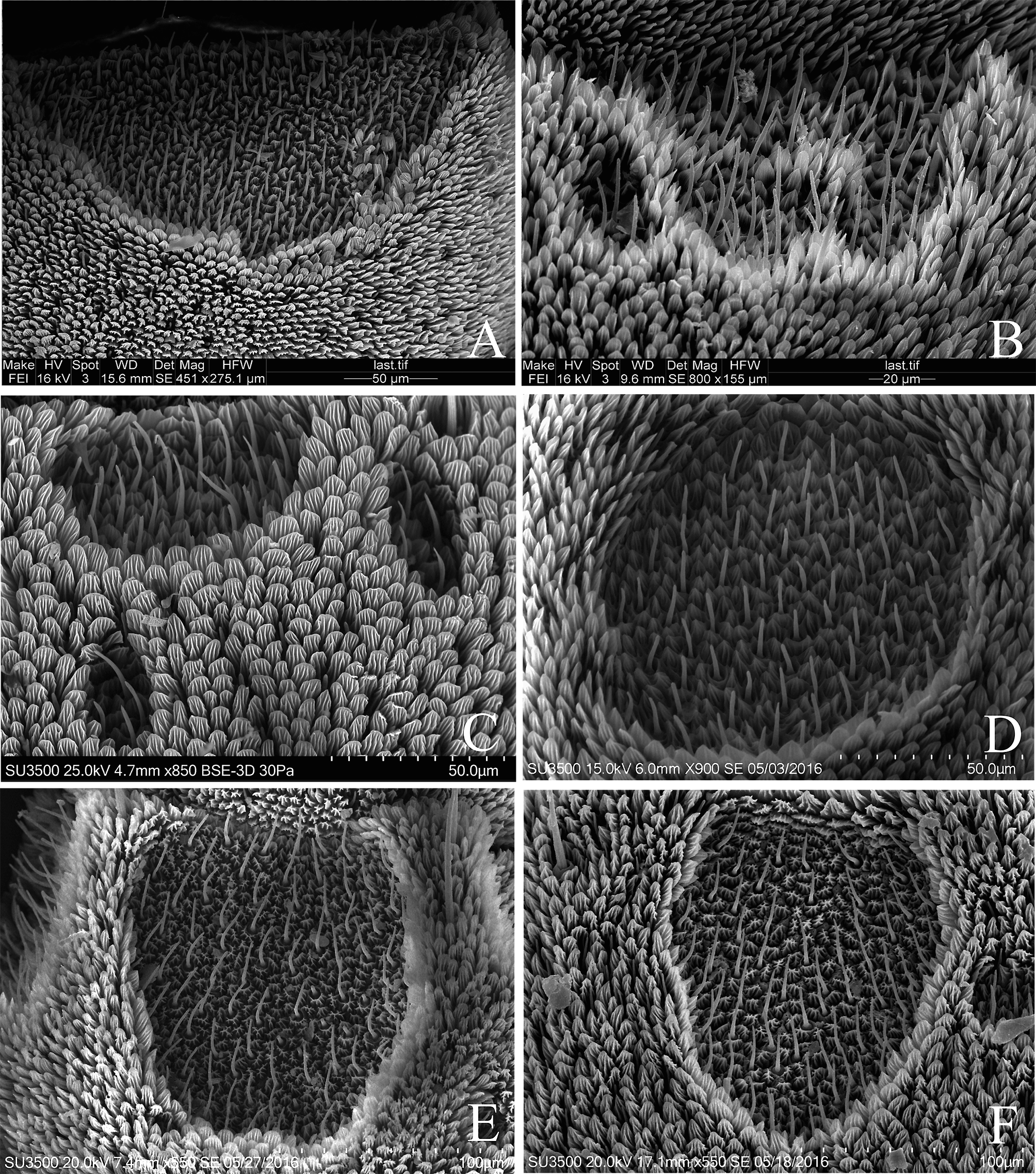
MICROTRICHIA: We observed m1 and m2 types. The ratio in the sulci is one sensillum for every three or four microtrichia.
TRICHOID SENSILLA: The stems of these sensilla are 19–22 µm in length. Apparently they do not have pores, and only in M. p. patricia were a few streaks observed in the cuticular wall, particularly toward the apex. The cuticular ring is independent of the microtrichia or they are partially fused.
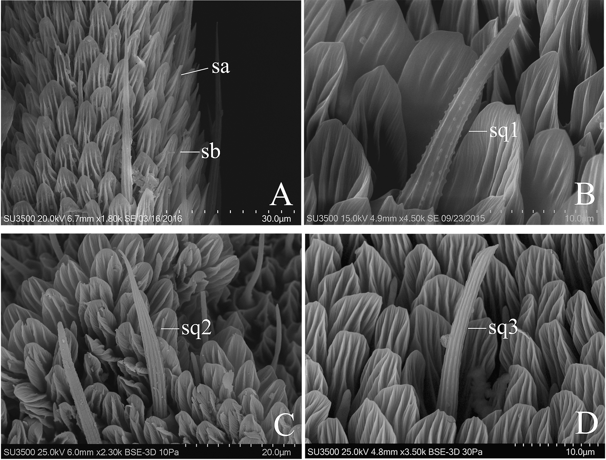
CHAETIC SENSILLA: We found sq1 and sq2 types; the sq1 are like those of Pseudopontia but longer; they are also thinner than those in other genera of the tribe. We note three chaetic sensilla sq1 next to the central sulcus (two near the distal edge and the other toward the medial antennomere) and one sensillum below each lateral sulcus for a total of five in the ventral surface. The length of the sq1 is 38–66 µm; M. e. xanthella has the longest and M. p. patricia has the shortest sensilla. The chaetic sq2 sensilla are present in the first antennomere in M. e. xanthella and M. i. ithomia ; in the first two in M. p. monica , and in almost all in M. p. patricia . The sq2 are shorter than sq1 and measure 29–32 µm ( Fig. 15C View FIGURE 15 ). The sq1 are concentrated on the apex of the distal antennomere ( M. p. monica ) and on the first antennomere in M. p. patricia . Their length is 45–66 µm. The sensilla sq1 are abundant in the antennomere before the first scaleless one, but show no order.
OTHER SENSILLA: Basiconic sensilla, especially auriculate, are very common and abundant. Coeloconic sensilla sc1 are present, particularly at the apex of the distal antennomere; these sensilla is longer than those in the other genera of the tribe. The ni3 are amid the lateral sulci and are very rare.
No known copyright restrictions apply. See Agosti, D., Egloff, W., 2009. Taxonomic information exchange and copyright: the Plazi approach. BMC Research Notes 2009, 2:53 for further explanation.