
Musserakis sulawesiensis, Hasegawa & Dewi & Asakawa, 2014
|
publication ID |
https://doi.org/ 10.11646/zootaxa.3881.2.4 |
|
publication LSID |
lsid:zoobank.org:pub:679028CD-1D89-4488-BD90-FD78956D1CAF |
|
DOI |
https://doi.org/10.5281/zenodo.5593183 |
|
persistent identifier |
https://treatment.plazi.org/id/674E87E4-FFAD-FFC8-D1FF-FA78FD7A4BED |
|
treatment provided by |
Felipe |
|
scientific name |
Musserakis sulawesiensis |
| status |
sp. nov. |
Musserakis sulawesiensis sp. n.
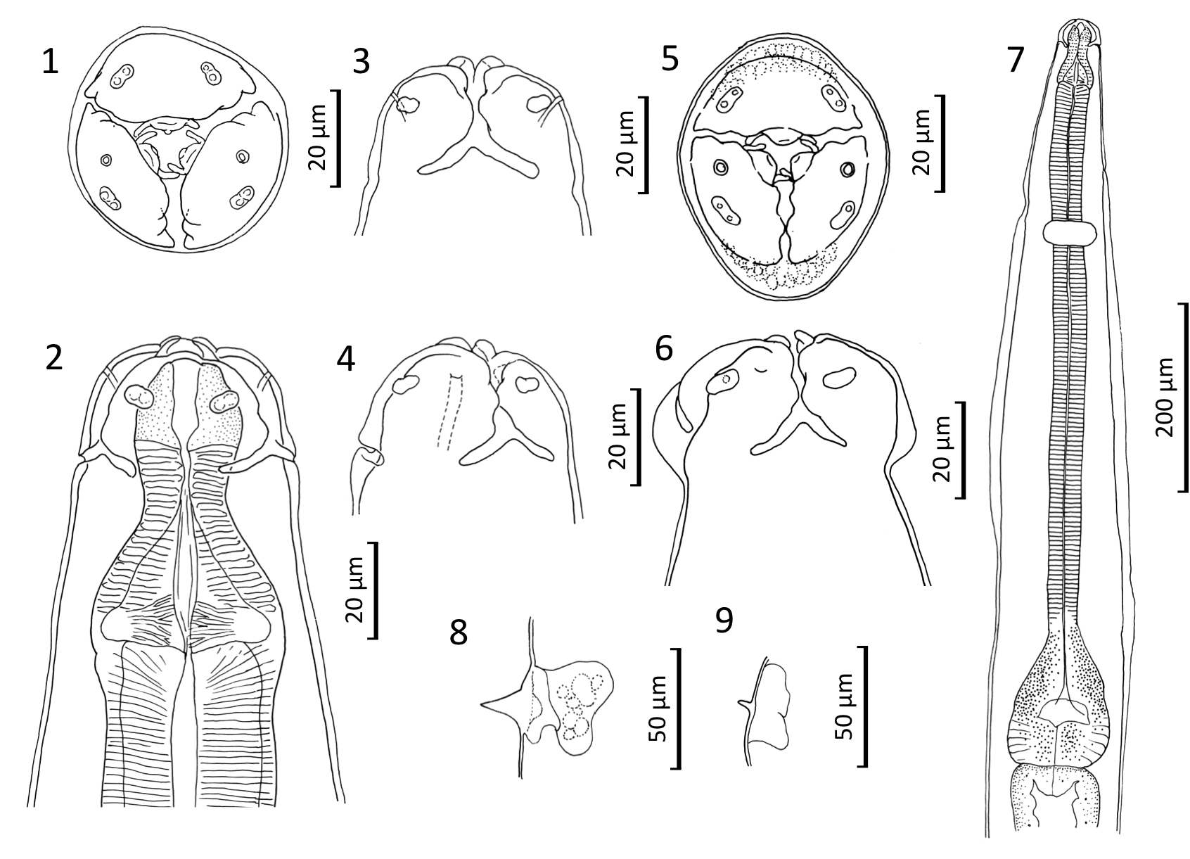
( Figs. 1–22 View FIGURES 1–6 View FIGURES 10–14 View FIGURES 19–22 )
General. Small nematodes. Cephalic extremity with characteristics defined above ( Figs. 1–4 View FIGURES 1–6 , 19–21 View FIGURES 19–22 ). Some worms with cephalic end dorso-ventrally elongated, giving inflated appearance in lateral view ( Figs. 5, 6 View FIGURES 1–6 , 22 View FIGURES 19–22 ). Anterior body usually bent dorsally ( Figs. 10 View FIGURES 10–14 , 15). Lips with weak lateral projections ( Figs. 1–6 View FIGURES 1–6 , 19–22 View FIGURES 19–22 ). Each proximal lobe of pharyngostome forming onchium with side projections ( Figs. 1, 5 View FIGURES 1–6 , 19–22 View FIGURES 19–22 ). Esophagus long and slender (Figs. 7, 10, 15). Isthmus not clearly defined. Esophageal bulb divided into anterior and posterior portions (Fig. 7). Cervical alae triangular, strongly pointed, commencing anterior to nerve ring, continuing to small, thin lateral alae (Figs. 7–9). Deirids not discernible. Somatic papillae absent.
Male (holotype and 10 paratypes): Worm length 2.91 (2.61–3.47) [3.02] mm, width in midbody 131 (115–131) [125]. Total esophagus including pharynx 712 (632–755) [722] long, i.e., 24.5 (21.5–26.2) [24.0] % of worm length (WL); pharynx 64 (56–70) [63] long, combined length of corpus and isthmus 520 (452–569) [530] long by 32 (30–38) [35] wide, bulb 128 (122–138) [128] long by 96 (83–96) [89] wide. Nerve ring 205 (202–234) [221] and excretory pore 320 (275–346) [312] from cephalic apex. Sucker, 32 (30–43) [35] in diameter, 85 (56–96) [86] in front of cloaca. Spicules equal, longitudinally striated faintly, variable in width and length, 227 (167–263) [218] long, i.e., 7.8 (5.2–9.3) [7.3] % of WL. In holotype and 5 paratypes, spicules stout, bent ventrally, flattened distally, often protruded from cloacal aperture ( Figs. 11–13 View FIGURES 10–14 ). In 5 paratypes spicules thin, tapered and winding distally, not projecting from cloacal aperture ( Fig. 14 View FIGURES 10–14 ). Ten pairs of caudal papillae present: 2 pairs slender, aside of sucker; 2 sessile large papillae lateral to cloacal aperture and 3 stout long papillae, forming cloacal group, supporting caudal alae; 3 small sessile pairs in middle of tail ( Figs. 11, 12 View FIGURES 10–14 ). Tail slender, ending in pointed tip, 237 (212–247) [228] long ( Figs. 11, 12 View FIGURES 10–14 ).
Female (Allotype and 10 paratypes): Worm length 3.3 (3.2–3.5) [3.4] mm, width in midbody 138 (128–147) [133]. Total esophagus including pharynx 731 (712–811) [759] long, i.e., 22.2 (21.4–24.1) [22.7] % of WL; pharynx 59 (58–67) [62] long, corpus 550 (517–628) [571] long by 40 (32–43) [37] wide, bulb 122 (122–134) [127] long by 88 (83–102) [96] wide. Nerve ring 218 (208–249) [222] and excretory pore 330 (282–335) [307] from cephalic apex. Vulva 1.92 (1.87–2.09) [2.00] mm from cephalic apex, i.e., 58.2 (57.5–62.2) [59.4] % of WL; opening in depression with small diverticulum; perivulval cuticle swollen, one low tubercle present at ca. 100 posterior to vulva (Figs. 15, 16). Vagina thick, muscular, strongly anteriorly curved, then abruptly recurved as ovejector, running posteriorly far beyond vulval level to join uteri (Fig. 16). Anterior and posterior ovaries ending at posterior to esophago-intestinal junction and anal level, respectively (Figs. 15, 17). Tail slender, ending in pointed tip, 448 (363–476) [429] long, i.e., 13.6 (10.9–14.1) [12.8] % of WL (Fig. 17). Eggs ellipsoidal, thick shelled, with round concave ornamentations on surface, unembryonated at deposition, 59–72 [68] by 40–48 [45] (Fig. 18).
Taxonomic summary.
Type host: Echiothrix centrosa Miller & Hollister, 1921 (large-bodied shrew rat)
Type locality: Kuala Navusu , Malakasa, Central Sulawesi, Indonesia .
Prevalence and intensity: All of 5 E. centrosa harbored numerous individuals.
Specimens deposited: USNM 1251671 View Materials (holotype male and allotype female), 1251672 (10 male and 10 female paratypes); Voucher specimens USNM 1251673–1251676 View Materials , MZB Na 646 .
Coparasites: Trichuris sp. (Site cecum; prevalence 4/5; intensity 1-5), Heligmonellidae gen. sp. (small intestine; 1/5; 1), Ascarididae gen. sp. (larva) (abdominal cavity; 1/5; 1) and Rhigonematidae gen. sp. (cecum; 1/5; 1).
Symbiotypes: AMNH 225678–225681, 225685.
Etymology. Generic name is dedicated to Dr. G. G. Musser, an outstanding mammalogist, who has made invaluable contributions on the murid rodents of Sulawesi for many years. The species epithet is named after the locality.
Remarks. By having 3 well defined lips, an esophagus with valved bulb, thick shelled eggs, a preanal sucker in male, Musserakis belongs to the superfamily Heterakoidea ( Chabaud, 1974) . Because the lips are round and not connected by lateral lobes, it is assigned to the family Heterakidae ( Chabaud, 1978) . By having interlabia and only limited number of sessile papillae on male tail, it is included in the subfamily Heterakinae ( Chabaud, 1978) . Five genera are currently recognized in this subfamily: Heterakis Dujardin, 1945 , Haroldakis Inglis, 1991 , Neoheterakis Kumar & Thienpoint, 1974 , Pseudoaspidodera Baylis & Daubney, 1922, Odontoterakis Skrjabin & Schikhobalova, 1947 ( Chabaud, 1978; Inglis, 1991a; Gibbons, 2010). Musserakis resembles Haroldakis and Odontoterakis by having non-recurrent and non-anastomosing cephalic cordons, whereas it differs clearly from Heterakis , which lacks cordons, and Neoheterakis and Pseudoaspidodera, which possess recurrent cephalic cordons ( Chabaud, 1978; Inglis, 1991a; Gibbons, 2010). Musserakis is readily distinguished from Haroldakis , which has 5 teeth on each proximal end of pharyngostome, transverse cushion with 2 sessile papillae between sucker and cloaca, and 2 sessile papillae on posterior cloacal lip in male ( Inglis, 1991a). It also differs from Odontoterakis , which has 1 pair of additional thin pedunculate papillae between papillae groups around sucker and cloaca ( Inglis, 1991a).
FIGURES 15–18. Female of Musserakis sulawesiensis gen. et sp. n. 15. Allotype, left lateral view. 16. Vulval portion, left lateral view. 17. Tail, left lateral view. 18. Uterine egg.


| MZB |
Museum Zoologicum Bogoriense |
No known copyright restrictions apply. See Agosti, D., Egloff, W., 2009. Taxonomic information exchange and copyright: the Plazi approach. BMC Research Notes 2009, 2:53 for further explanation.