
Mycale (Mycale) arenaria Hajdu & Desqueyroux-Faúndez, 1994
Van, Rob W. M., 2017, Sponges of the Guyana Shelf, Zootaxa 1, pp. 1-225 : 156-157
|
publication ID |
https://doi.org/ 10.5281/zenodo.272951 |
|
publication LSID |
lsid:zoobank.org:pub:6D68A019-6F63-4AA4-A8B3-92D351F1F69B |
|
DOI |
https://doi.org/10.5281/zenodo.5698716 |
|
persistent identifier |
https://treatment.plazi.org/id/03A80010-77CC-FF31-FF14-A75791FEFF62 |
|
treatment provided by |
Plazi |
|
scientific name |
Mycale (Mycale) arenaria Hajdu & Desqueyroux-Faúndez, 1994 |
| status |
|
Mycale (Mycale) arenaria Hajdu & Desqueyroux-Faúndez, 1994 View in CoL
Figures 97 View FIGURE 97 a–d, 98a–g
Mycale fusca View in CoL ; Solé-Cava et al. 1981: 132 (Not: Esperella fusca Ridley & Dendy, 1886 ).
Mycale arenosa Hajdu & Boury-Esnault, 1991: 506 View in CoL , figs 1–11 (Not: Mycale parasitica var. arenosa Hentschel, 1911 View in CoL ). Mycale arenaria Hajdu & Desqueyroux-Faúndez, 1994: 568 View in CoL (Not: Moraes 2011: 146, no sigmas, surface skeleton is characteristic for subgenus Aegogropila ).
Material examined. RMNH Por. 9994, Suriname, ‘ Snellius O.C.P.S. ’ Guyana Shelf Expedition, station F40, 7.0033°N 56.4417°W, depth 59 m, bottom sand, 6 May 1966 GoogleMaps .
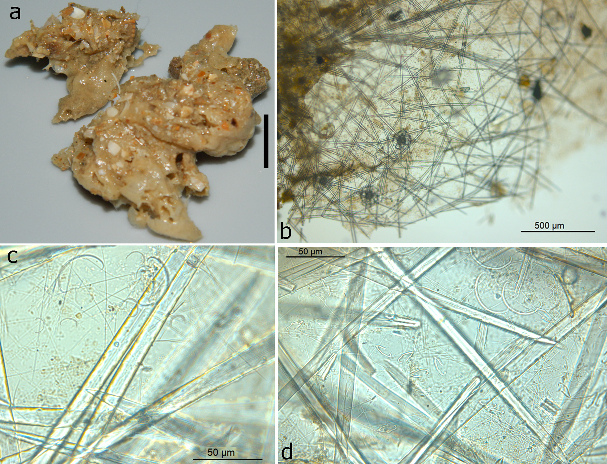
Description. Irregularly massive sponge ( Fig. 97 View FIGURE 97 a), encrusting and consolidating shell debris on soft bottom substratum. Size 4 x 4.5 x 2 cm. Surface smooth but irregularly grooved and lobate. Several oscular openings are present, flush with the surface, 2–4 mm in diameter. Consistency soft, cavernous, but resilient.
Skeleton. ( Figs 97 View FIGURE 97 b–d) The surface skeleton is a tangential layer of single intercrossing megascleres ( Fig. 97 View FIGURE 97 b). Rosettes of the largest anisochela category ( Fig. 97 View FIGURE 97 b) are common, approximately 150 µm in diameter, consisting of 20+ spicules. The surface membrane in between the megascleres is charged with with scattered sigmas and anisochelae ( Fig. 97 View FIGURE 97 c), and very numerous trichodragmas and single raphides ( Fig. 97 View FIGURE 97 d). The choanosomal skeleton consists of strong, plumose bundles of megascleres, which fan out in the subectosomal region carrying the surface skeleton. Megasclere bundles at the base approximately 1 mm in diameter, thinning out towards the surface where they comprise 3–7 spicules in cross section.
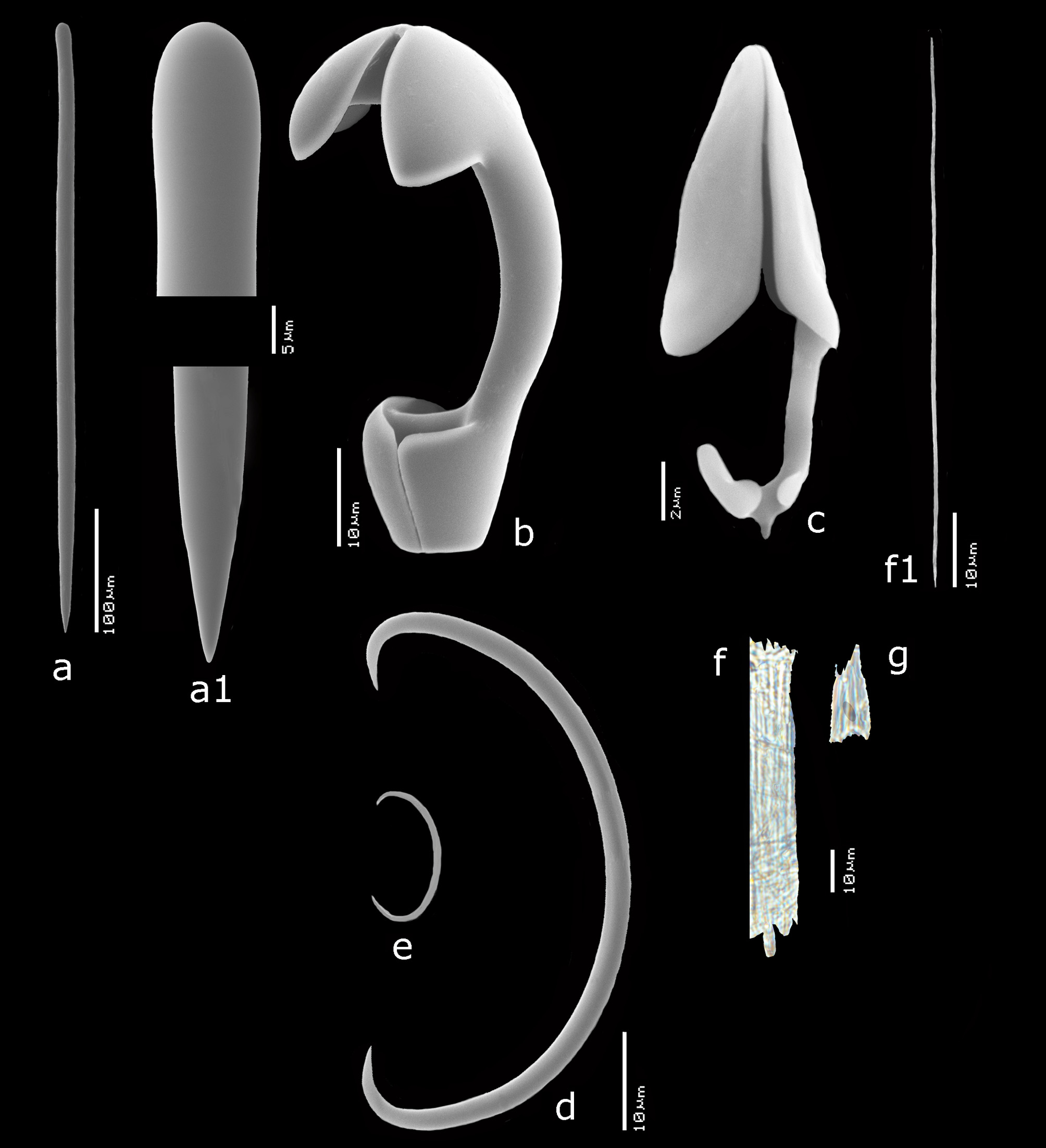
Spicules. ( Figs 98 View FIGURE 98 a–g) Styles, anisochelae, sigmas, trichodragmas.
Styles ( Figs 98 View FIGURE 98 a,a1), mycalostyles, fusiform, with faint constriction beneath the rounded heads, the opposite end sharply pointed, 468– 620 –744 x 12 – 15.4 –20 µm.
Anisochelae I ( Fig. 98 View FIGURE 98 b), robust, with short alae, 50– 55.9 –63 µm.
Anisochelae III ( Fig. 98 View FIGURE 98 c), spurred, with upper alae long, in a large size range, possibly divisible in a larger and a smaller size category, overall 16– 19.4 –24 µm.
Sigmas, thin, with slightly incurved apices, in two distinct size categories, (1) larger ( Fig. 98 View FIGURE 98 d), very common, 32– 40.4 –53 µm, and (2) smaller ( Fig. 98 View FIGURE 98 e), less common, 12– 13.2 –15 µm.
Raphides in trichodragmas, extremely common, in two size categories, (1) larger ( Fig. 98 View FIGURE 98 f), 61– 74.5 – 93 x 11 – 12.4 –15 µm (individual raphides ( Fig. 98 View FIGURE 98 f1) less than 0.5 µm thick), and (2) smaller ( Fig. 98 View FIGURE 98 g), 17– 26.1 – 33 x 9 – 10.8 –12 µm.
Distribution and ecology. Guyana Shelf, SE Brazil, soft substratum, 3– 59 m.
Remarks. The similarity to the Brazilian type material extensively described by Hajdu & Boury-Esnault (1991) and Hajdu & Desqueyrouz-Faúndez (1994) is convincing. However, there are a few differences: the type apparently has two categories of megascleres, which I cannot confirm from the present specimen; nevertheless the overall sizes given for the type are the same as for the present specimen. Hajdu & Boury-Esnault do not report the occurrence of two size categories of sigmas, but since these were fairly rare and very thin, it is possible that either category was overlooked. The presence of a rare anisochela II category of 40 µm is reported for the type, while in the present specimen there is at most a possible additional size category of anisochelae III of about 23–24 µm, which seems different from most other anisochelae by having wider alae. Finally, there is a distinct division into larger and smaller trichodragmas in the present specimen, not reported for the type material.
In view of the considerable overall similarity of the present material and Hajdu & Boury-Esnault’s material, I consider them both the same species for the time being.
Moraes’ (2011) record of this species is suspect as he did not mention sigmas and figured an Aegogropila surface skeleton.
A closely related species is Mycale (Mycale) alagona Cedro, Hajdu & Correia, 2011 from SE Brazil. It shows the same general similarity with my specimen as with the type of M. (M.) arenaria (curved, short alae in anisochela I, dense aggregation of trichodragmas). Differences are three instead of two anisochelae categories and three size categories of sigmas.
| RMNH |
National Museum of Natural History, Naturalis |
No known copyright restrictions apply. See Agosti, D., Egloff, W., 2009. Taxonomic information exchange and copyright: the Plazi approach. BMC Research Notes 2009, 2:53 for further explanation.
|
Kingdom |
|
|
Phylum |
|
|
Class |
|
|
Order |
|
|
Family |
|
|
Genus |
Mycale (Mycale) arenaria Hajdu & Desqueyroux-Faúndez, 1994
| Van, Rob W. M. 2017 |
Mycale arenosa
| Hajdu 1994: 568 |
| Hajdu 1991: 506 |
Mycale fusca
| Sole-Cava 1981: 132 |